. 2016 Jul 25;17(8):1200.
doi: 10.3390/ijms17081200.

Non-Ligand-Induced Dimerization is Sufficient to Initiate the Signalling and Endocytosis of EGF Receptor

Affiliations

Non-Ligand-Induced Dimerization is Sufficient to Initiate the Signalling and Endocytosis of EGF Receptor

George Kourouniotis et al. Int J Mol Sci. .

Abstract

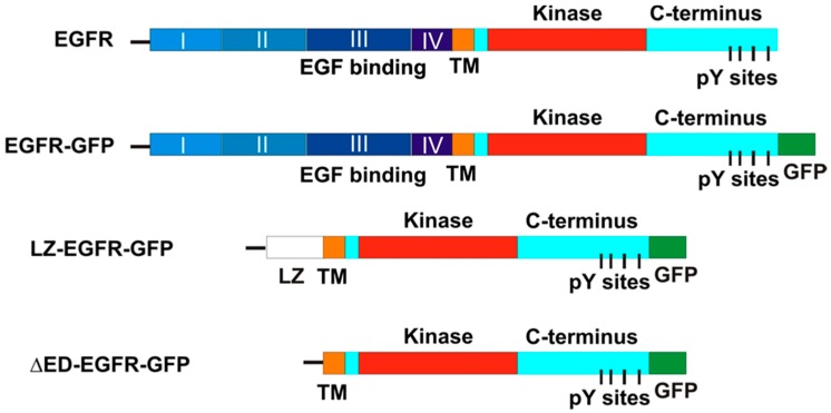
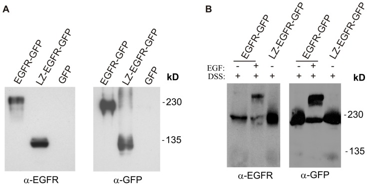
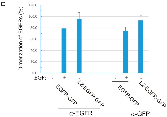
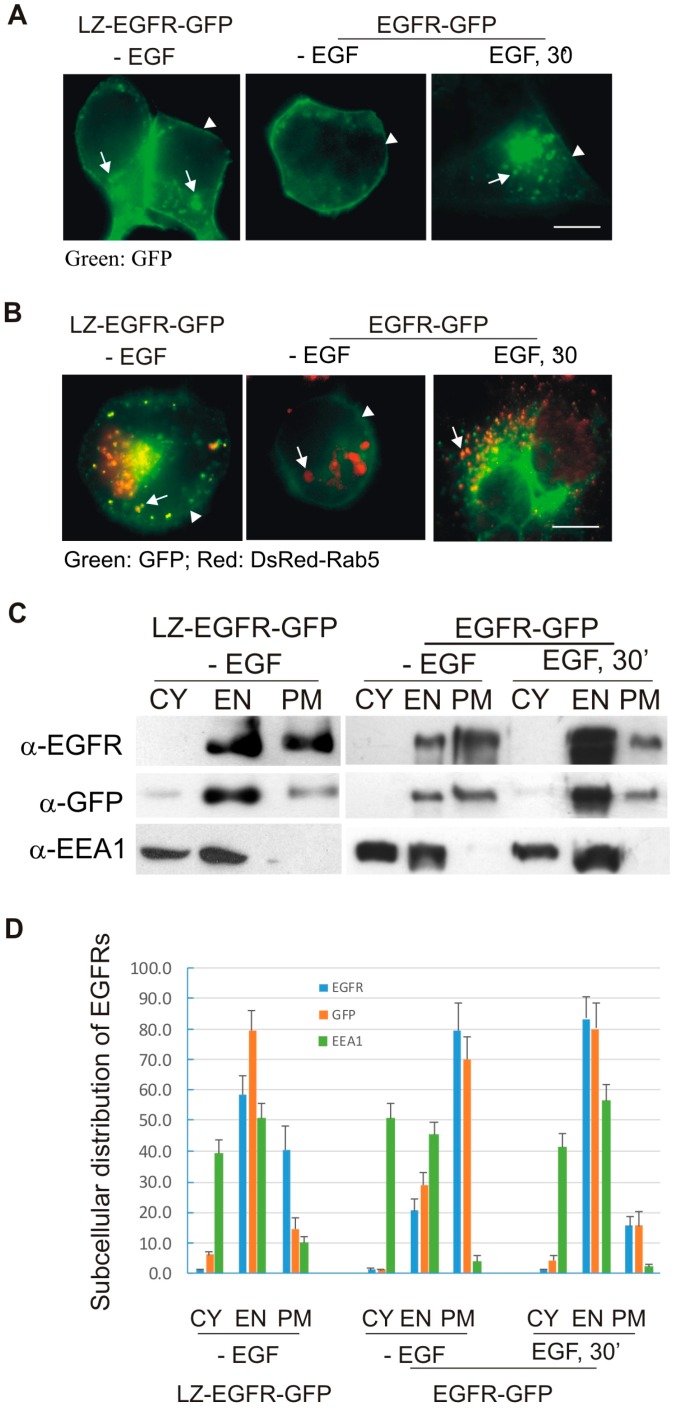
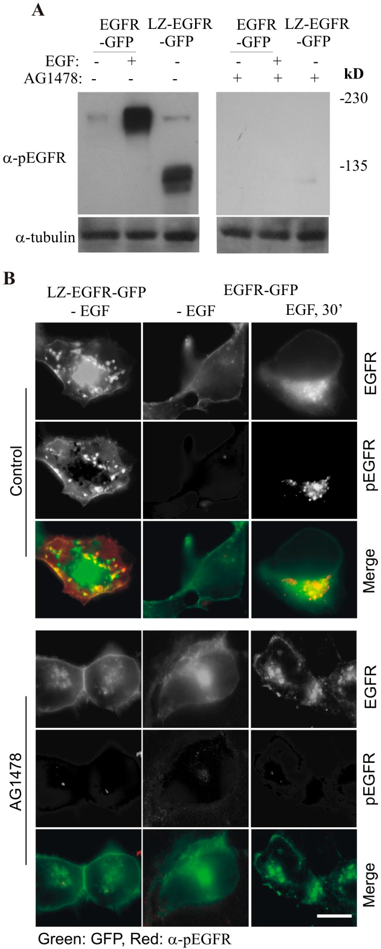
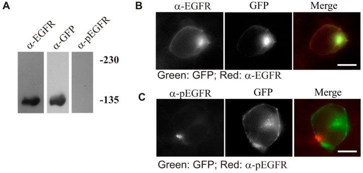
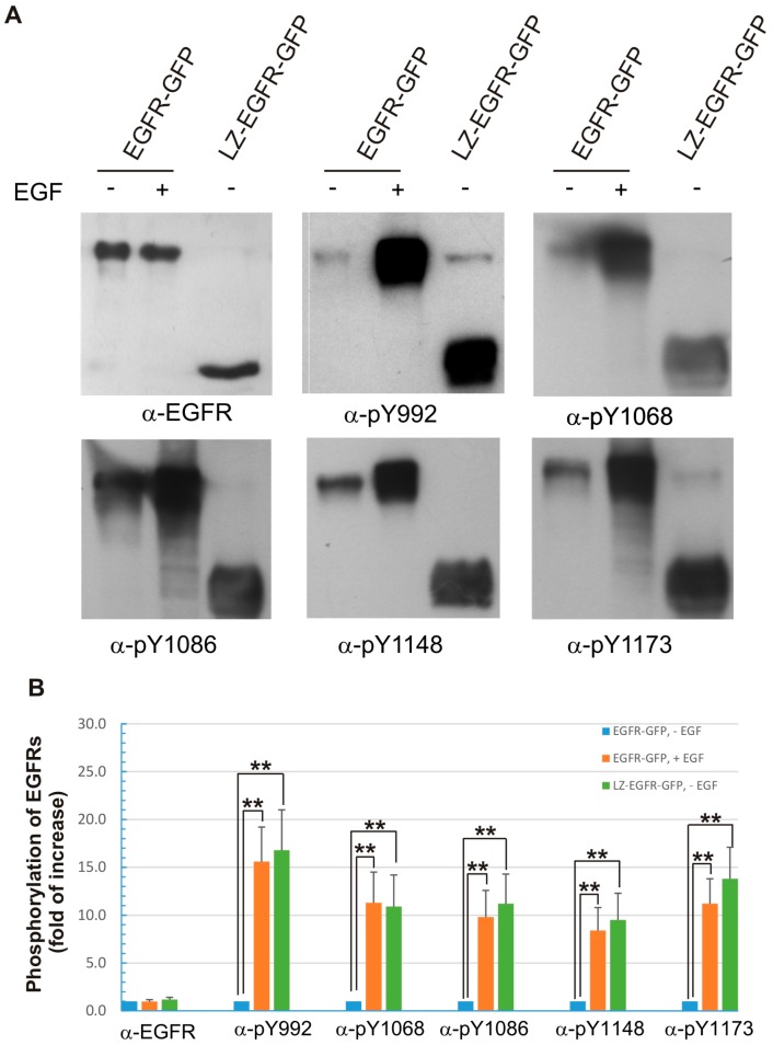
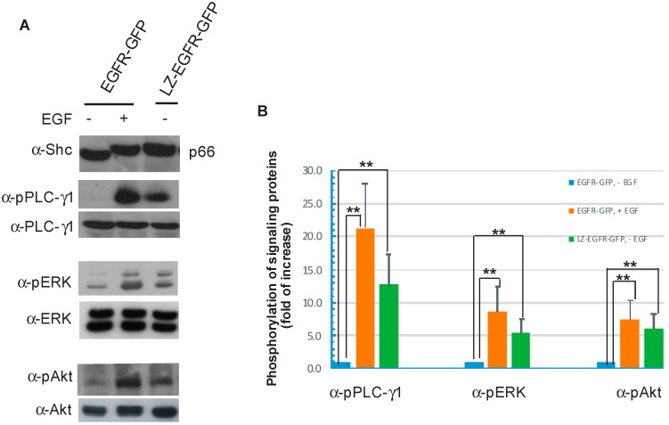
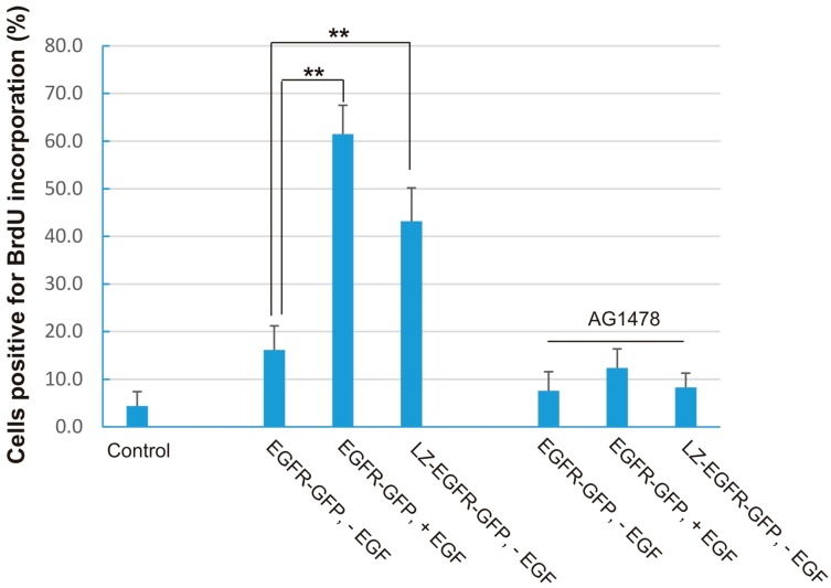
The binding of epidermal growth factor (EGF) to EGF receptor (EGFR) stimulates cell mitogenesis and survival through various signalling cascades. EGF also stimulates rapid EGFR endocytosis and its eventual degradation in lysosomes. The immediate events induced by ligand binding include receptor dimerization, activation of intrinsic tyrosine kinase and autophosphorylation. However, in spite of intensified efforts, the results regarding the roles of these events in EGFR signalling and internalization is still very controversial. In this study, we constructed a chimeric EGFR by replacing its extracellular domain with leucine zipper (LZ) and tagged a green fluorescent protein (GFP) at its C-terminus. We showed that the chimeric LZ-EGFR-GFP was constitutively dimerized. The LZ-EGFR-GFP dimer autophosphorylated each of its five well-defined C-terminal tyrosine residues as the ligand-induced EGFR dimer does. Phosphorylated LZ-EGFR-GFP was localized to both the plasma membrane and endosomes, suggesting it is capable of endocytosis. We also showed that LZ-EGFR-GFP activated major signalling proteins including Src homology collagen-like (Shc), extracellular signal-regulated kinase (ERK) and Akt. Moreover, LZ-EGFR-GFP was able to stimulate cell proliferation. These results indicate that non-ligand induced dimerization is sufficient to activate EGFR and initiate cell signalling and EGFR endocytosis. We conclude that receptor dimerization is a critical event in EGF-induced cell signalling and EGFR endocytosis.

Keywords: EGF receptors; dimerization; endocytosis; leucine zipper; signal transduction.


Figures

Figure 1
Figure 1
Schematic illustration of EGFR-GFP, LZ-EGFR-GFP and ΔED-(extracellular-domain deleted)-EGFR-GFP as compared to wild type EGFR (wtEGFR). The chimeric construct LZ-EGFR-GFP started with the signal sequence of wtEGFR (line), followed by a LZ sequence that replaced the complete extracellular domain of EGFR. ΔED-EGFR-GFP is a truncated EGFR with the deletion of its entire extracellular domain. The green fluorescent protein (GFP) is tagged to the C-terminus of EGFR.
Figure 2
Figure 2
Expression and dimerization of LZ-EGFR-GFP. 293T cells were transiently transfected with LZ-EGFR-GFP, EGFR-GFP or empty vector (GFP). (A) EGFR-GFP and LZ-EGFR-GFP were visualized by immunoblotting of the total lysates with antibodies to either EGFR or GFP; (B) Cells were crosslinked with disulfosuccinimidyl suberate (DSS). Both LZ-EGFR-GFP monomer (105 kD) and dimer (210 kD) were visible in the resulting immunoblots. EGFR-GFP dimerized only after EGF stimulation, as expected; (C) Quantification of the data from (B). The band is quantitated by densitometry with image J software (National Institute of Health, Bethesda, MD, USA) and the receptor dimerization was expressed as the percentage of dimers among the total receptor proteins. Each value is the mean of at least three independent experiments and the error bar represents the standard error.
Figure 2
Figure 2
Expression and dimerization of LZ-EGFR-GFP. 293T cells were transiently transfected with LZ-EGFR-GFP, EGFR-GFP or empty vector (GFP). (A) EGFR-GFP and LZ-EGFR-GFP were visualized by immunoblotting of the total lysates with antibodies to either EGFR or GFP; (B) Cells were crosslinked with disulfosuccinimidyl suberate (DSS). Both LZ-EGFR-GFP monomer (105 kD) and dimer (210 kD) were visible in the resulting immunoblots. EGFR-GFP dimerized only after EGF stimulation, as expected; (C) Quantification of the data from (B). The band is quantitated by densitometry with image J software (National Institute of Health, Bethesda, MD, USA) and the receptor dimerization was expressed as the percentage of dimers among the total receptor proteins. Each value is the mean of at least three independent experiments and the error bar represents the standard error.
Figure 3
Figure 3
Subcellular distribution of LZ-EGFR-GFP. (A,B) Fluorescence analysis of subcellular EGFR localization. 293T cells were transiently transfected with LZ-EGFR-GFP or EGFR-GFP (A), or co-transfected with DsRed-Rab5 and LZ-EGFR-GFP or EGFR-GFP (B). The cells were treated with or without EGF. The subcellular localization of EGFR and Rab5 was revealed by the intrinsic fluorescence of GFP and DsRed. Co-localization of EGFR and Rab5 was indicated by yellow. Arrows denote endosomes and arrowheads denote plasma membrane regions. Size bar = 20 µm; (C) Subcellular fractionation and immunoblotting analysis. 293T cells transiently expressing LZ-EGFR-GFP were homogenized and subcellularly fractionated into cytoplasmic (CY), endosomal (EN) and plasma membrane (PM) fractions, confirmed by immunobloting the cooresponding fraction lysates with antibodies to EGFR, GFP and early endosome antigen 1 (EEA-1), respectively. 293T cells transfected with EGFR-GFP were used as controls; (D) Quantification of the data from (C). Bands were quantitated by densitometry with image J software and subcellular distribution of the proteins among the three fractions (CY, EN, and PM) was expressed as percentage of the total protein content of all three fractions combined.Each value is the average of at least three independent experiments and the error bar is the standard error.
Figure 4
Figure 4
Phosphorylation of LZ-EGFR-GFP and the dependence on intrinsic tyrosine kinase activity. (A) Immunoblotting. 293T cells were transiently transfected with EGFR-GFP or LZ-EGFR-GFP. The cells were serum starved for 24 h and then were treated with EGF and/or AG1478 as indicated. Cell lysates were subjected to immunoblotting analysis with mouse anti-pEGFR antibody; (B) Immunofluorescence. 293T cells transiently transfected with either LZ-EGFR-GFP or EGFR-GFP were serum starved for 24 h. The cells were then treated with EGF and/or AG1478 as indicated. EGFR phosphorylation was examined by anti-pEGFR antibody followed by the secondary antibody conjugated with TRITC. Co-localization (yellow) of LZ-EGFR-GFP or EGFR-GFP (green) with p-EGFR (red) was determined by indirect immunofluorescence. Size bar = 20 µm.
Figure 5
Figure 5
Expression, phosphorylation and subcellular localization of ΔED-EGFR-GFP. 293T cells were transiently transfected with ΔED-EGFR-GFP. (A) The expression and phosphorylation of ΔED-EGFR-GFP. Following the transfection for 48 h, the cell were lysed and total cell lysates were used to determine the expression and phosphorylation of ΔED-EGFR-GFP by immunoblotting; (B) Subcellular distribution of ΔED-EGFR-GFP. Following the transfection for 48 h, the subcellular localization of ΔED-EGFR-GFP was revealed by the intrinsic GFP and by anti-EGFR antibody followed by TRITC-conjugated secondary antibody; (C) The phosphorylation of ΔED-EGFR-GFP. Following the transfection for 48 h, the phosphorylation of ΔED-EGFR-GFP was revealed by anti-pEGFR antibody followed by TRITC-conjugated secondary antibody. Size bar = 20 µm.
Figure 6
Figure 6
Phosphorylation of the five major C-terminal tyrosine residues of EGFR-GFP and LZ-EGFR-GFP. (A) 293T cells were transiently transfected with EGFR-GFP or LZ-EGFR-GFP. Following serum starvation for 24 h, cells were treated with or without EGF. The cell lysates were subjected to immunoblotting analysis with rabbit anti-pEGFR (pY992), anti-pEGFR (pY1068), anti-pEGFR (pY1086), anti-pEGFR (pY1148) and anti-pEGFR (pY1173) antibodies; (B) Quantification of the data from (A). The band is quantitated by densitometry with image J software. The phosphorylation level of the control (EGFR-GFP, without EGF treatment) was set to 1 and the phosphorylation of the receptors under other conditions was expressed as the fold increase compared to control. Each value is the average of at least three independent experiments and the error bar is the standard error. **: p < 0.01.
Figure 7
Figure 7
Stimulation of various signal transduction pathways by activation of EGFR-GFP or LZ-EGFR-GFP. (A) 293T cells were transiently transfected with EGFR-GFP or LZ-EGFR-GFP. Following serum starvation for 24 h, cells were treated with or without EGF. The cell lysates were subjected to immunoblotting analysis with rabbit anti-SHC, rabbit anti-phospho-PLC-γ1, rabbit anti-PLC-γ1, mouse anti-phospho-ERK1/2, mouse anti-Erk1/2, rabbit anti-phospho-Akt and rabbit anti-Akt antibodies; (B) Quantification of the data from (A). The band is quantitated by densitometry with image J software. The protein phosphorylation level of the control (EGFR-GFP, without EGF treatment) was set to 1 and the phosphorylation of the proteins under other conditions was expressed as the fold increase compared to control. Each value is the average of at least three independent experiments and the error bar is the standard error. **: p < 0.01.
Figure 8
Figure 8
Stimulation of DNA synthesis by LZ-EGFR-GFP. 293T cells were transiently transfected with EGFR-GFP or LZ-EGFR-GFP. Following serum starvation for 24 h, cells were treated with EGF and/or AG1478 as indicated. DNA synthesis was determined by BrdU incorporation as described in the Materials and methods. Cells were counted at 300 per sample and data was plotted as the mean of at least three experiments. The error bar is the standard error. **: p < 0.01.

References

    1. Sorkin A., Goh L.K. Endocytosis and intracellular trafficking of ErbBs. Exp. Cell Res. 2009;315:683–696. doi: 10.1016/j.yexcr.2008.07.029. - DOI - PubMed
    1. Lemmon M.A., Schlessinger J. Cell signaling by receptor tyrosine kinases 1. Cell. 2010;141:1117–1134. doi: 10.1016/j.cell.2010.06.011. - DOI - PMC - PubMed
    1. Pawson T. Protein modules and signalling networks. Nature. 1995;373:573–580. doi: 10.1038/373573a0. - DOI - PubMed
    1. Pawson T. New impressions of SRC and HCK [news; comment] Nature. 1997;385:582–583. doi: 10.1038/385582b0. - DOI - PubMed
    1. Yarden Y., Sliwkowski M.X. Untangling the ErbB signalling network. Nat. Rev. Mol. Cell Biol. 2001;2:127–137. doi: 10.1038/35052073. - DOI - PubMed

LinkOut - more resources