. 2016 Jul 27;12(7):e1005801.
doi: 10.1371/journal.ppat.1005801. eCollection 2016 Jul.

KSHV-Mediated Regulation of Par3 and SNAIL Contributes to B-Cell Proliferation

Affiliations

KSHV-Mediated Regulation of Par3 and SNAIL Contributes to B-Cell Proliferation

Hem C Jha et al. PLoS Pathog. .

Expression of concern in

Abstract

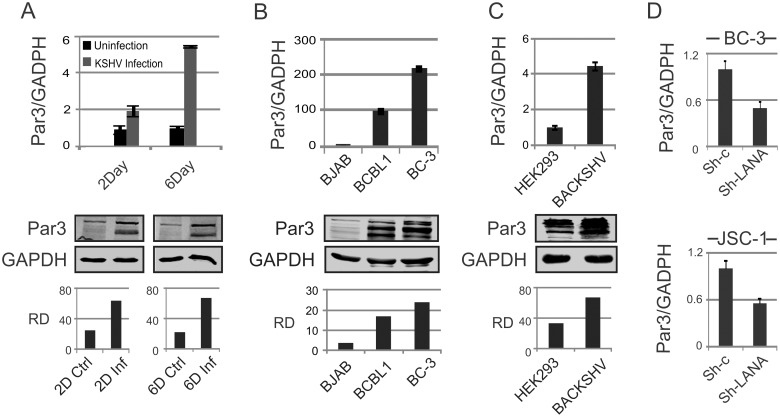
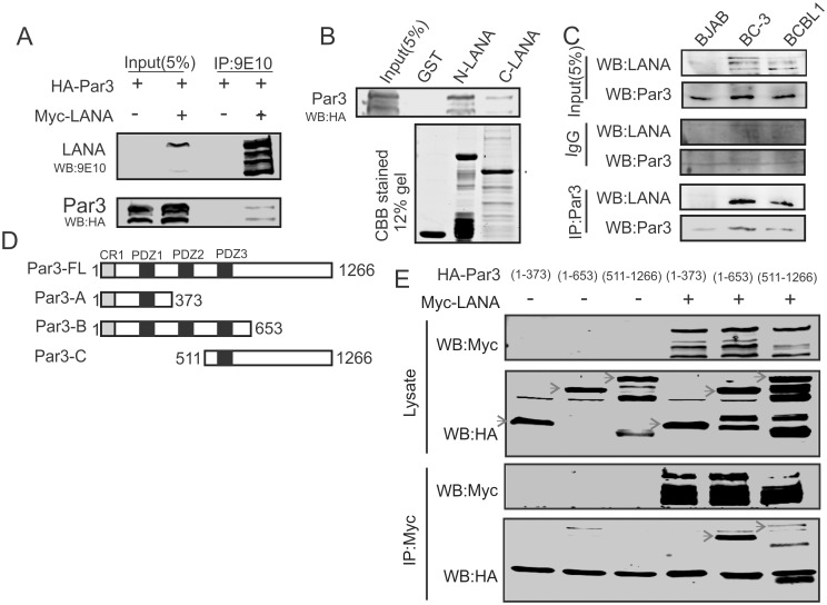
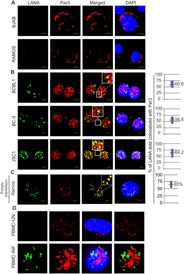
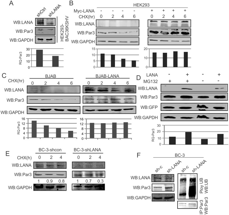
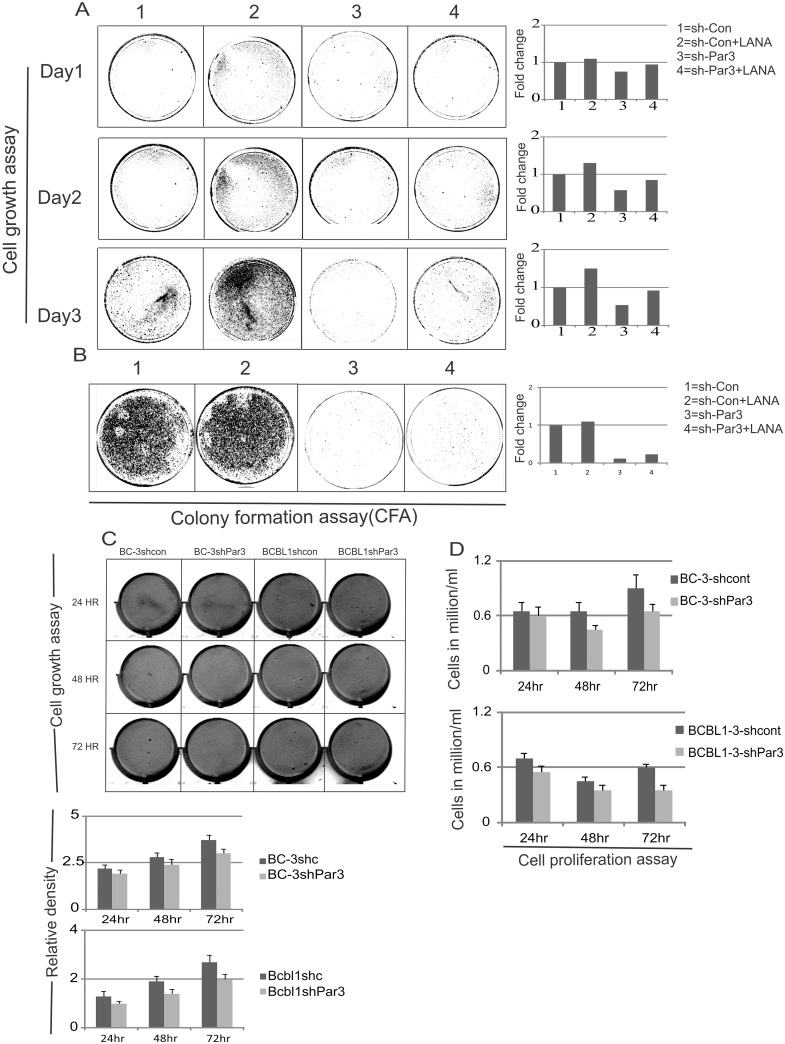
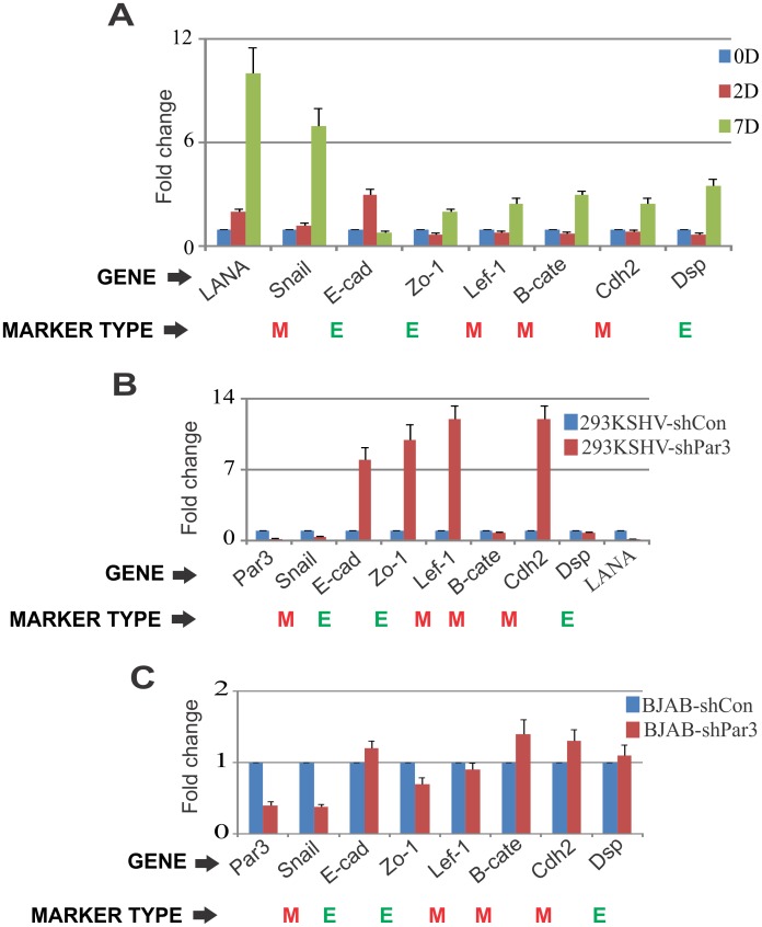
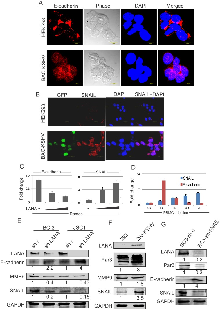
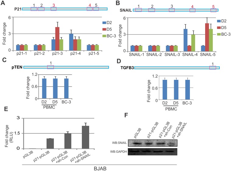
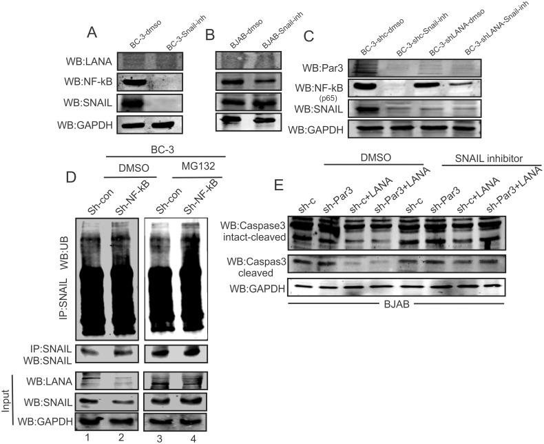
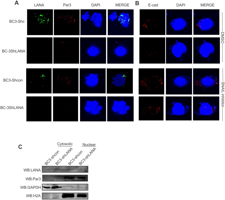
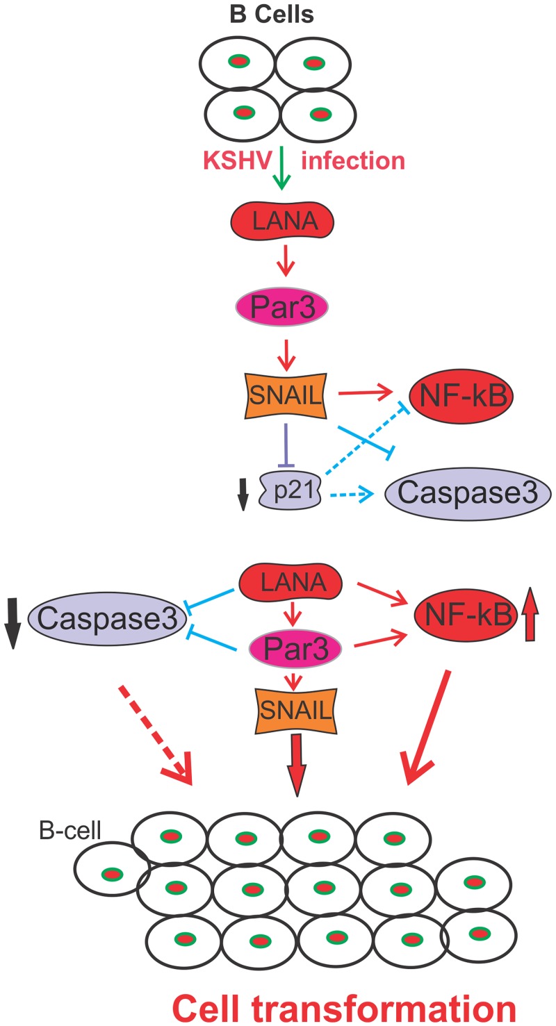
Studies have suggested that Epithelial-Mesenchymal Transition (EMT) and transformation is an important step in progression to cancer. Par3 (partitioning-defective protein) is a crucial factor in regulating epithelial cell polarity. However, the mechanism by which the latency associated nuclear antigen (LANA) encoded by Kaposi's Sarcoma associated herpesvirus (KSHV) regulates Par3 and EMTs markers (Epithelial-Mesenchymal Transition) during viral-mediated B-cell oncogenesis has not been fully explored. Moreover, several studies have demonstrated a crucial role for EMT markers during B-cell malignancies. In this study, we demonstrate that Par3 is significantly up-regulated in KSHV-infected primary B-cells. Further, Par3 interacted with LANA in KSHV positive and LANA expressing cells which led to translocation of Par3 from the cell periphery to a predominantly nuclear signal. Par3 knockdown led to reduced cell proliferation and increased apoptotic induction. Levels of SNAIL was elevated, and E-cadherin was reduced in the presence of LANA or Par3. Interestingly, KSHV infection in primary B-cells led to enhancement of SNAIL and down-regulation of E-cadherin in a temporal manner. Importantly, knockdown of SNAIL, a major EMT regulator, in KSHV cells resulted in reduced expression of LANA, Par3, and enhanced E-cadherin. Also, SNAIL bound to the promoter region of p21 and can regulate its activity. Further a SNAIL inhibitor diminished NF-kB signaling through upregulation of Caspase3 in KSHV positive cells in vitro. This was also supported by upregulation of SNAIL and Par3 in BC-3 transplanted NOD-SCID mice which has potential as a therapeutic target for KSHV-associated B-cell lymphomas.


Conflict of interest statement

The authors have declared that no competing interests exist.

Figures

Fig 1
Fig 1. KSHV infection leads to Par3 up-regulation.
(A) Expression of Par3 was examined at the transcript and protein levels by real-time PCR and Western blot in KSHV infected PBMCs at day 2 and 6. Here RQ and RD terms are using for relative quantification and relative density, respectively. (B) Par3 were measured at the transcript and protein levels in BJAB (KSHV negative) and BCBL1 and BC-3 (KSHV positive) cell lines. (C) Par3 levels were measured in HEK-293 and HEK-293-BACKSHV at the transcript and protein levels. (D) KSHV positive cells (BC-3 and JSC-1) were knockdown with LANA and compared with controls to assessed transcript of Par3.
Fig 2
Fig 2. LANA interacts and colocalizes with Par3.
(A) Co-Immunoprecipitation of Par3 with LANA was examined in HEK-293 cells. Myc tagged LANA and HA tagged Par3 constructs were used in this experiment. (B) GST pull-down assays shows binding of Par3 with N- (1–340 aa) and C-terminus (930–1162 aa) of LANA. GST-N-LANA and GST-C-LANA bound to Glutathione Sepharose beads were used to pull down Par3 from HA-Par3 transfected HEK-293 cell lysates. (C) Endogenous immunoprecipitation assays with Par3 antibody in KSHV negative BJAB and KSHV positive (BC-3 and BCBL1) cell line was performed. (D) Par3 domains and truncations used for mapping the interaction domain. (E) Co-Immunoprecipitation of Par3 truncations (1–1266, 1–373, 1–653 and 511–1266 aa) with LANA was examined in HEK-293 cells.
Fig 3
Fig 3. KSHV infection leads to Par3 nuclear localization.
(A) KSHV negative BJAB, Ramos, (B) KSHV positive BCBL1, BC-3, JSC-1 cell lines and (D) PBMCs uninfected and infected with KSHV at day 6 were used to determine the localization of Par3. LANA staining was used as positive control. In KSHV positive cells graphs were plotted with the percent of LANA foci colocalized with Par3. (C) Co-localization of LANA with Par3 was performed in KSHV negative Ramos cell line. The graphs represent the percent of LANA foci co-localized with Par3.
Fig 4
Fig 4. LANA stabilizes Par3 in KSHV positive cells.
(A) HEK-293-BAC-KSHV cells were transfected with sh-LANA and sh-Control. Endogenous Par3 expression was measured and presented in graph. GAPDH was used as a protein loading control. (B, C) Stabilization of Par3 was examined with cyclohexamide in HEK-293 and BJAB cells. Left panels and right panels were used as vector control and LANA expression, in a time dependent manner. (D) The proteosome inhibitor MG132 was used to determine if Par3 stability was linked to the proteosome degradation pathway. GFP and GAPDH were used as transfection and endogenous protein loading controls. (E) BC-3-shControl and BC-3-shLANA cells were treated with cyclohexamide and observed Par3 endogenous on hours dependent manner. (F) BC-3-shControl and BC-3shLANA cells were treated with MG132 and followed immunoprecipitation of Par3. Endogenous ubiquitin and Par3 were detected using specific antibodies in both the cell lines. GAPDH was monitored as an internal control for loading in the input section.
Fig 5
Fig 5. Par3 knockdown leads to delay in cell proliferation.
(A) Cell growth assay was carried out in HEK-293 cells. As indicated plasmids were transiently transfected in corresponding plates. 48 hour post transfection, 50,000 cells were plated for all groups into 100 mm dishes. Further cell density was scanned after fixing with 3% PFA and staining with crystal violet. Graphs are presented on the basis of intensity of colonies. Li-Cor Odyssey Scanner was used for scanning these plates. (B) Colony formation assays were carried out under puromycin and G418 antibiotic selection using HEK-293 cell lines. Representative graphs were also plotted for every set of experiments. Quantitation was done on the basis of intensity of colonies in every plates. (C) Cell growth assays were carried out in BC-3 and BCBL1 cells. As indicated plasmids were transiently transfected and transferred to corresponding flasks. 48 hour post transfection, 50,000 cells were plated for all groups into 6 well plates. Further cell densities were determined. Graphs are presented on the basis of intensity of cells. Li-Cor Odyssey Scanner was used for scanning the plates. Plotted graph based on cell density. (D) Cell proliferation assays were performed by using Trypan blue staining. 48 hour transfection, cells were counted for day1, 2 and 3 and plotted for live cells accordingly.
Fig 6
Fig 6. Status of EMTs in KSHV infected PBMCs and Par3 knockdown cells.
(A) PBMCs were subjected to infection with KSHV and analyzed using days post-infection for the screening of epithelial (E-cadherin, Zo-1, Dsp) and mesenchymal (Snail, Lef1, B-catenin, Cdh2). qRT-PCR was performed for the transcript analysis of EMTs (epithelial to mesenchymal markers). (B) HEK293-KSHV-shControl and HEK-293KSHV-shPar3 cells were used to study these EMTs. qRT-PCR was performed to determine the fold changes for EMTs and to confirm the Par3 knockdown in HEK-293KSHV stable cell lines. (C) BJAB-shContol and BJAB-shPar3 cells were generated to study these EMTs. qRT-PCR was performed to analyze the fold changes for EMTs and to confirm Par3 expression in transiently transfected BJAB cells.
Fig 7
Fig 7. KSHV can induce epithelial to mesenchymal transition markers in infected B-cells.
(A, B) HEK-293 and BAC-KHSV cells were probed with the antibodies against the EMT markers E-cadherin, and SNAIL. DAPI was used to stain the nucleus. (C) Ramos cells were transfected with an increasing dose of LANA to evaluate the transcript levels of E-cadherin and SNAIL. (D) KSHV infection was carried out in primary PBMCs and monitored up to 7 days post-infection. Control cells without KSHV infection was measured for days 1, 2, 4 and 7 for the measurement of SNAIL and E-cadherin transcripts. (E) Stable LANA knockdown cells were compared to controls for the measurement of Par3, E-cadherin, MMP9 and SNAIL. LANA blots were used as a positive control to monitor its expression in cells, and GAPDH was used as a protein loading control. (F) HEK-293-BAC-KSHV compared to HEK293 cells were evaluated at the protein levels for the expression of Par3, E-cadherin, MMP9 and SNAIL. LANA blots were used as a positive control for the expression of cells and GAPDH was used as a protein loading control. (G) SNAIL knockdown in BC-3 cells were evaluated with control vector for the measurement of the expression of LANA, Par3, and E-cadherin. GAPDH was used as a protein loading control.
Fig 8
Fig 8. Expression of SNAIL and Par3 in BJAB and BC-3 tumors in mouse xenograft model.
(A, B) NOD/SCID mice were injected with ten millions of BJAB and BC-3 cells intraperitoneal. (C, D). After 5 weeks, the mice were scarified and tumor dissected for analysis of transcripts and proteins of SNAIL and Par3 in these tumors. (E) Immunohistochemistry were performed on BJAB and BC-3 generated tumors. Here we used primary antibodies against LANA, E-cadherin and Par3. DAPI was used for nuclear staining. (F) H and E staining was shown for a similar group of tumor tissues capitalize to Fig 8F.
Fig 9
Fig 9. SNAIL can transcriptionally repress p21 in KSHV positive cells.
(A-D) SNAIL binding region "CACCTG" were mapped on the promoter region of known binding partner, p21, SNAIL, pTEN and TGFB3 respectively. (A-D) HEK-293-BACKSHV generated recombinant virus was used to infect PBMCs and ChIP performed for SNAIL at the selected binding regions on days 2 and 5 post infection. We also included BC-3 generated virus to infect PBMCs and also subjected to binding analysis on day 2 for p21, SNAIL, pTEN and TGFB3, respectively. (E) The Promoter activity of p21 was investigated in reporter assays in the presence of sh-SNAIL and sh-Control compared to pGL3-p21 alone in BJAB cells. (F) Western blot analysis was run for the confirmation of SNAIL knockdown compared to vector control.
Fig 10
Fig 10. NF-kB is regulated by SNAIL in KSHV positive cells.
(A) BC-3 and (B) BJAB cells were treated with DMSO and SNAIL inhibitor for 48 hours and assessed through Western blots for LANA, NF-kB, SNAIL and endogenous GAPDH. (C) BC-3 shControl and BC-3-ShLANA were treated with DMSO and SNAIL inhibitor for 48 hour and blotted for Par3, NF-kB (p65), SNAIL and endogenous control GAPDH. (D) Ubiquitination assays for SNAIL were performed in BC-3sh-control and sh-NF-kB cells treated with DMSO and MG132. (E) sh-Control and sh-Par3 in presence/absence of LANA were measured in the BJAB cell background. Left panel was observed in the presence of DMSO and the right panel was treated with SNAIL inhibitor at the same time. Caspase3 and GAPDH was observed in both panels.
Fig 11
Fig 11. E-Cadherin levels are induced by a SNAIL inhibitor in KSHV positive cells.
(A, B) BC-3 shControl and BC-3 sh-LANA cells were treated with DMSO and SNAIL inhibitor and immunostained with (A) LANA, Par3, and nuclear stain DAPI (B), E-Cadherin and nuclear stain with DAPI. (C) Cell fractionation assays were carried out in BC-3Shcontrol and BC-3shLANA cells. GAPDH was used as a control for cytosolic fraction and H2A was used as a control for nuclear fraction.
Fig 12
Fig 12. Schematic represents a putative model illustrating the contribution of Par3 and SNAIL to KSHV-associated cancers.
KSHV infected endothelial or B-cells expresses LANA important for establishment of latent infection. LANA up-regulates Par3 and SNAIL which leads to epithelial to mesenchymal transition through down-regulation of E-cadherin and enhanced expression of MMP9 in B-cells. This model suggests that KSHV-infection can regulate the EMTs important for progression of infected cells to an oncogenic and invasive phenotype.

Similar articles

Cited by

References

    1. Chang Y, Cesarman E, Pessin MS, Lee F, Culpepper J, et al. (1994) Identification of herpesvirus-like DNA sequences in AIDS-associated Kaposi's sarcoma. Science 266: 1865–1869. - PubMed
    1. McAllister SC, Moses AV (2007) Endothelial cell- and lymphocyte-based in vitro systems for understanding KSHV biology. Curr Top Microbiol Immunol 312: 211–244. - PubMed
    1. Nador RG, Cesarman E, Chadburn A, Dawson DB, Ansari MQ, et al. (1996) Primary effusion lymphoma: a distinct clinicopathologic entity associated with the Kaposi's sarcoma-associated herpes virus. Blood 88: 645–656. - PubMed
    1. Chen YB, Rahemtullah A, Hochberg E (2007) Primary effusion lymphoma. Oncologist 12: 569–576. - PubMed
    1. Elgui de Oliveira D (2007) DNA viruses in human cancer: an integrated overview on fundamental mechanisms of viral carcinogenesis. Cancer Lett 247: 182–196. - PubMed

MeSH terms