. 2017 Jun;55(6):894-901.
doi: 10.1002/mus.25267. Epub 2017 Apr 1.

Evidence for dying-back axonal degeneration in age-associated skeletal muscle decline

Affiliations

Evidence for dying-back axonal degeneration in age-associated skeletal muscle decline

Tae Chung et al. Muscle Nerve. 2017 Jun.

Abstract

Introduction: Age-associated muscle strength decline is a major contributing factor to increased late-life functional decline and comorbidity, and is strongly associated with early mortality. Although all parts of the neuromuscular system seem to be affected by aging, dying-back of motor axons likely plays a major role.

Methods: We compared the degeneration in ventral roots and neuromuscular junction denervation in young and aged mice and correlated the findings with strength and electrophysiological measures.

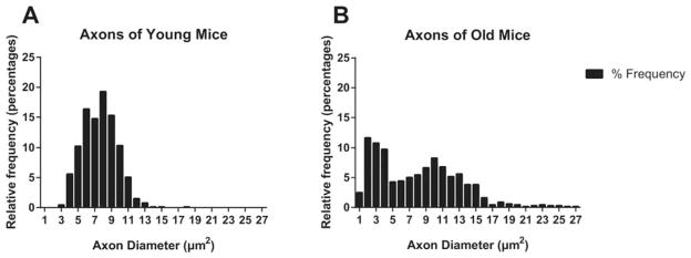
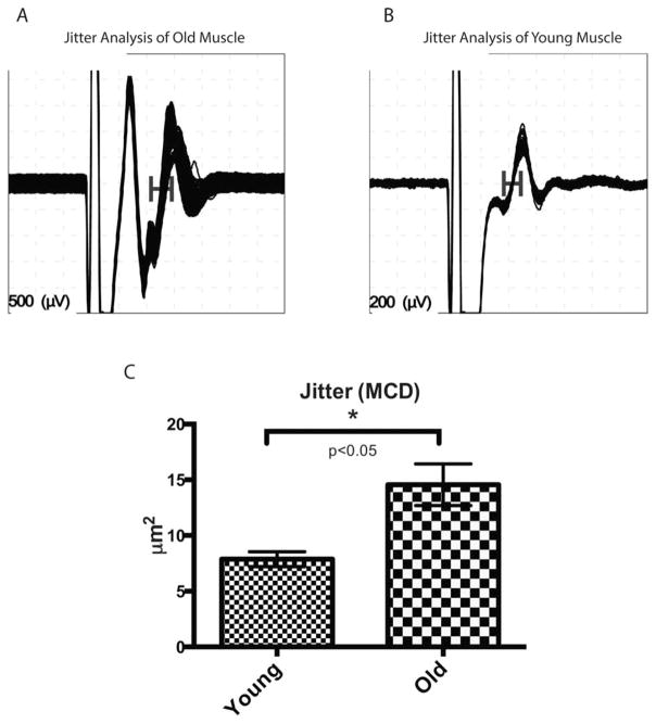
Results: With normal aging, there is little decline in motor axon numbers in the ventral roots, but the neuromuscular junctions show marked partial denervation that is associated with increased jitter on stimulated single fiber electromyography and a decrease in muscle strength.

Conclusions: These findings suggest that dying-back axonal degeneration may be partially responsible for the electrophysiological and strength changes observed with aging. Muscle Nerve 55: 894-901, 2017.

Keywords: SFEMG; denervation; distal axon; dying-back axonopathy; frailty; neuromuscular junction.


Figures

FIGURE 1
FIGURE 1
Comparison of grip strength between old and young mice. Grip strength of both forelimbs (A) and all limbs (B) are reduced in the old mice (N =5), compared with the young mice (N =4).
FIGURE 2
FIGURE 2
Partial denervation of NMJs in soleus muscles of young and old mice. Double immunofluorescence staining was performed (green =βIII-tubulin; red =bungarotoxin), and the confocal images were analyzed. (A) NMJ of soleus muscle in a young mouse. Note that postsynaptic motor endplates (yellow arrow) appear well-organized, distinct, and compact, with presynaptic axons covering wide areas of motor endplates. (B) NMJ of soleus muscle in an old mouse. Postsynaptic motor endplates are fragmented and disorganized (yellow arrow). Diameters of the motor endplates are larger, and presynaptic axons do not cover the entire areas of the motor end-plates, suggesting partial denervation (yellow arrow). (C) The percentage coverage of presynaptic axons over the postsynaptic motor endplate (AChR occupancy) was calculated using confocal imaging software; the results show significantly greater coverage in the young mice (N =5, 91.93%) than in the old mice (N =5, 40.97%). (D) Comparison of postsynaptic endplate area. The postsynaptic endplate areas are much greater in the old mice (N =5, average 497.74 μm2 than in the young mice (N =5, average 299.76 μm2. Representative images of completely denervated NMJs in old mice are seen in panels (E) and (F) (green =βIII-tubulin; red =bungarotoxin).
FIGURE 3
FIGURE 3
Comparison of L5 ventral roots between old and young mice. (A) Total counts of axons of cross-sections of L5 ventral roots are compared between young (N =7) and old (N =6) mice. Note that the standard deviation of the old group is greater than that of the young group, but there is no statistical difference in total axon counts between the 2 groups. (B–D) There were no statistical differences between the young and old groups for axon count per area, axon diameter, or G-ratio. (E) Cross-section of a young L5 ventral root. Note that the ratio of axonal diameter and myelin thickness is constant between different axons, and most axons have similar sizes. (F) Cross-section of an old L5 ventral root. Note the variation in ratio of axonal diameter and myelin thickness (yellow arrows, the axons with larger diameter have thinner myelin), with some axons showing early signs of myelin stripping. However, the average G-ratio between the 2 groups was not statistically different, although the standard deviation was greater in the old group.
FIGURE 4
FIGURE 4
Relative frequency histogram of axon diameter in young and old mice. (A) Axon diameters of young mice centered around the mean value (7.62 μm2, composing a Gaussian distribution. (B) Axon diameters of old mice show a bimodal distribution with some outliers toward both larger and smaller diameters.
FIGURE 5
FIGURE 5
Jitter analysis of SSFEMG. (A) Jitter analysis of old mice. Note the increased jitter (horizontal bar) in the second single fiber action potential. (B) Jitter analysis of young mice. (C) Jitter as represented by MCD (mean consecutive difference). Mean jitter of old mice (N =5) was significantly increased over that of young mice (N =4).

References

    1. Studenski S, Perera S, Patel K, Rosano C, Faulkner K, Inzitari M, et al. Gait speed and survival in older adults. JAMA. 2011;305:50–58. - PMC - PubMed
    1. Newman AB, Simonsick EM, Naydeck BL, Boudreau RM, Kritchevsky SB, Nevitt MC, et al. Association of long-distance corridor walk performance with mortality, cardiovascular disease, mobility limitation, and disability. JAMA. 2006;295:2018–2026. - PubMed
    1. Guralnik JM, Simonsick EM, Ferrucci L, Glynn RJ, Berkman LF, Blazer DG, et al. A short physical performance battery assessing lower extremity function: association with self-reported disability and prediction of mortality and nursing home admission. J Gerontol. 1994;49:M85–94. - PubMed
    1. Xue QL, Beamer BA, Chaves PH, Guralnik JM, Fried LP. Heterogeneity in rate of decline in grip, hip, and knee strength and the risk of all-cause mortality: the women’s health and aging study II. J Am Geriatr Soc. 2010;58:2076–2084. - PMC - PubMed
    1. Guralnik JM, Ferrucci L, Simonsick EM, Salive ME, Wallace RB. Lower-extremity function in persons over the age of 70 years as a predictor of subsequent disability. N Engl J Med. 1995;332:556–561. - PMC - PubMed

Publication types