

Multimodal Imaging Reveals Improvement of Blood Supply to an Artificial Cell Transplant Site Induced by Bioluminescent Mesenchymal Stem Cells
- PMID: 27464498
- PMCID: PMC5209399
- DOI: 10.1007/s11307-016-0986-1
Multimodal Imaging Reveals Improvement of Blood Supply to an Artificial Cell Transplant Site Induced by Bioluminescent Mesenchymal Stem Cells
Abstract
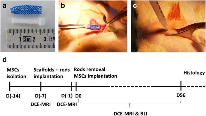
Purpose: An artificial site for cell or pancreatic islet transplantation can be created using a polymeric scaffold, even though it suffers subcutaneously from improper vascularisation. A sufficient blood supply is crucial for graft survival and function and can be enhanced by transplantation of mesenchymal stem cells (MSCs). The purpose of this study was to assess the effect of syngeneic MSCs on neoangiogenesis and cell engraftment in an artificial site by multimodal imaging.
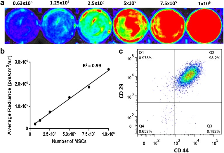
Procedures: MSCs expressing a gene for luciferase were injected into the artificial subcutaneous site 7 days after scaffold implantation. MRI experiments (anatomical and dynamic contrast-enhanced images) were performed on a 4.7-T scanner using gradient echo sequences. Bioluminescent images were acquired on an IVIS Lumina optical imager. Longitudinal examination was performed for 2 months, and one animal was monitored for 16 months.
Results: We confirmed the long-term presence (lasting more than 16 months) of viable donor cells inside the scaffolds using bioluminescence imaging with an optical signal peak appearing on day 3 after MSC implantation. When compared to controls, the tissue perfusion and vessel permeability in the scaffolds were significantly improved at the site with MSCs with a maximal peak on day 9 after MSC transplantation.
Conclusions: Our data suggest that the maximal signal obtained by bioluminescence and magnetic resonance imaging from an artificially created site between 3 and 9 days after MSC transplantation can predict the optimal time range for subsequent cellular or tissue transplantation, including pancreatic islets.
Keywords: Bioluminescence; DCE; Dynamic contrast-enhanced MRI; Magnetic resonance imaging; Mesenchymal stem cells; Vascularisation.
Conflict of interest statement
Compliance with Ethical Standards The care of all animals was in accordance with the European Convention of Animal Care and the Animal Care Committee of the Institute for Clinical and Experimental Medicine. The Ministry of Health of the Czech Republic also approved the protocols related to this study. Conflict of Interest The authors declare that they have no conflict of interest.
Figures


Similar articles
-
The Optimal Timing for Pancreatic Islet Transplantation into Subcutaneous Scaffolds Assessed by Multimodal Imaging.Contrast Media Mol Imaging. 2017 Dec 26;2017:5418495. doi: 10.1155/2017/5418495. eCollection 2017. Contrast Media Mol Imaging. 2017. PMID: 29440984 Free PMC article.
-
Effect of mesenchymal stem cells on the vascularization of the artificial site for islet transplantation in rats.Transplant Proc. 2014 Jul-Aug;46(6):1963-6. doi: 10.1016/j.transproceed.2014.05.074. Transplant Proc. 2014. PMID: 25131083
-
Vascularization of PLGA-based bio-artificial beds by hypoxia-preconditioned mesenchymal stem cells for subcutaneous xenogeneic islet transplantation.Xenotransplantation. 2019 Jan;26(1):e12441. doi: 10.1111/xen.12441. Epub 2018 Jul 28. Xenotransplantation. 2019. PMID: 30054954
-
Dynamic contrast-enhanced magnetic resonance imaging as a tool to monitor the blood supply to an artificial cavity used as a site for islet transplantation in rats.Transplant Proc. 2011 Nov;43(9):3226-30. doi: 10.1016/j.transproceed.2011.09.012. Transplant Proc. 2011. PMID: 22099763
-
A Trimodal Imaging Platform for Tracking Viable Transplanted Pancreatic Islets In Vivo: F-19 MR, Fluorescence, and Bioluminescence Imaging.Mol Imaging Biol. 2019 Jun;21(3):454-464. doi: 10.1007/s11307-018-1270-3. Mol Imaging Biol. 2019. PMID: 30167995 Free PMC article.
Cited by
-
Interplay of hypoxia, immune dysregulation, and metabolic stress in pathophysiology of type 1 diabetes.Front Immunol. 2025 Jun 4;16:1599321. doi: 10.3389/fimmu.2025.1599321. eCollection 2025. Front Immunol. 2025. PMID: 40534855 Free PMC article. Review.
-
The Optimal Timing for Pancreatic Islet Transplantation into Subcutaneous Scaffolds Assessed by Multimodal Imaging.Contrast Media Mol Imaging. 2017 Dec 26;2017:5418495. doi: 10.1155/2017/5418495. eCollection 2017. Contrast Media Mol Imaging. 2017. PMID: 29440984 Free PMC article.
-
Subpial transplantation of adipose-derived stem cells alleviates paraplegia in a rat model of aortic occlusion/reperfusion-induced spinal cord infarction.Regen Ther. 2024 Aug 21;26:611-619. doi: 10.1016/j.reth.2024.08.005. eCollection 2024 Jun. Regen Ther. 2024. PMID: 39263357 Free PMC article.
References
-
- Shapiro JA, Auchincloss H, Lindblad R, et al. (2006) International Trial of the Edmonton Protocol for Islet Transplantation 355:1318–1330. - PubMed
MeSH terms
Substances
LinkOut - more resources
Full Text Sources
Other Literature Sources
Research Materials
