. 2016 Oct;152(4):1059-1070.e2.
doi: 10.1016/j.jtcvs.2016.06.017. Epub 2016 Jun 25.

Disruption of desmin-mitochondrial architecture in patients with regurgitant mitral valves and preserved ventricular function

Affiliations

Disruption of desmin-mitochondrial architecture in patients with regurgitant mitral valves and preserved ventricular function

Mustafa I Ahmed et al. J Thorac Cardiovasc Surg. 2016 Oct.

Abstract

Objective: Recent studies have demonstrated improved outcomes in patients receiving early surgery for degenerative mitral regurgitation (MR) rather than adhering to conventional guidelines for surgical intervention. However, studies providing a mechanistic basis for these findings are limited.

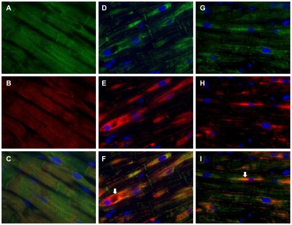
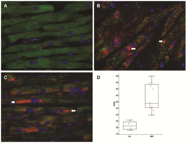
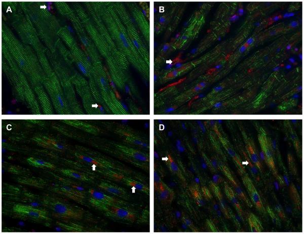
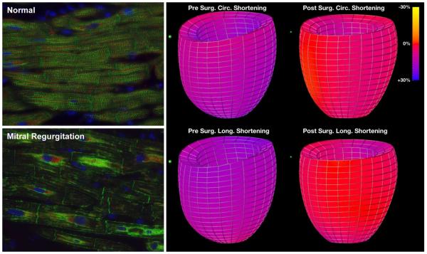
Methods: Left ventricular (LV) myocardium from 22 patients undergoing mitral valve repair for American Heart Association class I indications was evaluated for desmin, the voltage-dependent anion channel, α-B-crystallin, and α, β-unsaturated aldehyde 4-hydroxynonenal by fluorescence microscopy. The same was evaluated in 6 normal control LV autopsy specimens. Cardiomyocyte ultrastructure was examined by transmission electron microscopy. Magnetic resonance imaging with tissue tagging was performed in 55 normal subjects and 22 MR patients before and 6 months after mitral valve repair.

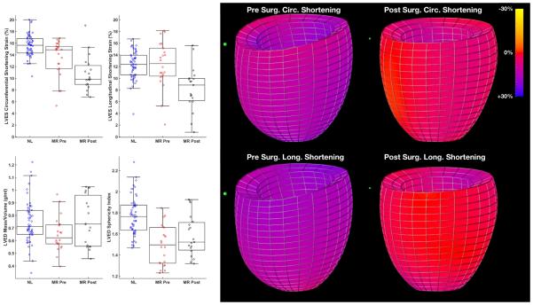
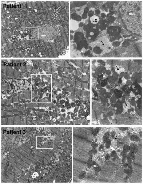
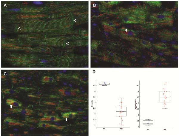
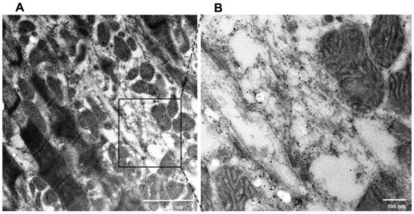
Results: LV end-diastolic volume was 1.5-fold (P < .0001) higher and LV mass-to-volume ratio was lower in MR (P = .004) hearts versus normal hearts and showed improvement 6 months after mitral valve surgery. However, LV ejection fraction decreased from 65% ± 7% to 52% ± 9% (P < .0001) and LV circumferential (P < .0001) and longitudinal strain decreased significantly below normal values (P = .002) after surgery. Hearts with MR had a 53% decrease in desmin (P < .0001) and a 2.6-fold increase in desmin aggregates (P < .0001) versus normal, along with substantial, intense perinuclear staining of α, β-unsaturated aldehyde 4-hydroxynonenal in areas of mitochondrial breakdown and clustering. Transmission electron microscopy demonstrated numerous electron-dense deposits, myofibrillar loss, Z-disc abnormalities, and extensive granulofilamentous debris identified as desmin-positive by immunogold transmission electron microscopy.

Conclusions: Despite well-preserved preoperative LV ejection fraction, severe oxidative stress and disruption of cardiomyocyte desmin-mitochondrial sarcomeric architecture may explain postoperative LV functional decline and further supports the move toward earlier surgical intervention.

Keywords: cardiomyocyte; heart failure; mitochondria; mitral regurgitation.


Figures

Figure 1
Figure 1. LVED mass/volume, LVED sphericity index and LV circumferential and longitudinal shortening strains in MR patients pre- and six month post-mitral valve (MV) repair with color coded strain maps in MR subject pre- and post-mitral valve repair
Box and whisker plots demonstrate that LVED sphericity index (LVED length/LVED inner diameter) pre- and post-surgery were significantly different from normal controls (p <0.0001 and p=0.001 respectively) but did improve post-surgery. LVED mass to volume ratio pre-surgery was significantly different from normal controls (p=0.0042) but did improve and did not differ from normal post-surgery (p=0.0013). LV longitudinal systolic shortening strains were not different from normal controls before surgery, but were decreased following MV repair vs before surgery (p=0.0013) and relative to normal control subjects (p=0.0020). The color-coded strain map demonstrates the significant global decrease in circumferential and longitudinal strains in a representative MR patient post mitral valve repair.
Figure 2
Figure 2. Transmission electron microscopy of LV from 3 patients with MR and LVEF > 60%
In Patients 1-3, 4,500X LV images (left panels) demonstrate extensive myofibrillar breakdown amid mitochondrial (mt) clustering of various small sizes. There is extensive granulofilamentous protein aggregates (right panels, black arrows) representing cytoskeletal and myofibrillar degeneration. In Patients 1 and 2, white boxes (left panels) outline the 16,000X images (right panels) of perinuclear areas with electron dense bodies consistent with lipofuscin also containing lipid droplets (LD) in interfibrillar areas and in electron dense bodies (white arrow Patient 2). Patient 3 demonstrates extensive granulo-filamentous protein aggregates (light gray) in areas of myofibrillar and cytoskeletal degeneration being engulfed by autophagic vacuole (right panel, black arrow in 16,000X image in right panel).
Figure 3
Figure 3. Immunohistochemistry of desmin and VDAC in normal human heart (A) and in patients with MR and LVEF > 60%
Normal LV (A) demonstrates a regular pattern of desmin (green, arrowheads) along Z-lines in between intercalated discs (arrowheads). Co-immunostaining with VDAC (red) show that mitochondria are in linear register in the interfibrillar areas and grouped in perinuclear (DAPI, blue) locations. MR hearts (B and C) demonstrate loss of desmin interspersed with areas of intense desmin staining that co-stains with VDAC (arrows, B and C), especially in the perinuclear areas (arrows). Box-and-whisker plots with the data points superimposed (D) shows the quantitation of desmin loss and aggregate as percent of area of cardiomyocytes. Desmin loss as per cent of cardiomyocytes: Grade 1: 100% loss; Grade 2: 75% loss, 25% intact; Grade 3: 50% loss, 50% intact; Grade 4: 25% loss, 75% intact; and Grade 5: 100% intact. N=22 patients. Desmin staining vs. normal hearts (p<0.0001) Desmin aggregates as percent of cardiomyocytes: Grade 1: no aggregates; Grade 2: 25% aggregates; Grade 3: 50% aggregates; Grade 4: 75%; Grade 5 100% aggregates. N=22 patients. Desmin aggregates vs. normal (p<0.0001)
Figure 4
Figure 4. Transmission electron microscopy immunogold labeling for desmin in MR heart
TEM of MR heart of patient with LVEF of 65% demonstrates marked disruption and degradation of the mitochondrial sarcomeric units (left panel) and higher magnification (black box, right panel) of immunogold labeling demonstrates desmin (black dots) in areas of sarcomere/mitchondrial breakdown. Note the desmin in the areas of myofibrillar degradation near the interfibrillar disorganization of mitochondria.
Figure 5
Figure 5. Immunohistochemistry for desmin (green) and αβ-crystallin (red) in MR and normal heart
In the normal heart (A) desmin (green) and α, β-crystallin (red, B) are co-localized to the Z-disc as seen in merged image (C). In MR hearts of patients with LVEF 68% (D-F) and LVEF 62% (G-I) there is extensive desmin loss along with areas of aggregation most prominent in the perinuclear region (DAPI, blue). αβ-crystallin in MR hearts (D-F and G-I) relocates to the perinuclear region (arrows) with desmin aggregates as seen in merged images (F and I).
Figure 6
Figure 6. Immunohistochemistry for desmin (green) and 4-hydroxy-2-nonenal (HNE, red) in normal and MR hearts
Normal heart (A) has striated pattern of desmin (green) with very little HNE staining (red). In MR hearts (B, C) there is extensive desmin loss along with increased HNE staining, especially in the perinuclear areas (arrows) that is increased two-fold in MR hearts in bar graph, measured as HNE intensity as percent area of cardiomyocyte as demonstrated in box and whisker plot (D). HNE intensity in MR vs. normal p=0.012)
Figure 7
Figure 7. Immunohistochemistry for desmin (green) and catalase (red) in normal and MR hearts
The normal heart (A) has the striated desmin with very little catalase staining except in some interstitial cells (A, arrows). In three MR hearts (B-D) there is extensive desmin loss along with increased catalase staining particularly in the perinuclear regions (D, arrows).
Central Figure
Central Figure
Breakdown of desmin (green) and aggregation in mitral regurgitation underpins decrease in LV shortening strains below normal (purple to red on color strain map) post-surgery.

Comment in

Similar articles

Cited by

References

    1. Enriquez-Sarano M, Akins CW, Vahanian A. Mitral regurgitation. Lancet. 2009;373:1382–94. - PubMed
    1. Ahmed MI, Aban I, Lloyd SG, Gupta H, Howard G, Inusah S, Peri K, et al. A randomized controlled Phase IIb trial of Beta-1 receptor blockade in isolated degenerative mitral regurgitation. J Am Coll Cardiol. 2012;60:833–38. - PMC - PubMed
    1. Suri RM, Aviernos J-F, Dearani JA, Mahoney DW, Michelena HI, Schaff HV, Enriquez-Sarano M. Management of less-than-severe mitral regurgitation: should guidelines recommend earlier surgical intervention. Eur J Cardiovasc Surg. 2011;40:496–502. - PubMed
    1. Enriquez-Sarano M, Avierinos JF, Messika-Zeitoun D, Detaint D, Capps M, Nkomo V, et al. Quantitative determinants of the outcome of asymptomatic mitral regurgitation. N Eng J Med. 2005;352:875–83. - PubMed
    1. Nishimura RA, Otto CM, Bonow RO, Carabello BA, Erwin JP, III, Guyton RA, et al. 2014 AHA/ACC Guideline for the management of patients with valvular heart disease: report of the American College of Cardiology/American Heart Association Task Force on Practice Guidelines. Circulation. 2014;129:2440–92. - PubMed

Publication types

MeSH terms