

Form and function of the human and chimpanzee forefoot: implications for early hominin bipedalism
- PMID: 27464580
- PMCID: PMC4964565
- DOI: 10.1038/srep30532
Form and function of the human and chimpanzee forefoot: implications for early hominin bipedalism
Abstract
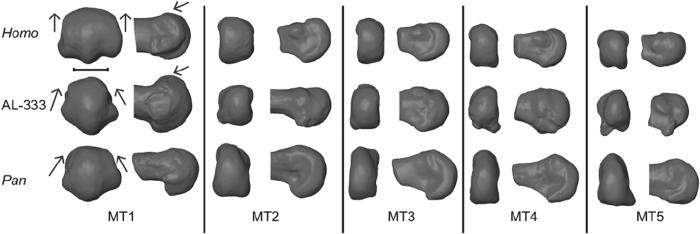
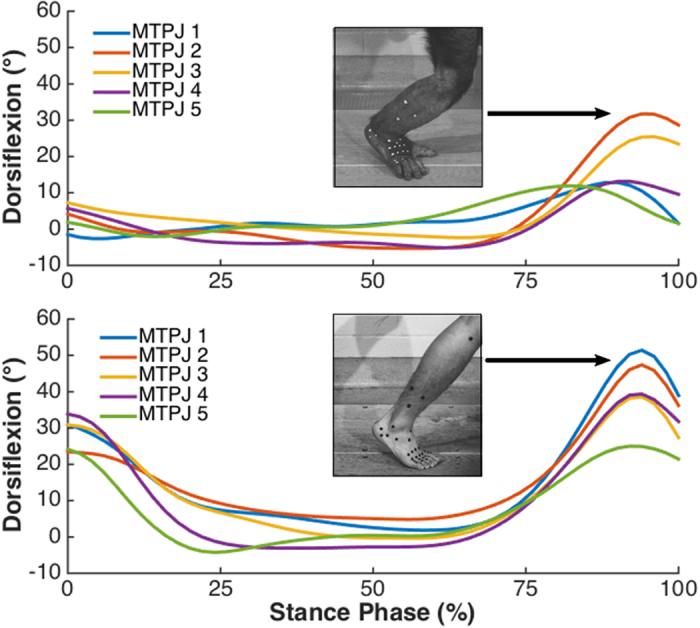
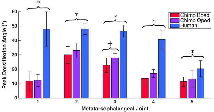
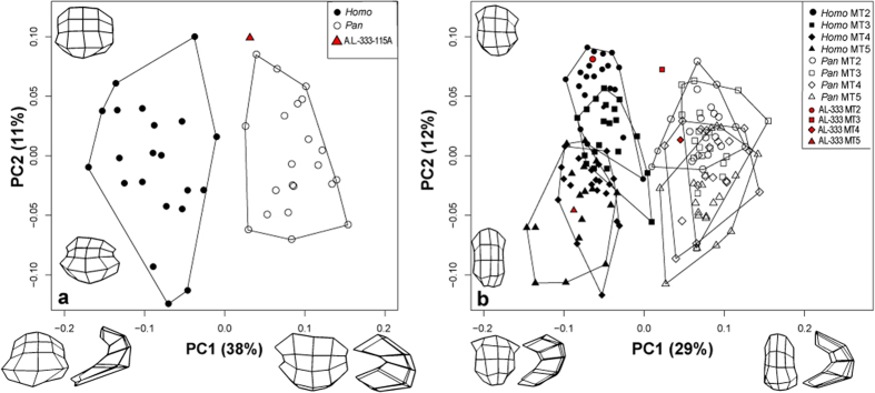
During bipedal walking, modern humans dorsiflex their forefoot at the metatarsophalangeal joints (MTPJs) prior to push off, which tightens the plantar soft tissues to convert the foot into a stiff propulsive lever. Particular features of metatarsal head morphology such as "dorsal doming" are thought to facilitate this stiffening mechanism. In contrast, chimpanzees are believed to possess MTPJ morphology that precludes high dorsiflexion excursions during terrestrial locomotion. The morphological affinity of the metatarsal heads has been used to reconstruct locomotor behavior in fossil hominins, but few studies have provided detailed empirical data to validate the assumed link between morphology and function at the MTPJs. Using three-dimensional kinematic and morphometric analyses, we show that humans push off with greater peak dorsiflexion angles at all MTPJs than do chimpanzees during bipedal and quadrupedal walking, with the greatest disparity occurring at MTPJ 1. Among MTPJs 2-5, both species exhibit decreasing peak angles from medial to lateral. This kinematic pattern is mirrored in the morphometric analyses of metatarsal head shape. Analyses of Australopithecus afarensis metatarsals reveal morphology intermediate between humans and chimpanzees, suggesting that this species used different bipedal push-off kinematics than modern humans, perhaps resulting in a less efficient form of bipedalism.
Figures
References
-
- Susman R. L. Evolution of the human foot: evidence from Plio-Pleistocene hominids. Foot Ankle 3, 365–376 (1983). - PubMed
-
- Latimer B. M. & Lovejoy C. O. Hallucal tarsometatarsal joint in Australopithecus afarensis. Am. J. Phys. Anthropol. 82, 125–133 (1990). - PubMed
-
- Griffin N. L., D’Août K., Richmond B., Gordon A. & Aerts P. Comparative in vivo forefoot kinematics of Homo sapiens and Pan paniscus. J. Hum. Evol. 59, 608–619 (2010). - PubMed
-
- Proctor D. J., Broadfield D. & Proctor K. Quantitative three-dimensional shape analysis of the proximal hallucial metatarsal articular surface in Homo, Pan, Gorilla, and Hylobates. Am. J. Phys. Anthropol. 135, 216–224 (2008). - PubMed
-
- Proctor D. J. Brief communication: shape analysis of the MT 1 proximal articular surface in fossil hominins and shod and unshod Homo. Am. J. Phys. Anthropol. 143, 631–637 (2010). - PubMed
Publication types
MeSH terms
LinkOut - more resources
Full Text Sources
Other Literature Sources
Medical
