. 2016 Dec;5(12):1607-1619.
doi: 10.5966/sctm.2016-0030. Epub 2016 Jul 27.

Clinical-Grade Human Multipotent Adult Progenitor Cells Block CD8+ Cytotoxic T Lymphocytes

Affiliations

Clinical-Grade Human Multipotent Adult Progenitor Cells Block CD8+ Cytotoxic T Lymphocytes

Jeroen Plessers et al. Stem Cells Transl Med. 2016 Dec.

Abstract

: MultiStem cells are clinical-grade multipotent adult bone marrow-derived progenitor cells (MAPCs), with extensive replication potential and broader differentiation capacity compared with mesenchymal stem cells. Human MAPCs suppress T-cell proliferation induced by alloantigens and mutually interact with allogeneic natural killer cells. In this study, the interaction between MultiStem and CD8+ cytotoxic T lymphocytes (CTLs) was addressed for the first time. In an in vitro setting, the immunogenicity of MultiStem, the susceptibility of MultiStem toward CTL-mediated lysis, and its effects on CTL function were investigated. MultiStem was nonimmunogenic for alloreactive CTL induction and was-even after major histocompatibility complex class I upregulation-insensitive to alloantigen-specific CTL-mediated lysis. Furthermore, MultiStem reduced CTL proliferation and significantly decreased perforin expression during the T-cell activation phase. As a consequence, MultiStem dose-dependently impaired the induction of CTL function. These effects of MultiStem were mediated predominantly through contact-dependent mechanisms. Moreover, MultiStem cells considerably influenced the expression of T-cell activation markers CD25, CD69, and human leukocyte antigen-DR. The MultiStem-induced CD8-CD69+ T-cell population displayed a suppressive effect on the induction of CTL function during a subsequent mixed-lymphocyte culture. Finally, the killer activity of activated antigen-specific CTLs during their cytolytic effector phase was also diminished in the presence of MultiStem. This study confirms that these clinical-grade MAPCs are an immune-modulating population that inhibits CTL activation and effector responses and are, consequently, a highly valuable cell population for adoptive immunosuppressive therapy in diseases where damage is induced by CTLs.

Significance: Because multipotent adult progenitor cells (MAPCs) are among the noteworthy adult mesenchymal stem cell populations for immune therapy and have the advantage over mesenchymal stem cells (MSCs) of large-scale manufacturing and banking potential and thus prompt availability, it is important to understand how MAPCs interact with immune cells to validate their widespread therapeutic applicability. Cytotoxic immune effector cells play a crucial role in immune homeostasis and in the pathogenesis of some autoimmune diseases. This study assessed for the first time the in vitro influence of a clinical-grade human MAPC product (MultiStem) on the cytotoxic function of CD8+ T cells (CTLs) by evaluating the immunogenicity of MAPCs and the susceptibility of MAPCs toward CTL-mediated lysis and by analyzing the mechanism of MAPC-mediated modulation of CTL functionality. These results may represent a highly relevant contribution to the current knowledge and, in combination with the results of future phase II/III trials using MultiStem, could lead to an intriguing continuation of stem cell-based research for immunotherapy.

Keywords: Clinical-grade human multipotent adult progenitor cells; Cytotoxic T cells; MultiStem; Stem cell-based immune modulation; T cell-mediated cytotoxicity.


Figures

Figure 1.
Figure 1.
MultiStem does not induce cytotoxic activity in T cells. Freshly isolated responder CD3+ T cells were stimulated with either allogeneic-irradiated (30 Gy) peripheral blood mononuclear cells or allogeneic-irradiated (30 Gy) MultiStem (PBMCs and MultiStem were from the same donor) at a stimulator:responder ratio of 1:2 for 7 days. Coculture was followed by an assessment of anti-CD3-redirected cytotoxic activity against murine P815 mastocytoma target cells (A) or alloantigen-specific cytotoxic activity against Epstein-Barr virus-transformed B cells (B) at an effector:target ratio of 10:1 in a standard 51Cr-release assay. Data are expressed as mean ± SEM percentage of anti-CD3-dependent specific 51Cr-release (% SR) of five independent experiments with four different T cell donors and three different PBMC/MultiStem donors (donors 1, 2, and 3) (A) and mean ± SEM % 51Cr-release of three independent experiments with two different T cell donors and two different PBMC/MultiStem/B cell donors (donors 1 and 2) (B). Statistical significance was calculated with the unpaired t test. ∗∗∗, p < .001. Abbreviations: EBV, Epstein-Barr virus; P815, murine P815 mastocytoma target cells; PBMCs, peripheral blood mononuclear cells; SR, specific 51Cr-release.
Figure 2.
Figure 2.
Activated T cells do not lyse allogeneic MultiStem cells. Results of anti-CD3-redirected and alloantigen-specific cytotoxic activity of irradiated (40 Gy) EBV+ B cell-stimulated CD8+ cytotoxic T lymphocytes (CTLs) (stimulator:responder ratio of 1:10 for 7 days) against respectively anti-CD3-coated P815 target cells, MultiStem, or EBV+ B cells at an effector:target ratio of 10:1. MultiStem cells and EBV+ B target cells were from the same donor as the EBV+ B cells used for CTL activation. Target cell killing is expressed as mean ± SEM percentage of 51Cr-release of n independent experiments (P815 targets, n = 6; MultiStem targets, n = 6; EBV+ B targets, n = 3). Four different CTL donors and three different MultiStem/B-cell donors (donors 1, 2, and 3) were used. Statistical significance was calculated with the paired t test. ∗, p < .05; ∗∗∗, p < .001. Abbreviations: EBV, Epstein-Barr virus; P815, murine P815 mastocytoma target cells.
Figure 3.
Figure 3.
MultiStem impairs the proliferation and (induction of) cytotoxicity of CD8+ T cells. (A): Purified CD8+ cytotoxic T lymphocytes (CTLs) were stimulated with irradiated allogeneic EBV-transformed B cells (stimulator:responder [S:R] ratio of 1:10) during 7 days with or without irradiated third-party MultiStem at a suppressor:responder ratio of 1:2. Proliferation was measured on day 6 with [3H]thymidine incorporation. Results are expressed as mean ± SEM % proliferation relative to control (average counts per minute: 46,801) of quadruplicates in five experiments with five CTL and two MultiStem donors (donors 2 and 5). (B): Results of anti-CD3-redirected cytotoxicity of B cell-stimulated CD8+ CTLs against P815 targets (effector:target [E:T] ratio of 10:1; left) or alloantigen-specific activity against target B cells (right) with or without MultiStem (E:T ratio of 1:2) during the 7-day activation phase. Data are expressed as mean ± SEM percentage of anti-CD3-dependent specific 51Cr-release (% SR) of 32 experiments with 19 CTL and 4 MultiStem donors (donors 1–4; left) or percentage of 51Cr-release of 9 experiments with 6 CTL and 3 MultiStem donors (donors 1–3; right). (C): Same experimental setup as (B), but MultiStem was only present during the 4-hour cytotoxic effector phase. Left: A total of 18 experiments with 10 CTL donors and 4 MultiStem donors (donors 1–4). Right: A total of 15 experiments with 8 CTL donors and 4 MultiStem donors (donors 1–4). (D): Results of anti-CD3-redirected cytotoxicity of B cell-stimulated CD3+ T cells (S:R ratio of 1:20) against P815 targets with or without MultiStem at different ratios during the 7-day activation phase. Data are pooled from three experiments with two T-cell and two MultiStem donors (donors 2 and 5). Statistical significance was calculated with the paired t test. ∗, p < .05; ∗∗, p < .01; ∗∗∗, p < .001. Abbreviations: EBV, Epstein-Barr virus; P815, murine P815 mastocytoma target cells; SR, specific 51Cr-release.
Figure 4.
Figure 4.
MultiStem impairs the expression of perforin in CD8+ T cells. Flow-cytometric analysis of irradiated (40 Gy) Epstein-Barr virus-positive B cell-stimulated CD3+ T cells (stimulator:responder ratio of 1:20) for intracellular perforin expression after a 7-day stimulation period in the absence (upper) or presence (lower) of irradiated (30 Gy) third-party MultiStem cells (suppressor:responder ratio of 1:2). Results are expressed as percentage of positive cells in the CD3+ lymphocyte gate of one representative experiment with one T-cell donor and two different MultiStem donors (donors 2 and 4), out of three independent experiments. Abbreviations: Comp, compensation; cy5, cyanine 5; FITC-A, fluorescein isothiocyanate-A; PerCP, peridinin chlorophyll protein; Q1, quarter 1.
Figure 5.
Figure 5.
MultiStem-mediated suppression of T-cell cytotoxicity is contact-dependent. (A): Purified CD8+ cytotoxic T lymphocytes (CTLs) were stimulated with irradiated allogeneic Epstein-Barr virus-positive B cells (stimulator:responder [S:R] ratio of 1:10) with or without irradiated third-party MultiStem (ratio of 1:2) and with or without interleukin-2 (IL-2) for 7 days. Data are expressed as mean ± SEM percentage of SR of P815 targets (effector:target ratio of 10:1) and pooled from four experiments with three CTL and two MultiStem donors (donors 2 and 4). (B): Standard experimental setup was used (left). Right: After a 3-day resting period, CTLs from the primary mixed-lymphocyte culture, cultured with or without MultiStem, were restimulated during 4 days with the same alloantigens in the absence of MultiStem. Results are expressed as mean ± SEM percentage of SR against P815 targets of three experiments with two CTL and two MultiStem donors (donors 2 and 4). (C): Results of anti-CD3-redirected cytotoxicity of B cell-stimulated CD3+ T cells (S:R ratio of 1:20) against P815 targets with or without MultiStem, in direct contact or separated by a transwell system, during the 7-day activation phase. Data are pooled from six experiments with five T-cell and two MultiStem donors (donors 2 and 5). (D): Flow-cytometric analysis of MultiStem, pretreated or not with interferon-γ, for the PD-1 receptor and its ligands programmed death ligands 1 and 2 and for the ligand of the Fas pathway. Average mean fluorescence intensity values ± SD of three experiments (MultiStem donors 2, 4, and 5) are shown. The background signal of unstained cells is included. Statistical significance was calculated with the paired t test. ∗, p < .05. Abbreviations: FasL, Fas ligand; IFN-γ, interferon-γ; IL-2, interleukin 2; ns, not significant; PD-1, programmed death-1; SR, specific 51Cr-release; w/, with; w/o, without.
Figure 6.
Figure 6.
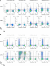
T-cell activation marker expression is altered in the presence of MultiStem. (A–C): Flow-cytometric analysis of CD3+ T lymphocytes for expression of T-cell activation markers CD69 (A), CD25 (B), and HLA-DR (C) on days 2 and 6 of a 6-day stimulation period with irradiated (40 Gy) allogeneic Epstein-Barr virus-positive (EBV+) B cells (stimulator:responder [S:R] ratio of 1:20) in the absence [(un)stimulated T cells] or presence (+MultiStem donor 2/4) of irradiated (30 Gy) third-party MultiStem cells (suppressor:responder ratio of 1:1). Data are presented as percentage of cells within the CD3+ lymphocyte gate. One representative experiment with one T-cell donor and two different MultiStem donors (donors 2 and 4) out of three independent experiments is shown. (D): Freshly isolated CD3+ T cells were primed with irradiated (40 Gy) allogeneic EBV+ B cells (S:R ratio of 1:20) in the presence of irradiated (30 Gy) third-party MultiStem cells (ratio of 1:2). After 6 days of stimulation, two populations were sorted from the coculture (CD45+CD3+CD8CD69+ T cells and CD45+CD3+CD8CD69 T cells). These fractions were added at a ratio of 1:2 to a subsequent mixed-lymphocyte culture with freshly isolated responder T cells and irradiated allogeneic EBV+ B cells (S:R ratio of 1:20). After 7 days, anti-CD3-redirected cytotoxicity was analyzed. Data are expressed as mean ± SEM percentage of anti-CD3-dependent specific 51Cr-release (% SR) of P815 target cells (effector:target ratio 10:1). Results are pooled from two independent experiments, in which two different T-cell donors and two MultiStem donors (donors 2 and 3) were used. Abbreviations: APC, allophycocyanin; Comp, compensation; cy5, cyanine 5; HLA-DR, human leukocyte antigen-DR; PE, phycoerythrin; SR, specific 51Cr-release.
Figure 6.
Figure 6.
T-cell activation marker expression is altered in the presence of MultiStem. (A–C): Flow-cytometric analysis of CD3+ T lymphocytes for expression of T-cell activation markers CD69 (A), CD25 (B), and HLA-DR (C) on days 2 and 6 of a 6-day stimulation period with irradiated (40 Gy) allogeneic Epstein-Barr virus-positive (EBV+) B cells (stimulator:responder [S:R] ratio of 1:20) in the absence [(un)stimulated T cells] or presence (+MultiStem donor 2/4) of irradiated (30 Gy) third-party MultiStem cells (suppressor:responder ratio of 1:1). Data are presented as percentage of cells within the CD3+ lymphocyte gate. One representative experiment with one T-cell donor and two different MultiStem donors (donors 2 and 4) out of three independent experiments is shown. (D): Freshly isolated CD3+ T cells were primed with irradiated (40 Gy) allogeneic EBV+ B cells (S:R ratio of 1:20) in the presence of irradiated (30 Gy) third-party MultiStem cells (ratio of 1:2). After 6 days of stimulation, two populations were sorted from the coculture (CD45+CD3+CD8CD69+ T cells and CD45+CD3+CD8CD69 T cells). These fractions were added at a ratio of 1:2 to a subsequent mixed-lymphocyte culture with freshly isolated responder T cells and irradiated allogeneic EBV+ B cells (S:R ratio of 1:20). After 7 days, anti-CD3-redirected cytotoxicity was analyzed. Data are expressed as mean ± SEM percentage of anti-CD3-dependent specific 51Cr-release (% SR) of P815 target cells (effector:target ratio 10:1). Results are pooled from two independent experiments, in which two different T-cell donors and two MultiStem donors (donors 2 and 3) were used. Abbreviations: APC, allophycocyanin; Comp, compensation; cy5, cyanine 5; HLA-DR, human leukocyte antigen-DR; PE, phycoerythrin; SR, specific 51Cr-release.
Figure 7.
Figure 7.
MultiStem cells express Gal-1. (A): Fluorescent microscopic analysis of MultiStem (donor 4) for galectin-1 (yellow). Nuclear control staining with 4′,6-diamidino-2-phenylindole (blue) is included. Magnification, ×400. (B): Purified CD3+ T cells were stimulated with irradiated (40 Gy) allogeneic Epstein-Barr virus-transformed B cells (stimulator:responder ratio of 1:20) during 7 days in the absence (control) or presence of irradiated (30 Gy) third-party MultiStem cells at suppressor:responder ratio of 1:1. MultiStem cells were transfected with scrambled small interfering RNA or with siRNA targeted against Gal-1 and were added to the culture at day 4 after transfection. After 7 days, anti-CD3-redirected cytotoxicity was analyzed. Data are expressed as mean ± SEM percentage of anti-CD3-dependent specific 51Cr-release (% SR) of P815 target cells (effector:target ratio of 10:1). Results are pooled from four independent experiments with two different T cell donors and two different MultiStem donors (donors 2 and 4). Statistical significance was calculated with the paired t test. Abbreviations: Gal-1, galectin-1; ns, not significant; siRNA, small interfering RNA; SR, specific 51Cr-release.

Similar articles

Cited by

References

    1. Bongso A, Lee EH. Stem cells: Their definition, classification and sources. In: Bongso A, Lee EH, eds. Stem Cells: From Bench to Bedside. Toh Tuck Link, Singapore: World Scientific Publishing, 2005:1–13
    1. Friedenstein AJ, Deriglasova UF, Kulagina NN, et al. Precursors for fibroblasts in different populations of hematopoietic cells as detected by the in vitro colony assay method. Exp Hematol. 1974;2:83–92. - PubMed
    1. Prockop DJ. Marrow stromal cells as stem cells for nonhematopoietic tissues. Science. 1997;276:71–74. - PubMed
    1. Pittenger MF, Mackay AM, Beck SC, et al. Multilineage potential of adult human mesenchymal stem cells. Science. 1999;284:143–147. - PubMed
    1. da Silva Meirelles L, Chagastelles PC, Nardi NB. Mesenchymal stem cells reside in virtually all post-natal organs and tissues. J Cell Sci. 2006;119:2204–2213. - PubMed

Publication types

MeSH terms