. 2016 Sep;95(3):60.
doi: 10.1095/biolreprod.116.141671. Epub 2016 Jul 27.

Effects of Aging and Oxidative Stress on Spermatozoa of Superoxide-Dismutase 1- and Catalase-Null Mice

Affiliations

Effects of Aging and Oxidative Stress on Spermatozoa of Superoxide-Dismutase 1- and Catalase-Null Mice

Johanna S Selvaratnam et al. Biol Reprod. 2016 Sep.

Abstract

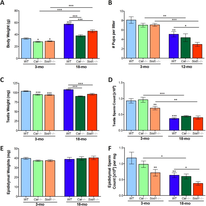
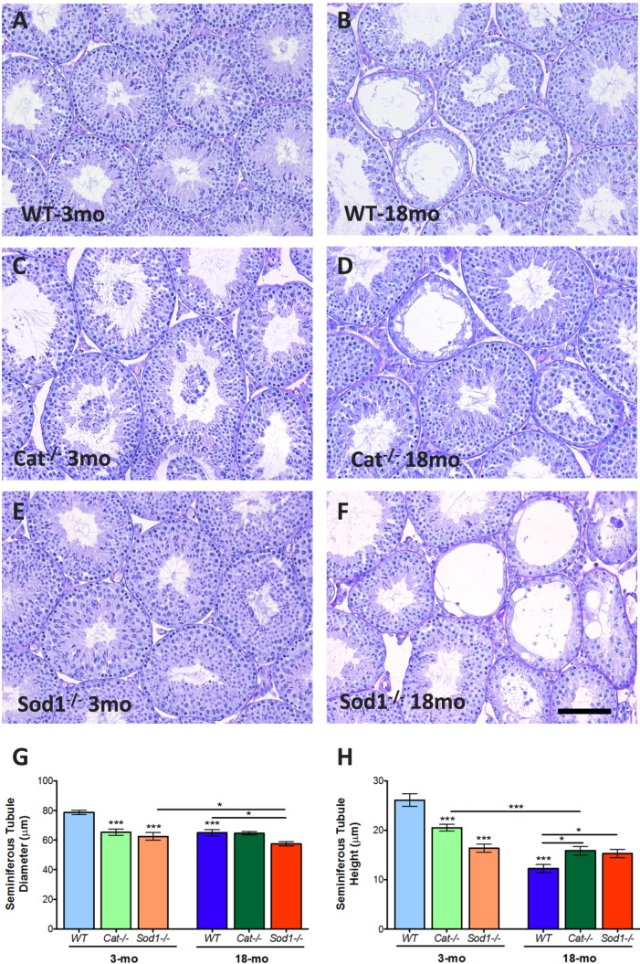
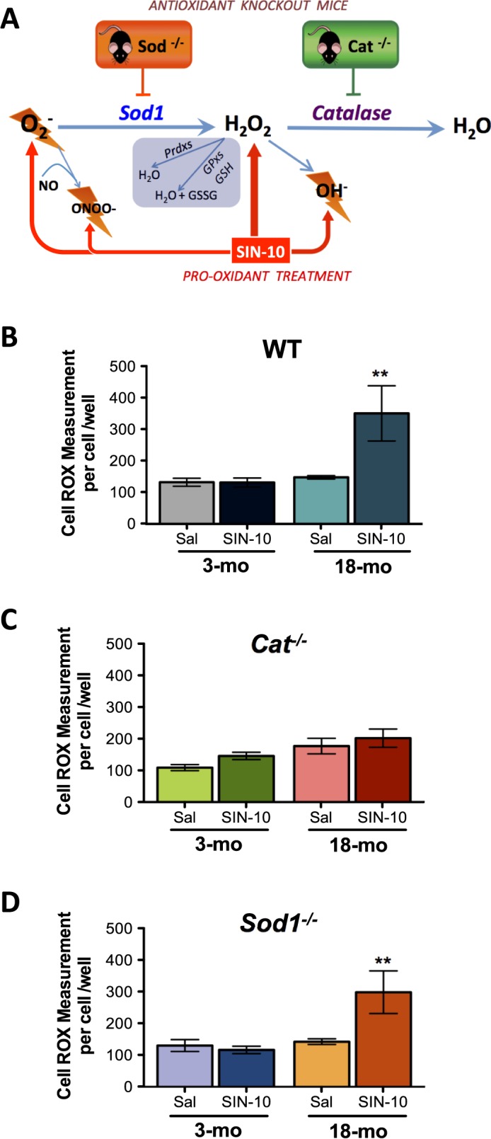
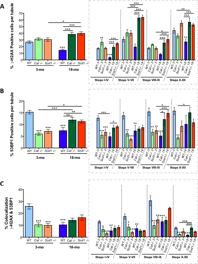
Advanced paternal age is linked to complications in pregnancy and genetic diseases in offspring. Aging results in excess reactive oxygen species (ROS) and DNA damage in spermatozoa; this damage can be transmitted to progeny with detrimental consequences. Although there is a loss of antioxidants with aging, the impact on aging male germ cells of the complete absence of either catalase (CAT) or superoxide dismutase 1 (SOD1) has not been investigated. We used CAT-null (Cat(-/-)) and SOD1-null (Sod(-/-)) mice to determine whether loss of these antioxidants increases germ cell susceptibility to redox dysfunction with aging. Aging reduced fertility and the numbers of Sertoli and germ cells in all mice. Aged Sod(-/-) mice displayed an increased loss of fertility compared to aged wild-type mice. Treatment with the pro-oxidant SIN-10 increased ROS in spermatocytes of aged wild-type and Sod(-/-) mice, while aged Cat(-/-) mice were able to neutralize this ROS. The antioxidant peroxiredoxin 1 (PRDX1) increased with age in wild-type and Cat(-/-) mice but was consistently low in young and aged Sod(-/-) mice. DNA damage and repair markers (γ-H2AX and 53BP1) were reduced with aging and lower in young Sod(-/-) and Cat(-/-) mice. Colocalization of γ-H2AX and 53BP1 suggested active repair in young wild-type mice but reduced in young Cat(-/-) and in Sod(-/-) mice and with age. Oxidative DNA damage (8-oxodG) increased in young Sod(-/-) mice and with age in all mice. These studies show that aged Sod(-/-) mice display severe redox dysfunction, while wild-type and Cat(-/-) mice have compensatory mechanisms to partially alleviate oxidative stress and reduce age-related DNA damage in spermatozoa. Thus, SOD1 but not CAT is critical to the maintenance of germ cell quality with aging.

Keywords: Sertoli cells; aging; oxidative stress; spermatogenesis.


Figures

FIG. 1
FIG. 1
Reproductive organ weights and testis and epididymal sperm counts in aged wild-type (WT), Cat−/− and Sod−/− mice. Body weights increased with age in WT, Cat−/−, and Sod−/− mice (A), while Cat−/−, and Sod−/− mice remained consistently lower in weight than WT mice. The numbers of pups per litter decreased with aging (B) and were significantly lower in aged Sod−/− mice. Testis weights (C) were reduced in Cat−/− and Sod−/− compared to WT mice. Testis sperm counts (D) were lower in young Sod−/− mice and all aged mice. Epididymal weights were not significantly changed (E); sperm count in the epididymis (F) reflected the changes observed in the testis sperm counts. Mean values with SEM error bars; two-way ANOVA with Bonferroni test; *P < 0.05; **P < 0.01; ***P < 0.001, n = 10–14 (A, B, D, F), n = 5–8 (E, G), n = 4–5 (C, F).
FIG. 2
FIG. 2
Histological analyses of young and aged Cat−/− and Sod−/− mouse testes. Testis histology in young and aged mice stained with PAS-hematoxylin (AF). Seminiferous tubule diameters (G) were reduced in knockout (KO) mice and in all mice with aging. The seminiferous tubule epithelium height (H) was reduced in KO mice, with the most extreme loss in height in Sod−/− mice. Loss of tubule height was observed with aging in all mice. Mean values with SEM error bars; two-way ANOVA with Bonferroni test; *P < 0.05; **P < 0.01; ***P < 0.001, n = 6 (AF); n = 8 (G, H). Bar = 100 μm.
FIG. 3
FIG. 3
Increased Sertoli cell loss in Sod−/− mice and with aging in all mice. Representative image with DAPI (nuclear stain), ZBTB16 (spermatogonial marker), and vimentin (Sertoli marker in the testis epithelium) (A). Imaris quantification of the number of germ cells per tubule (B) and the number of Sertoli cells per tubule (C) are also shown sorted by stage groupings. Mean values with error bars (±SEM); two-way ANOVA with Bonferroni test; *P < 0.05; **P < 0.01; ***P < 0.001; n = 4. Bar = 20 μm.
FIG. 4
FIG. 4
Redox dysfunction with aging in WT and Sod−/− spermatocytes and efficient management of oxidative stress in spermatocytes from Cat−/− mice following SIN-10 induced oxidative insult. Schematic of SIN-10 induced ROS generation (A). Overall ROS, measured using CellROX, showed efficient removal of ROS in all young animals, WT (B), Cat−/− (C), and Sod−/− (D). While aged Cat−/− mice responded to SIN-10 with removal of ROS, aged WT and Sod−/− did not efficiently remove ROS following SIN-10 treatment (D). Mean values shown with error bars (±SEM); one-way ANOVA with Newman-Keuls; **P < 0.01, n = 4–5.
FIG. 5
FIG. 5
Immunolocalization of PRDX1 to the developing acrosome and inhibition of the increase in PRDX1 with aging in Sod−/− mice. PRDX1 was localized in the seminiferous tubules to the cytoplasm of spermatocytes (portions of the images showing this are magnified in insets) and within the acrosome of spermatids (A) (see insets). Imaris quantified percentage of PRDX1-positive cells per tubule showed increased PRDX1 with age in WT and Cat−/− mice; however, Sod−/− mice did not display this increase in PRDX1 with age (B). Mean values shown with error bars (±SEM); two-way ANOVA with Bonferroni test; *P < 0.05; **P < 0.01; ***P < 0.001; n = 5. Bar = 20 μm.
FIG. 6
FIG. 6
Reduced DNA damage repair in seminiferous tubules of young Cat−/− and Sod−/− mice and in all aged mice. The percentage of γ-H2AX-positive cells per seminiferous tubule increased with age in knockout (KO) mice (A). The percentage of 53BP1-positive cells per tubule was reduced in young KO mice and increased in aged KO mice, while the opposite was seen in aged WT mice (B). Colocalization of γ-H2AX- and 53BP1-positive cells showed decreased DNA damage repair in young KO mice and in all aged mice (C). Mean values shown with error bars (±SEM); two-way ANOVA with Bonferroni test; *P < 0.05; **P < 0.01; ***P < 0.001; n = 5.
FIG. 7
FIG. 7
Elevated oxidative stress-induced DNA damage in young and aged Sod−/− mice. Sample immunofluorescence images of sperm heads, with 8-oxodG (green) counterstained with DAPI (blue); inset shows negative control (A). The quantified percentage of 8-oxodG-positive sperm heads showed an increase in 8-oxodG with aging in WT and an increased 8-oxodG in Cat−/− mice that remained at the same level with aging. The highest amounts of 8-oxodG were observed in Sod−/− mice in both the young and the aged groups. Mean values shown with error bars (±SEM); two-way ANOVA with Bonferroni multiple comparison test; *P < 0.05; ***P < 0.001; n = 5. Bar = 2.5 μm.

References

    1. Lawson G, Fletcher R. Delayed fatherhood. J Fam Plan Reprod Health Care. 2014;40:283–288. - PubMed
    1. Hassan MA, Killick SR. Effect of male age on fertility: evidence for the decline in male fertility with increasing age. Fertil Steril. 2003;79(suppl 3):1520–1527. - PubMed
    1. Serre V, Robaire B. Paternal age affects fertility and progeny outcome in the Brown Norway rat. Fertil Steril. 1998;70:625–631. - PubMed
    1. Kleinhaus K, Perrin M, Friedlander Y, Paltiel O, Malaspina D, Harlap S. Paternal age and spontaneous abortion. Obstet Gynecol. 2006;108:369–377. - PubMed
    1. Martin AK, Robinson G, Reutens D, Mowry B. Clinical and parental age characteristics of rare copy number variant burden in patients with schizophrenia. Am J Med Genet B Neuropsychiatr Genet. 2015;168B:374–382. - PubMed

Publication types

MeSH terms