. 2016 Jul 28:6:30405.
doi: 10.1038/srep30405.

Melanoma dormancy in a mouse model is linked to GILZ/FOXO3A-dependent quiescence of disseminated stem-like cells

Affiliations

Melanoma dormancy in a mouse model is linked to GILZ/FOXO3A-dependent quiescence of disseminated stem-like cells

Yasmine Touil et al. Sci Rep. .

Abstract

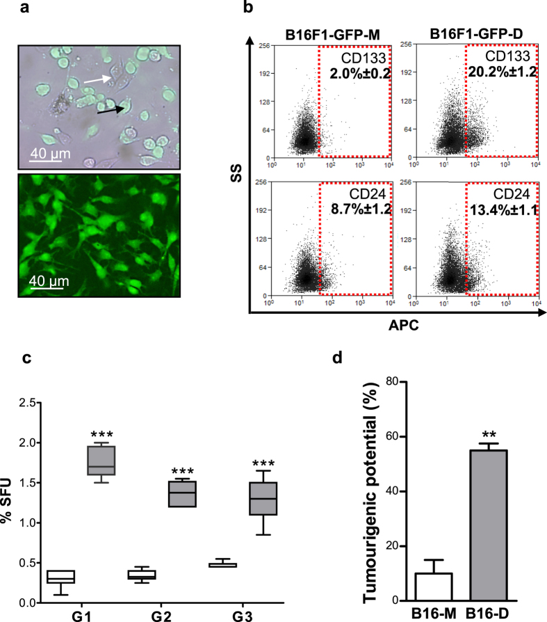
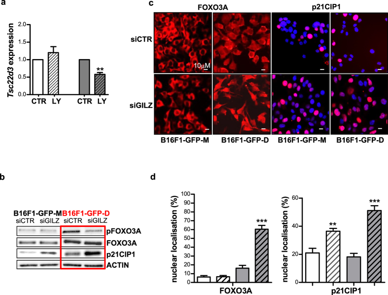
Metastatic cancer relapses following the reactivation of dormant, disseminated tumour cells; however, the cells and factors involved in this reactivation are just beginning to be identified. Using an immunotherapy-based syngeneic model of melanoma dormancy and GFP-labelled dormant cell-derived cell lines, we determined that vaccination against melanoma prevented tumour growth but did not prevent tumour cell dissemination or eliminate all tumour cells. The persistent disseminated melanoma tumour cells were quiescent and asymptomatic for one year. The quiescence/activation of these cells in vitro and the dormancy of melanoma in vivo appeared to be regulated by glucocorticoid-induced leucine zipper (GILZ)-mediated immunosuppression. GILZ expression was low in dormant cell-derived cultures, and re-expression of GILZ inactivated FOXO3A and its downstream target, p21CIP1. The ability of dormancy-competent cells to re-enter the cell cycle increased after a second round of cellular dormancy in vivo in association with shortened tumour dormancy period and faster and more aggressive melanoma relapse. Our data indicate that future cancer treatments should be adjusted according to the stage of disease progression.


Figures

Figure 1
Figure 1. Vaccination with irradiated B16F1 murine melanoma cells expressing GM-CSF elicited anti-melanoma activity and improved survival of C57BL6/rj mice.
(a) Schema of the vaccination protocol: C57BL6/rj mice were injected i.d. twice a week for 19 days (red arrows) with 1 × 106 irradiated B16F1 cells either expressing GM-CSF (B16F1-GM-CSF) or carrying an empty pcDNA3.1-Zeo vector, (not shown for clarity). The mice were challenged (green arrow) with B16F1 cells expressing GFP (B16F1-GFP, s.c. injection) at one week after initiation of the vaccination protocol, and the organs of surviving mice were analysed 350 days later. (b) Survival rates of 40 mice injected with 1 × 106 irradiated B16F1-pcDNA3.1-Zeo control cells (black) and 55 mice injected with 1 × 106 irradiated B16F1-GM-CSF cells (red) after challenge with 2 × 103 (▲) or 5 × 103 (●) B16F1-GFP cells. The results are presented as the means ± SEM of 3 independent experiments. The red arrows indicate the days when the mice were vaccinated (7 and 3 days before the challenge, indicated as “0”, and 4, 7 and 12 days after the challenge).
Figure 2
Figure 2. The dormant B16F1-GFP cell line was enriched in melanoma cells with stem cell properties.
(a) Fluorescent microscopic images of FACS-sorted GFP-positive dormant cells isolated from organs of tumour-free mice, cultured for 23 (up) days and then cloned (down). The white and black arrows indicate GFP-negative and GFP-positive cells, respectively. (b) Flow cytometry dot plot profiles of the stem cell surface markers, CD133 (up) and CD24 (down), expressed by both the maternal B16F1-GFP-M (left) and dormant DMC-derived B16F1-GFP-D (right) clonal cell lines. The numbers represent the mean percentage ± SEM. (c) Box plot of sphere-forming units (SFUs) formed by B16F1-GFP-M (white) and B16F1-GFP-D (grey) cells plated at a clonal density of 1000 cells/ml. SFUs were calculated according to the following formula: % SFU = number of spheres/number of plated cellsx100. Self-renewal ability was evaluated by dissociating the spheres and then re-plating the sphere cell suspensions for 3 successive sphere generations (G1–G3). The graphs represent 3 independent experiments performed in quadruplicate. ***p < 0.001. (d) Dormant DMC-derived B16F1-GFP-D cells (B16-D) had a higher tumourigenic potential than maternal B16F1-GFP-M cells (B16-M). The mice were injected s.c. with 200 B16F1-GFP-M or B16F1-GFP-D cells. The results are expressed as the per cent of mice bearing tumours to the total number of injected mice. The histograms represent the means ± SEM of 2 independent experiments. **p < 0.01.
Figure 3
Figure 3. The brain microenvironment preselected dormant DMC-derived cells with a new phenotype.
Dormant DMC-derived B16F1-GFP-D cells persisting asymptomatically in the brains of mice bearing primary tumours overexpressed factors that potentially facilitated their in vitro propagation. RT-PCR was performed to analyse Ccnd2, Rxrb, Nupr1, Cdc25a and Id3 expression in B16F1-GFP-DB #1, #2 and #3 brain-derived GFP+ cells and in their respective primary tumours #1, #2 and #3. The values represent the levels of each transcript in the brain-derived B16F1-GFP-DB cells relative to the average levels of the corresponding transcripts (set to 1) in three B16F1-GFP-D-initiated tumours.
Figure 4
Figure 4. Down-regulation of GILZ expression induced quiescence of dormant DMC-derived B16F1-GFP-D cells in the G0 phase.
(a) Cell cycle analysis of dormant DMC-derived B16F1-GFP-D (lower panel) and maternal B16F1-GFP-M (upper panel) cells transfected with control (left) or GILZ-specific (right) siRNA revealed that the dormant DMC-derived cells possessed the novel, GILZ-dependent ability to control the G0-to-G1 transition in addition to the known GILZ-dependent ability to control the G1-to-S transition of the cell cycle. A fraction of cells in the G0 phase of the cell cycle are shown in red. (b) Sphere-forming units (SFUs) formed by B16F1-GFP-D (grey) or B16F1-GFP-M (white) cells transfected with control and GILZ-specific siRNA. (c) qRT-PCR assay of Tsc22d3 expression in spheres obtained from B16F1-GFP-M and B16F1-GFP-D cells transfected with either control or GILZ-specific siRNA and cultured for 7 days. The values indicate the level of GILZ-encoding mRNA relative to the control (B16-F1GFP-M adherent cells) (ΔΔCt); siCTR (control siRNA), siGILZ (GILZ-specific siRNA). The results represent 3 independent experiments. *p < 0.05; **p < 0.01; ***p < 0.001.
Figure 5
Figure 5. GILZ knockdown delayed tumour formation and increased tumourigenicity in syngeneic mice.
Twenty C57BL6/rj mice were injected (s.c.) with 5 × 103 B16F1-GFP-M or B16F1-GFP-D cells after transient transfection with control siRNA (siCTR) or GILZ-specific siRNA (siGILZ). (a) Delayed tumour appearance (mean ± SEM) was observed under the indicated conditions (n = 5). ***p < 0.001 vs. B16F1-GFP-M siCTR cells; §§§p < 0.001 vs. B16F1-GFP-D siCTR cells. (b) Graph showing the tumour sizes at each time point after transient transfection with control (●) or GILZ-specific (■) siRNA in B16F1-GFP-M (black) and B16F1-GFP-D (red) cells. The numbers in the upper left corner of the graph indicate the number of mice with tumours/the total number of mice; C = control siRNA, and G = GILZ-specific siRNA. Tumour size was evaluated as described in the Methods. (c) qRT-PCR assay of Tsc22d3 expression in B16F1-GFP-M (white) and B16F1-GFP-D cells (grey) transfected with control (siCTR) or GILZ-specific (siGILZ, stripes) siRNA. **p < 0.01; *p < 0.05 (n = 5). (d) The percentage of mice bearing human HBL melanoma tumours and the delay in tumour appearance. CB17-SCID mice were injected (s.c.) with 5 × 106 HBL H2B-GFP cells transfected with either control or GILZ-specific siRNA (5 mice per condition).
Figure 6
Figure 6. Down-regulation of GILZ induced regulators of quiescence.
(a) Inactivation of the PI3K/AKT pathway via incubation in10 μM LY294002 (LY) for 24 h inhibited Tsc22d3 expression in dormant DMC-derived B16-F1-GFP-D (right) and B16-F1-GFP-M cells (left) **p < 0.01 (n = 3). (b) Western blot analysis of relevant proteins affected by GILZ down-regulation in the maternal and dormant cell-derived cell lines; pFOXO3A denotes phosphorylated FOXO3A. (c) Down-regulation of GILZ affects the subcellular localisation of quiescence-related factors. Representative immunofluorescence images generated using anti-p21CIP1 and anti-FOXO3A (red) antibodies are shown. Hoechst 33342 (blue) was used for nuclear counterstaining. (d) Quantification by counting of the immunostained nuclei shown in (c); maternal cells (white), dormant cells (grey), GILZ KD (stripes); 4–15 fields were counted by 3 independent investigators. *p < 0.05; ***p < 0.001.
Figure 7
Figure 7. Down-regulation of GILZ induced cellular quiescence of human melanoma in vitro and in vivo.
Human melanoma HBL-H2B-GFP cells were treated with tetracycline to induce the cell division-tracking H2B-GFP protein, a marker of quiescence, and were subsequently transfected transiently with control (siCTR) or GILZ-specific (siGILZ) siRNA. (a) qRT-PCR analysis of GILZ expression in FACS-sorted melanoma GFPlow fast-cycling cells and GFPhigh quiescent cells. *p < 0.05 (n = 2). (b) Flow cytometry analysis of the cell cycle in control (siCTR) and GILZ down-regulated (siGILZ) human melanoma cells. (c) Immunoblots with an anti-phosphorylated (S253) FOXO3A (pFOXO3A) antibody after transfection of human melanoma HBL-H2B-GFP cells with control or human GILZ-specific siRNA. (d) Graphs showing the changes in the sphere-forming units (SFUs) and colony-forming units (CFUs) of human melanoma HBL cells after the down-regulation of GILZ expression. ***p < 0.001, **p < 0.01 (n = 3). (e) qRT-PCR analysis of relative TYROSINASE and TSC22D3 expression in 6 human melanoma samples obtained from patients and in human melanoma HBL H2B-GFP cells treated with siCTR and siGILZ.

References

    1. Bissell M. J. & Hines W. C. Why don’t we get more cancer? A proposed role of the microenvironment in restraining cancer progression. Nat. Med. 17, 320–329 (2011). - PMC - PubMed
    1. Ghajar C. Metastasis prevention by targeting the dormant niche. Nat. Rev. Cancer 15, 238–247 (2015). - PMC - PubMed
    1. Giancotti F. G. Mechanisms governing metastatic dormancy and reactivation. Cell 155, 750–764 (2013). - PMC - PubMed
    1. Ossowski L. & Aguirre-Ghiso J. A. Dormancy of metastatic melanoma. Pigment Cell Melanoma Res. 23, 41–56 (2010). - PMC - PubMed
    1. Kleffel S. & Schatton T. Tumor dormancy and cancer stem cells: two sides of the same coin? Adv. Exp. Med. Biol. 734, 145–179 (2013). - PubMed

Publication types

MeSH terms