. 2016 Jul 28:6:30568.
doi: 10.1038/srep30568.

Magnetic Resonance Imaging of Phosphocreatine and Determination of BOLD Kinetics in Lower Extremity Muscles using a Dual-Frequency Coil Array

Affiliations

Magnetic Resonance Imaging of Phosphocreatine and Determination of BOLD Kinetics in Lower Extremity Muscles using a Dual-Frequency Coil Array

Ryan Brown et al. Sci Rep. .

Abstract

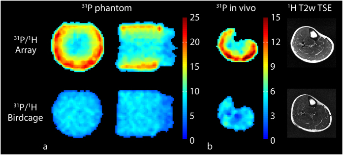
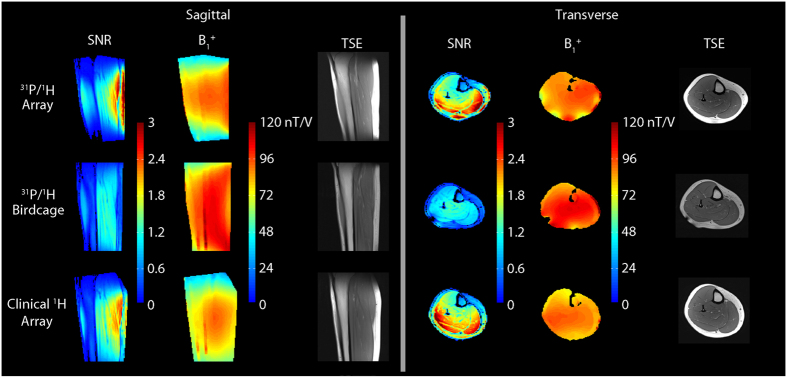
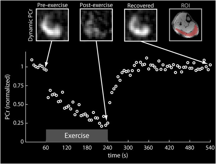
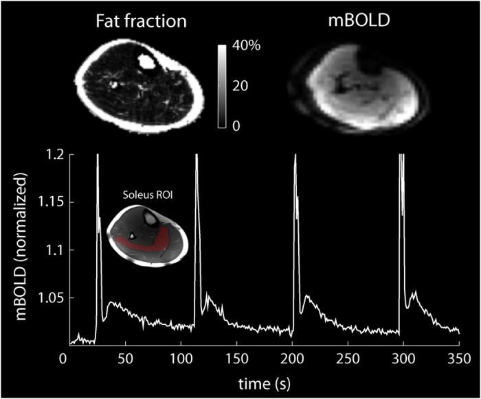
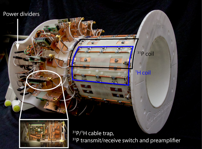
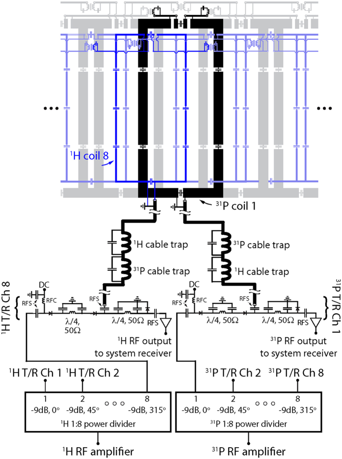
Magnetic resonance imaging (MRI) provides the unique ability to study metabolic and microvasculature functions in skeletal muscle using phosphorus and proton measurements. However, the low sensitivity of these techniques can make it difficult to capture dynamic muscle activity due to the temporal resolution required for kinetic measurements during and after exercise tasks. Here, we report the design of a dual-nuclei coil array that enables proton and phosphorus MRI of the human lower extremities with high spatial and temporal resolution. We developed an array with whole-volume coverage of the calf and a phosphorus signal-to-noise ratio of more than double that of a birdcage coil in the gastrocnemius muscles. This enabled the local assessment of phosphocreatine recovery kinetics following a plantar flexion exercise using an efficient sampling scheme with a 6 s temporal resolution. The integrated proton array demonstrated image quality approximately equal to that of a clinical state-of-the-art knee coil, which enabled fat quantification and dynamic blood oxygen level-dependent measurements that reflect microvasculature function. The developed array and time-efficient pulse sequences were combined to create a localized assessment of calf metabolism using phosphorus measurements and vasculature function using proton measurements, which could provide new insights into muscle function.


Conflict of interest statement

RB discloses the US patent, “Multi-Nuclei MRI Coil,” 13/866,728,2013, which is related to this work. PP and OK declare no financial conflicts of interest.

Figures

Figure 1
Figure 1. Characterization of the eight-channel 31P module of the developed array.
The array provided a similar 31P SNR in the center and greater than 2-fold gain in the periphery over the birdcage in the 42 mM Pi phantom (a). Similar results were observed in vivo (b). The measurements are summarized in Table 1.
Figure 2
Figure 2. Normalized 1H SNR maps (first and fourth columns), B1+ maps (second and fifth columns), and TSE images (third and last columns) acquired with the eight-channel 1H module (top row) of the developed array.
The array showed favorable performance over the dual-frequency birdcage coil (middle) and similar performance to the clinical 15-channel 1H array (bottom). The measurements are summarized in Table 2.
Figure 3
Figure 3. Dynamic 31P PCr (top row) images acquired using the developed coil array in the calf muscle at different time points during the plantar flexion exercise protocol with the PCr signal time course (bottom) from the segmented gastrocnemius muscle (top right).
Figure 4
Figure 4. In vivo fat fraction and mBOLD images (top row) and mBOLD signal evolution in the soleus muscle (bottom row, ROI inset) following maximum voluntary isometric plantar flexions.
Figure 5
Figure 5. Photograph of the developed 31P/1H array with the protective cover removed.
Overlays highlight an interface board that accommodates the cable trap, transmit/receive switch, and the preamplifier for each coil as well as the power dividers, a 31P coil (black), and an 1H coil (blue).
Figure 6
Figure 6. Unrolled schematic diagram of the 31P/1H coil array and interface.
For simplicity, single 31P (black) and 1H (blue) coils of the 16-channel nested array are highlighted whereas neighboring elements are displayed in a semi-transparent fashion. Abbreviations: RFC = radio frequency choke and RFS = radio frequency short.

Similar articles

Cited by

References

    1. Kemp G., Ahmad R., Nicolay K. & Prompers J. Quantification of skeletal muscle mitochondrial function by 31P magnetic resonance spectroscopy techniques: a quantitative review. Acta Physiologica 213, 107–144 (2015). - PubMed
    1. Arnold D. L., Matthews P. M. & Radda G. K. Metabolic recovery after exercise and the assessment of mitochondrial function in vivo in human skeletal-muscle by means of 31P NMR. Magnetic Resonance in Medicine 1, 307–315, 10.1002/mrm.1910010303 (1984). - DOI - PubMed
    1. Kemp G. J., Taylor D. J. & Radda G. K. Control of phosphocreatine resynthesis during recovery from exercise in human skeletal-muscle. Nmr in Biomedicine 6, 66–72, 10.1002/nbm.1940060111 (1993). - DOI - PubMed
    1. Prompers J. J. et al.. Dynamic MRS and MRI of skeletal muscle function and biomechanics. NMR in Biomedicine 19, 927–953, 10.1002/nbm.1095 (2006). - DOI - PubMed
    1. Kemp G. J., Meyerspeer M. & Moser E. Absolute quantification of phosphorus metabolite concentrations in human muscle in vivo by P-31 MRS: a quantitative review. Nmr in Biomedicine 20, 555–565, 10.1002/nbm.1192 (2007). - DOI - PubMed

Publication types

Substances