

Size and dose dependent effects of silver nanoparticle exposure on intestinal permeability in an in vitro model of the human gut epithelium
- PMID: 27465730
- PMCID: PMC4963959
- DOI: 10.1186/s12951-016-0214-9
Size and dose dependent effects of silver nanoparticle exposure on intestinal permeability in an in vitro model of the human gut epithelium
Abstract
Background: The antimicrobial activity of silver nanoparticles (AgNP) has led to interest in their use in consumer products such as food contact materials, utensils, and storage containers. Incorporation of these materials into items intended for food processing and storage suggests that consumer use of these products could result in gastrointestinal exposure to AgNP, should the nanoparticles migrate from the product. The health impact of AgNP exposure is unknown, especially effects related to intestinal epithelial permeability and barrier function. This study examined the effects of AgNP exposure of different sizes (10, 20, 75 and 110 nm) and doses (20 and 100 µg/mL) on the permeability of T84 human colonic epithelial cells, which serve as an in vitro model of the human gut epithelium.
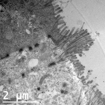
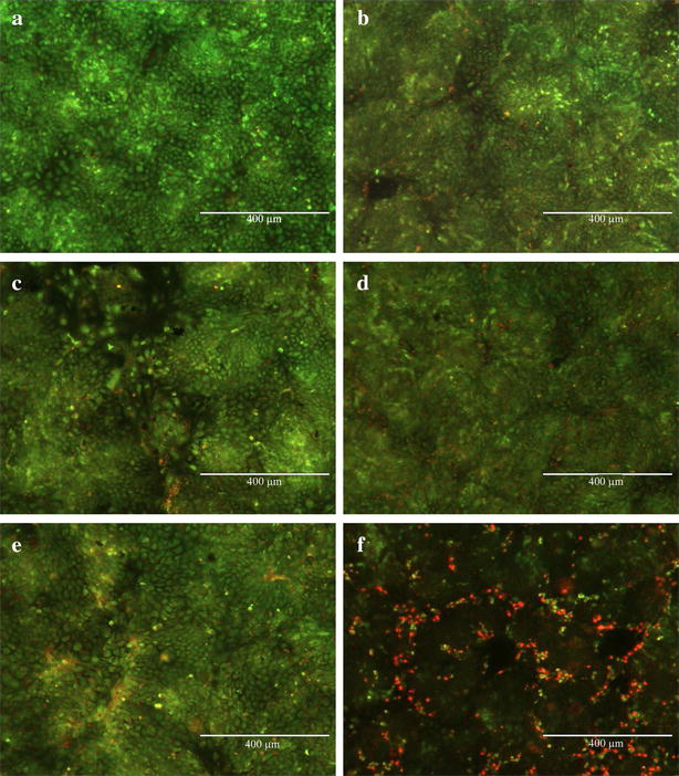
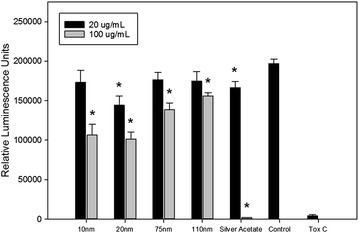
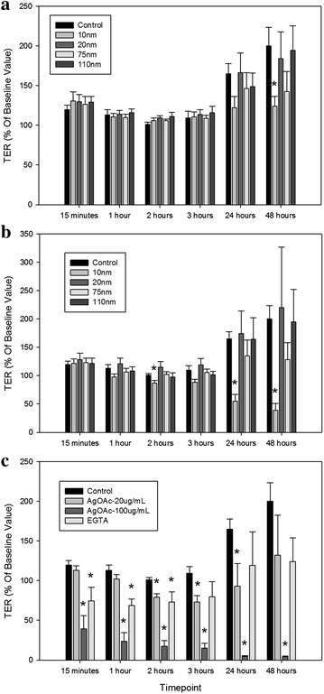
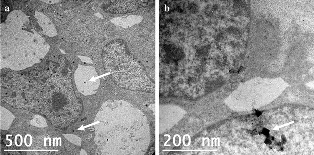
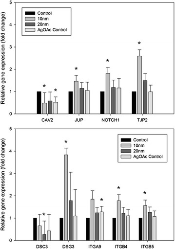
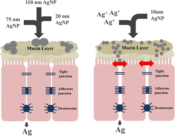
Results: Results showed that effects of AgNP on the T84 epithelial cells were size- and dose-dependent, with the 10 nm AgNP causing the most significant changes. Changes in permeability of the epithelial cell monolayer, as measured by transepithelial electrical resistance, after exposure to 10 nm AgNP were most dramatic at the highest dose (100 µg/mL), but also observed at the lower dose (20 µg/mL). AgNP could be visualized inside cells using transmission electron microscopy and silver was detected in basal wells using inductively coupled plasma-mass spectrometry. Exposure to AgNP significantly affected the expression of genes involved in anchoring tight junctions, cellular proliferation and signaling, endocytosis, and cell-cell adhesion, with the 10 nm AgNP having the greatest effect.
Conclusions: The results of this study show that small-size AgNP have significant effects on intestinal permeability in an in vitro model of the human gastrointestinal epithelium. Such effects have the potential to compromise the integrity of the intestinal epithelium and this disruption of barrier function could have health consequences for the gastrointestinal tract.
Keywords: Barrier function; Cell junctions; Intestinal permeability; Silver nanoparticles.
Figures

Similar articles
-
Alteration in the mRNA expression of genes associated with gastrointestinal permeability and ileal TNF-α secretion due to the exposure of silver nanoparticles in Sprague-Dawley rats.J Nanobiotechnology. 2019 May 13;17(1):63. doi: 10.1186/s12951-019-0499-6. J Nanobiotechnology. 2019. PMID: 31084603 Free PMC article.
-
Effects of subchronic exposure of silver nanoparticles on intestinal microbiota and gut-associated immune responses in the ileum of Sprague-Dawley rats.Nanotoxicology. 2015 May;9(3):279-89. doi: 10.3109/17435390.2014.921346. Epub 2014 May 30. Nanotoxicology. 2015. PMID: 24877679
-
Human Intestinal Tissue Explant Exposure to Silver Nanoparticles Reveals Sex Dependent Alterations in Inflammatory Responses and Epithelial Cell Permeability.Int J Mol Sci. 2020 Dec 22;22(1):9. doi: 10.3390/ijms22010009. Int J Mol Sci. 2020. PMID: 33374948 Free PMC article.
-
Pro-inflammatory effects of silver nanoparticles in the intestine.Arch Toxicol. 2022 Jun;96(6):1551-1571. doi: 10.1007/s00204-022-03270-w. Epub 2022 Mar 16. Arch Toxicol. 2022. PMID: 35296919 Review.
-
Mucus and microbiota as emerging players in gut nanotoxicology: The example of dietary silver and titanium dioxide nanoparticles.Crit Rev Food Sci Nutr. 2018 Apr 13;58(6):1023-1032. doi: 10.1080/10408398.2016.1243088. Epub 2017 Jun 12. Crit Rev Food Sci Nutr. 2018. PMID: 27740849 Review.
Cited by
-
Influence of Physicochemical Characteristics and Stability of Gold and Silver Nanoparticles on Biological Effects and Translocation across an Intestinal Barrier-A Case Study from In Vitro to In Silico.Nanomaterials (Basel). 2021 May 21;11(6):1358. doi: 10.3390/nano11061358. Nanomaterials (Basel). 2021. PMID: 34063963 Free PMC article.
-
Responses of intestinal virome to silver nanoparticles: safety assessment by classical virology, whole-genome sequencing and bioinformatics approaches.Int J Nanomedicine. 2018 May 16;13:2857-2867. doi: 10.2147/IJN.S161379. eCollection 2018. Int J Nanomedicine. 2018. PMID: 29844669 Free PMC article.
-
Alteration in the mRNA expression of genes associated with gastrointestinal permeability and ileal TNF-α secretion due to the exposure of silver nanoparticles in Sprague-Dawley rats.J Nanobiotechnology. 2019 May 13;17(1):63. doi: 10.1186/s12951-019-0499-6. J Nanobiotechnology. 2019. PMID: 31084603 Free PMC article.
-
Biotransformation of Silver Nanoparticles into Oro-Gastrointestinal Tract by Integrated In Vitro Testing Assay: Generation of Exposure-Dependent Physical Descriptors for Nanomaterial Grouping.Nanomaterials (Basel). 2021 Jun 17;11(6):1587. doi: 10.3390/nano11061587. Nanomaterials (Basel). 2021. PMID: 34204296 Free PMC article.
-
Glutathione-Stabilized Silver Nanoparticles: Antibacterial Activity against Periodontal Bacteria, and Cytotoxicity and Inflammatory Response in Oral Cells.Biomedicines. 2020 Sep 23;8(10):375. doi: 10.3390/biomedicines8100375. Biomedicines. 2020. PMID: 32977686 Free PMC article.
References
-
- Schafer B, Brocke JV, Epp A, Gotz M, Herzberg F, Kneuer C, Sommer Y, Tentschert J, Noll M, Gunther I, et al. State of the art in human risk assessment of silver compounds in consumer products: a conference report on silver and nanosilver held at the BfR in 2012. Arch Toxicol. 2013;87:2249–2262. doi: 10.1007/s00204-013-1083-8. - DOI - PMC - PubMed
MeSH terms
Substances
LinkOut - more resources
Full Text Sources
Other Literature Sources
