. 2016 Sep 15;25(18):3908-3924.
doi: 10.1093/hmg/ddw232. Epub 2016 Jul 27.

The chaperone HSPB8 reduces the accumulation of truncated TDP-43 species in cells and protects against TDP-43-mediated toxicity

Affiliations

The chaperone HSPB8 reduces the accumulation of truncated TDP-43 species in cells and protects against TDP-43-mediated toxicity

Valeria Crippa et al. Hum Mol Genet. .

Abstract

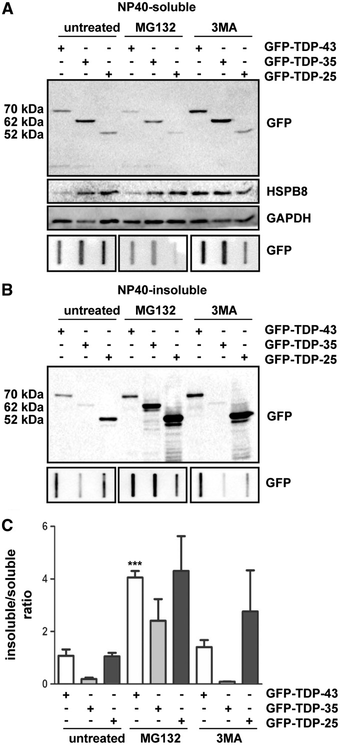
Aggregation of TAR-DNA-binding protein 43 (TDP-43) and of its fragments TDP-25 and TDP-35 occurs in amyotrophic lateral sclerosis (ALS). TDP-25 and TDP-35 act as seeds for TDP-43 aggregation, altering its function and exerting toxicity. Thus, inhibition of TDP-25 and TDP-35 aggregation and promotion of their degradation may protect against cellular damage. Upregulation of HSPB8 is one possible approach for this purpose, since this chaperone promotes the clearance of an ALS associated fragments of TDP-43 and is upregulated in the surviving motor neurones of transgenic ALS mice and human patients. We report that overexpression of HSPB8 in immortalized motor neurones decreased the accumulation of TDP-25 and TDP-35 and that protection against mislocalized/truncated TDP-43 was observed for HSPB8 in Drosophila melanogaster Overexpression of HSP67Bc, the functional ortholog of human HSPB8, suppressed the eye degeneration caused by the cytoplasmic accumulation of a TDP-43 variant with a mutation in the nuclear localization signal (TDP-43-NLS). TDP-43-NLS accumulation in retinal cells was counteracted by HSP67Bc overexpression. According with this finding, downregulation of HSP67Bc increased eye degeneration, an effect that is consistent with the accumulation of high molecular weight TDP-43 species and ubiquitinated proteins. Moreover, we report a novel Drosophila model expressing TDP-35, and show that while TDP-43 and TDP-25 expression in the fly eyes causes a mild degeneration, TDP-35 expression leads to severe neurodegeneration as revealed by pupae lethality; the latter effect could be rescued by HSP67Bc overexpression. Collectively, our data demonstrate that HSPB8 upregulation mitigates TDP-43 fragment mediated toxicity, in mammalian neuronal cells and flies.


Figures

Figure 1.
Figure 1.
GFP-tagged truncated TDP-35 and TDP-25 show different biochemical behaviour from that of full-length TDP-43. (A) Confocal microscopy analysis (63× magnification) of mouse motor neurone NSC34 cells transiently overexpressing GFP-TDP-43, GFP-TDP-35, GFP-TDP-25, or GFP alone (pEGFP-N1, used as a control). DAPI: nuclei staining. A 2.5x magnification of selected areas is shown. (B): Western Blot (WB; upper insets) and filter retardation assay (FRA, lower inset) of the PBS extracts of mouse motor neurone NSC34 cells transiently overexpressing the GFP-TDP-43, GFP-TDP-35 or GFP-TDP-25. (C) Quantification of the PBS extract by FRA (***P < 0.001 vs GFP-TDP-43). The data represent in each case the mean ± sem of n = 3 independent samples. (D): WB (upper insets) and FRA (lower insets) of the NP40 extracts of cells transiently overexpressing GFP-TDP-43, GFP-TDP-35 or GFP-TDP-25 (left insets, NP40-soluble fraction; right insets, NP40-insoluble fraction). (E): Quantification of FRA NP40 insoluble/soluble ratio (°°°P < 0.001 vs GFP-TDP-43; * P < 0.05 vs GFP-TDP-43; ### P < 0.001 vs GFP-TDP-35). The data again represent the mean ± sem of n=3 independent samples.
Figure 2.
Figure 2.
GFP-TDP-35 and GFP-TDP-25 show different clearance from that of full-length GFP-TDP-43. (A) WB (upper insets) and FRA (lower insets) of the NP40-soluble extracts of mouse motor neurone NSC34 cells transiently overexpressing GFP-TDP-43, GFP-TDP-35 or GFP-TDP-25 and treated with 10 µM MG132 for 16 h or 10 mM 3-methyladenine (3-MA) for 32 h. (B) WB (upper insets) and FRA (lower insets) of NP40-insoluble extracts. (C) Quantification of FRA NP40 insoluble/soluble ratios (***P < 0.001 vs GFP-TDP-43). The data represent the mean ± sem of n=3 independent samples.
Figure 3.
Figure 3.
HSPB8 affects the accumulation of GFP-TDP-35 and GFP-TDP-25 fragments. (A,B) Confocal microscopy analysis (63× magnification) of NSC34 cells transiently co-transfected with plasmids coding for GFP-TDP-43, GFP-TDP-35 or GFP-TDP-25 and pCDNA3 (A) or hHSPB8 plasmid (B). DAPI: nuclei staining. RED: murine endogenous HSPB8 (mHSPB8) or human transfected HSPB8 (hHSPB8). A 2.5× magnification of selected areas is shown. (C) WB of the NP40-soluble extracts of mouse motor neurone NSC34 cells transiently overexpressing GFP-TDP-43, GFP-TDP-35 or GFP-TDP-25 and human HSPB8. (D): WB of the NP40-insoluble extracts. E-G: Representative FRA (upper panels) and FRA quantification (lower panels) of the NP40-soluble and NP40-insoluble extracts of mouse motor neurone NSC34 cells stably overexpressing GFP-TDP-43 (E), GFP-TDP-35 (F) or GFP-TDP-25 (G), induced for 72 h with 1 μg/ml doxycycline and transfected with scramble or siRNA-HSPB8; the cells were treated with 10 μM MG132 and 10 mM 3-MA for the last 16 h prior to extraction. The optical densities of GFP-TDPs were expressed relative to the mean optical densities of the corresponding samples treated with doxycycline and a scramble RNAi sequence, taken as internal references. The data represent the mean ± sem of n = 3 independent samples. (*P < 0.05 vs scramble; ** P < 0.01 vs scramble).
Figure 4.
Figure 4.
Accumulation and clearance of FLAG-tagged TDP-43, TDP-35 and TDP-25. (A) Confocal microscopy analysis (63× magnification) of mouse motor neurone NSC34 control cells or cells transiently overexpressing 2xFLAG-TDP-43, 2xFLAG-TDP-35, 2xFLAG-TDP-25. DAPI: nucleic acid staining. A 2.5x magnification image of selected areas is shown. (B) WB (upper insets) and FRA (lower insets and FRA quantification, bar graph) of the PBS extracts of mouse motor neurone NSC34 cells transiently overexpressing the 2xFLAG-TDP-43, 2xFLAG-35 or 2xFLAG-TDP-25. The data represent the mean ± sem of n = 3 independent samples. ** P < 0.001 vs 2xFLAG-TDP-43; ° P < 0.05 vs 2xFLAG-TDP-35. (C-E): WB (upper insets) and FRA (lower insets and FRA quantification, bar graph) of the PBS extracts of mouse motor neurone NSC34 cells transiently overexpressing 2xFLAG-TDP-43 (C), 2xFLAG-35 (D) or 2xFLAG-TDP-25 (E) and treated with 10 µM MG132 for 16 h or 10 mM 3-methyladenine (3-MA) for 32 h (**P < 0.001 or * P < 0.01 vs untreated controls). The data represent the mean ± sem of n = 3 independent samples. (F-I): NSC34 cells transiently co-transfected with plasmids coding for 2xFLAG-TDP-43, 2xFLAG-TDP-35 or 2xFLAG-TDP-25 and pCDNA3 or hHSPB8. WB and representative FRA (F). FRA quantification (G, TDP-43; H, TDP-35; I, TDP-25) (**P < 0.001 vs pCDNA3 transfected cells). The data represent the mean ± sem of n=3 independent samples.
Figure 5.
Figure 5.
HSP67Bc rescues mutant eye degeneration mediated by TDP-43-NLS. (A) Representative pictures showing eyes from 1-day-old flies expressing human TDP-43 under the control of the gmr-GAL4 driver alone (TDP-43 wt: GMR-TDP-43 wt/w1118) or with V5-HSP67Bc. (B) WB showing expression levels of TDP-43 and V5-HSP67Bc (*corresponds to V5-tagged HSP67Bc). α-tubulin was used as the loading control. (C) Top row, Representative pictures showing eyes from control flies (Control: GMR-GAL4/w1118) or flies expressing human NLS-mutant TDP-43 under the control of the gmr-GAL4 driver alone (TDP-43 NLS: GMR-TDP-43 NLS/w1118) or with V5-HSP67Bc. The eyes of flies expressing mutant TDP-43 NLS show partial loss of ommatidia, a rough phenotype and depigmentation, while co-expression with HSP67Bc rescues the eye pigmentation and ommatidia organization. Bottom row, corresponding Richardson's stained frontal sections showing mutation-dependent internal degeneration that is rescued by overexpression of HSP67Bc. The quantification of the extent of eye degeneration (partially re-pigmented eyes and rough eyes) is reported (***P<0.001; V5-HSP67Bc expressing flies versus control flies). Between 91 and 126 eyes/fly genotype were scored (n = 3 ± sem). (D) WB showing expression levels of mutant TDP-43 NLS and HS67Bc in the eyes of 1-day-old flies. α-tubulin served as the loading control. The quantification of TDP-43 NLS protein levels is shown (***P<0.001; V5-HSP67Bc expressing flies versus control flies). For the analysis of TDP-43-NLS protein, n = 3–5 independent samples ± sem; each sample was obtained from 10 to 12 fly heads.
Figure 6.
Figure 6.
Downregulation of HSP67Bc increases TDP-43-mediated toxicity and impairs protein homeostasis. (A) Top and middle row, representative pictures showing eyes from flies expressing human NLS-mutant TDP-43 under the control of the gmr-GAL4 driver alone (TDP-43 NLS: GMR-TDP-43 NLS/w1118) or with two independent sequences silencing endogenous HSP67Bc (RNAi#1 or RNAi#2). Bottom row, corresponding Richardson's stained frontal sections showing internal degeneration in flies with HSP67Bc depletion. The quantification of the extent of eye degeneration (rough eye, dark brown eye or eye with patches) is shown (***P < 0.001; ** P < 0.01; HSP67Bc-depleted flies versus control flies). 50–100 eyes/fly genotypes were scored (n = 3 ± sem). (B): WB and data quantitation showing expression levels of mutant NLS TDP-43, high molecular weight (MW) species and endogenous ubiquitin in the eyes of 1-day-old flies. α-tubulin was used as the loading control (***P < 0.001; ** P < 0.01; * P < 0.05; HSP67Bc-depleted flies versus control). For the analysis of monomeric TDP-43, high molecular weight (MW) TDP-43 and ubiquitinated protein, n=3–6 independent samples ± sem; each sample was obtained from 10 to 12 fly heads. (C) WB and data quantitation showing expression levels of endogenous ubiquitin and HSP67Bc in the eyes of 1-day-old control flies or flies with ubiquitous expression (Act5C driver) of two independent sequences silencing endogenous HSP67Bc (RNAi#1 or RNAi#2). α-tubulin served as the loading control. n=3–5 independent samples ± sem; each sample was obtained from 5 whole flies.
Figure 7.
Figure 7.
HSP67Bc rescues pupae lethality caused by expression of TDP-35. (A–J) Top and bottom row, representative pictures showing eyes from 1-day-old flies expressing human TDP-43 or TDP-35 under the control of the gmr-GAL4 driver alone (no chaperone) or with V5-tagged HSP67Bc (HSP67Bc). Control: gmr/51D (no chaperone) and gmr/51D;HSP67Bc (HSP67Bc alone). For gmr/TDP-35;HSP67Bc (TDP-35/HSP67Bc) flies, ca. 40 eyes were scored from 3 independent experiments. (K, L) Western blot showing expression levels of TDP-43 wt (K) or TDP-35 (L) and V5-HSP67Bc in the eyes of 1-day-old flies (* corresponds to V5-tagged HSP67Bc). UAS-TDP-43 fly extracts were used as control. α-tubulin served as the loading control. 10 fly heads/sample were used for the analysis of TDP-43 and TDP-35 protein levels.
Figure 8.
Figure 8.
HSP67Bc significantly rescues the eye degeneration induced by the ALS-causing mutant M337V TDP-43. (A) Representative pictures showing eyes from 1-day-old control flies (Control: GMR/w1118) or flies expressing human M337V TDP-43 under the control of the gmr-GAL4 driver alone (TDP-43 M337V: GMR-TDP-43 M337V/w1118) or with V5-HSP67Bc. (B) Quantitation of the eye phenotype shown in (A). For this experiment the following eye phenotypic score was assessed: eye degeneration score of 4 for area less than 10%; eye degeneration score of 5 for area between 30 and 50%; eye degeneration score of 6 for area between 50 and 70%. In each case 20–39 eyes/fly genotype were scored. ***P < 0.0001.

References

    1. Robberecht W., Philips T. (2013) The changing scene of amyotrophic lateral sclerosis. Nat. Rev. Neurosci., 14, 248–264. - PubMed
    1. Strong M.J., Yang W. (2011) The frontotemporal syndromes of ALS. Clinicopathological correlates. J. Mol. Neurosci.: MN, 45, 648–655. - PubMed
    1. Geser F., Lee V.M., Trojanowski J.Q. (2010) Amyotrophic lateral sclerosis and frontotemporal lobar degeneration: a spectrum of TDP-43 proteinopathies. Neuropathology, 30, 103–112. - PMC - PubMed
    1. Haapasalo A., Viswanathan J., Bertram L., Soininen H., Tanzi R.E., Hiltunen M. (2010) Emerging role of Alzheimer's disease-associated ubiquilin-1 in protein aggregation. Biochem. Soc. Trans., 38, 150–155. - PubMed
    1. Lagier-Tourenne C., Polymenidou M., Cleveland D.W. (2010) TDP-43 and FUS/TLS: emerging roles in RNA processing and neurodegeneration. Hum. Mol. Genet., 19, R46–R64. - PMC - PubMed

MeSH terms