. 2016 Oct;204(2):531-541.
doi: 10.1534/genetics.116.192534. Epub 2016 Jul 27.

Biochemical Activities and Genetic Functions of the Drosophila melanogaster Fancm Helicase in DNA Repair

Affiliations

Biochemical Activities and Genetic Functions of the Drosophila melanogaster Fancm Helicase in DNA Repair

Noelle-Erin Romero et al. Genetics. 2016 Oct.

Abstract

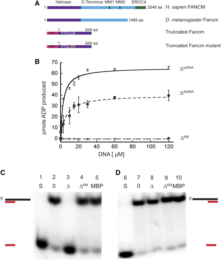
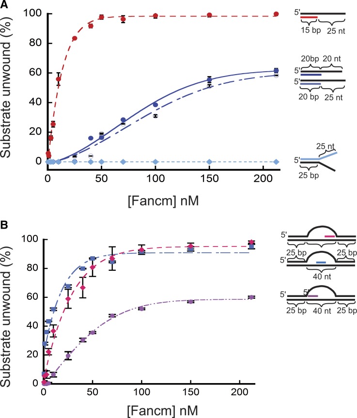
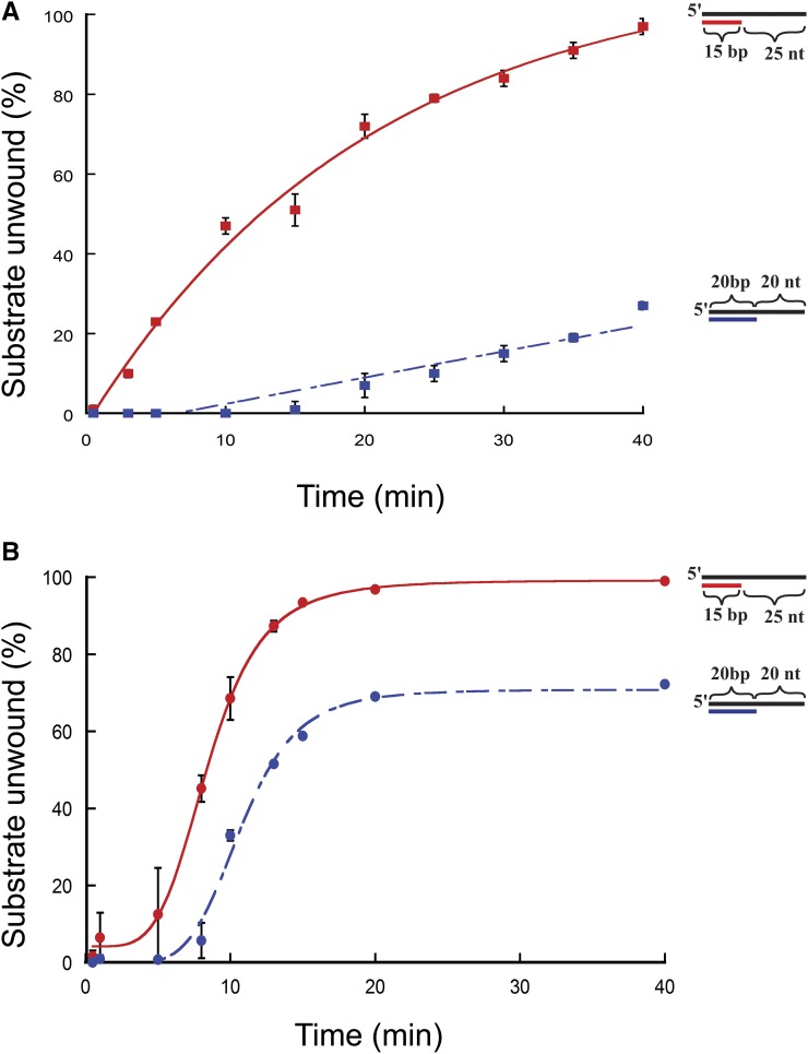
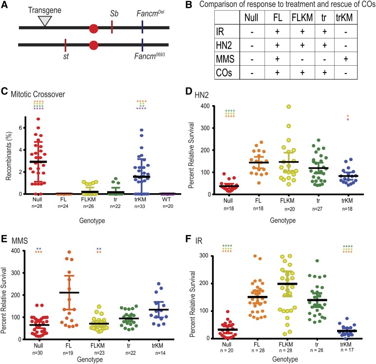
Repair of DNA damage is essential to the preservation of genomic stability. During repair of double-strand breaks, several helicases function to promote accurate repair and prevent the formation of crossovers through homologous recombination. Among these helicases is the Fanconi anemia group M (FANCM) protein. FANCM is important in the response to various types of DNA damage and has been suggested to prevent mitotic crossovers during double-strand break repair. The helicase activity of FANCM is believed to be important in these functions, but no helicase activity has been detected in vitro We report here a genetic and biochemical study of Drosophila melanogaster Fancm. We show that purified Fancm is a 3' to 5' ATP-dependent helicase that can disassemble recombination intermediates, but only through limited lengths of duplex DNA. Using transgenic flies expressing full-length or truncated Fancm, each with either a wild-type or mutated helicase domain, we found that there are helicase-independent and C-terminal-independent functions in responding to DNA damage and in preventing mitotic crossovers.

Keywords: ATP activity; DNA helicase; biochemistry; crossing over; genetics; homologous recombination; synthesis-dependent strand annealing.


Figures

Figure 1
Figure 1
Drosophila Fancm is a 3′ to 5′ DNA helicase dependent on ATP hydrolysis. (A) Schematic of Fancm. Domains and motifs present in human FANCM are marked. Conserved domains or motifs in D. melanogaster are noted. Truncated forms depicted are with an N-terminal MBP tag. (B) ATP hydrolysis by Fancm. Fancm ATPase activity was examined as a function of DNA concentration using either M13mp18 ssDNA (□⋄) or dsDNA (▪♦) as the DNA cofactor. All reactions were incubated at 37° for 5 min. □ ▪ 212 nM Fancm∆ on ssDNA (∆ssDNA); ⋄♦ 212 nM Fancm∆KM (∆KM). The average values from at least three independent experiments were plotted. Error bars represent SEM (ssDNA) or SD of the mean (dsDNA). (C) Fancm unwinds duplex DNA. Protein (212 nM) was incubated with a 5′ radiolabeled 15-bp partial duplex with a 25-nt 3′ overhang (15/40). (D) Fancm is a 3′ to 5′ DNA helicase. Protein (212 nM) was incubated with a 5′ radiolabeled 15-bp partial duplex with a 25-nt 5′ overhang (−15/40). Lane 1 and 6 (S) are boiled loading controls indicating ssDNA. Lanes 2 and 7 (0) are no-protein controls. Fancm∆ in lane 3 and 8 (∆), Fancm∆KM in lane 4 and 9 (∆KM), and MBP in lane 5 and 10 (MBP). Colored strand represents radiolabeled strand. Substrate oligonucleotides are in Table S1.
Figure 2
Figure 2
Unwinding of partial duplex DNA substrates by Fancm. Helicase reactions were performed as described in Materials and Methods. The indicated concentrations of Fancm were incubated with 0.1 nM of the indicated substrate for 15 min. Colored strand on each substrate represents radiolabeled 5′ strand. Quantitative data from at least three experiments were plotted as the average for each protein concentration. Error bars represent the SEM. Oligonucleotides used to make these substrates can be found in Table S1. (A) Comparison of the fraction of substrate unwound with partial duplex substrates of different duplex lengths. Pink ●, 15-bp duplex region with a 25-nt overhang. Blue ●, 20-bp duplex region with a 20-nt overhang. Blue ○, 20-bp duplex region with a 25-nt overhang; Blue ♦, 25-bp duplex region with 25-nt single-stranded arms. (B) Unwinding of D-loop intermediate substrates by Fancm. Pink ●, front; blue ▪, middle; pink ♦, end. Bubble structures were made using a two 90-nt oligonucleotides with 25 bp of complementary ends with a 40-nt noncomplementary middle (A1/A2). Substrate oligonucleotides are in Table S1.
Figure 3
Figure 3
Time-course unwinding of partial-duplex DNA substrates by Fancm. Helicase reactions were performed as described in Materials and Methods. The indicated concentrations of Fancm were incubated with 0.1 nM of the indicated substrate for the indicated time. Colored strand on each substrate represents radiolabeled 5′ strand. Quantitative data from at least three experiments were plotted as the average for each protein concentration. Error bars represent the SEM. Oligonucleotides used to make these substrates can be found in Table S1. (A and B) Comparison of the fraction of substrate unwound with 10-nm FancmΔ on partial-duplex substrates at the indicated time points of different duplex lengths. (A) Red ▪, 15-bp duplex region with a 25-nt overhang (15/40); blue ▪, 20-bp duplex region with a 20-nt overhang (20/40). (B) Red ●, 15-bp duplex region with a 25-nt overhang (15/40); blue ●, 20-bp duplex region with a 20-nt overhang (20/40). Substrate oligonucleotides are in Table S1.
Figure 4
Figure 4
Fancm has genetically separable functions. (A) Map of Fancm null allele (Fancm0693), CRISPR deletion (Fancmdel), transgene landing site (▾), and st and Sb genes. Schematic of transgenes generated are as seen in Figure 1A, without tags. (B) Table comparison of all transgenic Fancm genotypes and null genotype. − indicates no rescue of the null phenotype, + indicates rescue. (C) Spontaneous mitotic CO rates were measured between st and Sb. (D–F) Comparison of sensitivities of Fancm. Plots show the survival of the indicated phenotype relative to wild-type control flies in the same vial after exposure to (D) 0.002% HN2 (0.1 M), (E) 0.05% MMS (3.23 mM), or (F) IR (1500 rad). Red ●, null; Orange ●, FL; yellow ●, FLKM; green ●, tr, light blue ●, trKM; dark blue ●, wild type (WT). Each dot represents one vial, n measures number of vials. Mean percentage of progeny is represented by black horizontal bar. 95% C.I.s are represented by colored error bars. Statistical comparisons were done for Fancm compared to each other genotype. Statistically significant comparisons are indicated above error bars; **** P < 0.0001 by Kruskal–Wallace test, corrected for multiple comparisons.

References

    1. Adams M. D., McVey M., Sekelsky J., 2003. Drosophila BLM in double-strand break repair by synthesis-dependent strand annealing. Science 299: 265–267. - PubMed
    1. Andersen S. L., Sekelsky J., 2010. Meiotic vs. mitotic recombination: two different routes for double-strand break repair: the different functions of meiotic vs. mitotic DSB repair are reflected in different pathway usage and different outcomes. BioEssays 32: 1058–1066. - PMC - PubMed
    1. Bassett A. R., Tibbit C., Ponting C. P., Liu J. L., 2013. Highly efficient targeted mutagenesis of Drosophila with the CRISPR/Cas9 system. Cell Reports 4: 220–228. - PMC - PubMed
    1. Brosh R. M., Jr, 2013. DNA helicases involved in DNA repair and their roles in cancer. Nat. Rev. Cancer 13: 542–558. - PMC - PubMed
    1. Ciccia A., Ling C., Coulthard R., Yan Z., Xue Y., et al. , 2007. Identification of FAAP24, a Fanconi anemia core complex protein that interacts with FANCM. Mol. Cell 25: 331–343. - PubMed

MeSH terms