. 2016 Jul 27;36(30):8026-36.
doi: 10.1523/JNEUROSCI.1038-16.2016.

Increased Amplitude of Thalamocortical Low-Frequency Oscillations in Patients with Migraine

Affiliations

Increased Amplitude of Thalamocortical Low-Frequency Oscillations in Patients with Migraine

Duncan J Hodkinson et al. J Neurosci. .

Abstract

For many years, neurobiological theories have emphasized the importance of neuronal oscillations in the emergence of brain function. At the same time, clinical studies have shown that disturbances or irregularities in brain rhythms may relate to various common neurological conditions, including migraine. Increasing evidence suggests that the CNS plays a fundamental role in the predisposition to develop different forms of headache. Here, we present human imaging data that strongly support the presence of abnormal low-frequency oscillations (LFOs) in thalamocortical networks of patients in the interictal phase of migraine. Our results show that the main source of arrhythmic activity was localized to the higher-order thalamic relays of the medial dorsal nucleus. In addition, spontaneous LFOs in the thalamus were selectively associated with the headache attack frequency, meaning that the varying amplitude of dysrhythmia could predispose patients to recurrent attacks. Rhythmic cortical feedback to the thalamus is a major factor in the amplification of thalamocortical oscillations, making it a strong candidate for influencing neuronal excitability. We further speculate that the intrinsic dynamics of thalamocortical network oscillations are crucial for early sensory processing and therefore could underlie important pathophysiological processes involved in multisensory integration.

Significance statement: In many cases, migraine attacks are thought to begin centrally. A major obstacle to studying intrinsic brain activity has been the identification of the precise anatomical structures and functional networks that are involved in migraine. Here, we present imaging data that strongly support the presence of abnormal low-frequency oscillations in thalamocortical networks of patients in the interictal phase of migraine. This arrhythmic activity was localized to the higher-order thalamic relays of the medial dorsal nucleus and was selectively associated with headache attack frequency. Rhythmic cortical feedback to the thalamus is a major factor in the amplification of thalamocortical oscillations, making it a strong candidate for influencing neuronal excitability and higher-level processes involved in multisensory integration.

Keywords: frequency; headache; migraine; oscillations; pain; thalamocortical.


Figures

Figure 1.
Figure 1.
Amplitude of spontaneous LFOs in the resting brain. The statistical maps illustrate the detectable fractional amplitude of f/ALFFs across all subjects in the sample (n = 80). Data are separated into 4 frequency bands: slow-5 (0.01–0.027 Hz), slow-4 (0.027–0.073 Hz), slow-3 (0.073–0.198 Hz), and slow-2 (0.198–0.250 Hz). All statistical images are displayed with a cluster probability threshold of p < 0.05, corrected for multiple comparisons (familywise error, FWE). The sagittal image on the right displays the position of slices in standard MNI space (gray lines). R (right) and L (left) indicate image orientations for slices in the Z plane.
Figure 2.
Figure 2.
Frequency-dependent changes in the amplitude of LFOs in migraine. The statistical maps illustrate the differences in fALFF between migraine patients and healthy controls. Data are separated into 4 frequency bands: slow-5 (0.01–0.027 Hz), slow-4 (0.027–0.073 Hz), slow-3 (0.073–0.198 Hz), and slow-2 (0.198–0.250 Hz). All statistical images are displayed with a cluster probability threshold of p < 0.05, corrected for multiple comparisons (familywise error, FWE). The sagittal image on the right displays the position of slices in standard MNI space (gray lines). R (right) and L (left) indicate image orientations for slices in the Z plane.
Figure 3.
Figure 3.
A, Increased amplitude of the LFOs in thalamocortical circuits. The statistical maps illustrate the differences in fALFF between migraine patients and healthy controls in the frequency band slow-4 (0.027–0.073 Hz). B, Increased amplitude of the LFOs in higher-order thalamic relays. Localization of the thalamus according to the Harvard–Oxford subcortical atlas (green line). An axial section based on a histological atlas of the human thalamus is shown with nuclei outlined by black lines (Morel et al., 1997). All statistical images are displayed with a cluster probability threshold of p < 0.05, corrected for multiple comparisons (familywise error, FWE). R (right) and L (left) indicate image orientations for slices in the Z plane. VD, Ventral diencephalon; Thal, thalamus; Ins/OP, insula/operculum; dACC, dorsal anterior cingulate cortex; MDThal, medial dorsal thalamus.
Figure 4.
Figure 4.
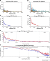
Power spectra analysis of LFOs in the thalamus. A, B, Individual PSD of LFOs in the thalamus of migraine patients (n = 40) and healthy controls (n = 40). C, D, Group averages (±SEM) of PSD in the thalamus. E, F, Expanded PSD covering the four frequency bands for linear and log scaled data. Red line indicates migraine; blue line, controls).
Figure 5.
Figure 5.
Correlations comparing clinical reported variables and PSD in the thalamus. Each patient's average PSD was calculated according to the four frequency bands and plotted against their age, current frequency of attacks, and disease duration. Pearson's correlation coefficient (R) and associated p-values (P) are reported for comparison.

References

    1. Akerman S, Holland PR, Goadsby PJ. Diencephalic and brainstem mechanisms in migraine. Nat Rev Neurosci. 2011;12:570–584. doi: 10.1038/nrn3057. - DOI - PubMed
    1. Alshelh Z, Di Pietro F, Youssef AM, Reeves JM, Macey PM, Vickers ER, Peck CC, Murray GM, Henderson LA. Chronic neuropathic pain: it's about the rhythm. J Neurosci. 2016;36:1008–1018. doi: 10.1523/JNEUROSCI.2768-15.2016. - DOI - PMC - PubMed
    1. Andreou AP, Goadsby PJ. Topiramate in the treatment of migraine: a kainate (glutamate) receptor antagonist within the trigeminothalamic pathway. Cephalalgia. 2011;31:1343–1358. doi: 10.1177/0333102411418259. - DOI - PubMed
    1. Andreou AP, Shields KG, Goadsby PJ. GABA and valproate modulate trigeminovascular nociceptive transmission in the thalamus. Neurobiol Dis. 2010;37:314–323. doi: 10.1016/j.nbd.2009.10.007. - DOI - PubMed
    1. Antal A, Kriener N, Lang N, Boros K, Paulus W. Cathodal transcranial direct current stimulation of the visual cortex in the prophylactic treatment of migraine. Cephalalgia. 2011;31:820–828. doi: 10.1177/0333102411399349. - DOI - PubMed

Publication types