. 2016 Oct;87(10):1084-90.
doi: 10.1136/jnnp-2015-312906. Epub 2016 Jul 27.

Misinterpretation of ischaemic infarct location in relationship to the cerebrovascular territories

Affiliations

Misinterpretation of ischaemic infarct location in relationship to the cerebrovascular territories

Nolan S Hartkamp et al. J Neurol Neurosurg Psychiatry. 2016 Oct.

Abstract

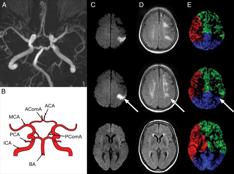
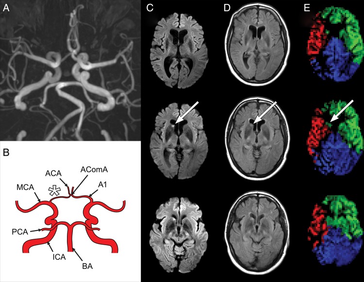
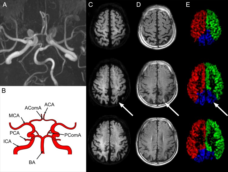
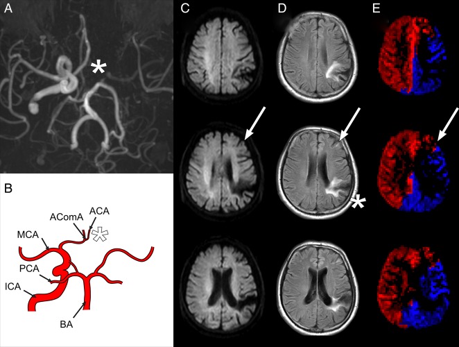
Purpose: Cerebral perfusion territories are known to vary widely among individuals. This may lead to misinterpretation of the symptomatic artery in patients with ischaemic stroke to a wrong assumption of the underlying aetiology being thromboembolic or hypoperfusion. The aim of the present study was to investigate such potential misinterpretation with territorial arterial spin labelling (T-ASL) by correlating infarct location with imaging of the perfusion territory of the carotid arteries or basilar artery.

Materials and methods: 223 patients with subacute stroke underwent MRI including structural imaging scans to determine infarct location, time-of-flight MR angiography (MRA) to determine the morphology of the circle of Willis and T-ASL to identify the perfusion territories of the internal carotid arteries, and basilar artery. Infarct location and the perfusion territory of its feeding artery were classified with standard MRI and MRA according to a perfusion atlas, and were compared to the classification made according to T-ASL.

Results: A total of 149 infarctions were detected in 87 of 223 patients. 15 out of 149 (10%) infarcts were erroneously attributed to a single perfusion territory; these infarcts were partly located in the originally determined perfusion territory but proved to be localised in the border zone with the adjacent perfusion territory instead. 12 out of 149 (8%) infarcts were misclassified with standard assessments and were not located in the original perfusion territory.

Conclusions: T-ASL with territorial perfusion imaging may provide important additional information for classifying the symptomatic brain-feeding artery when compared to expert evaluation with MRI and MRA.


Figures

Figure 1
Figure 1
Example of a 53-year-old female with left internal carotid artery (ICA) stenosis >70%. Time-of-flight magnetic resonance angiography (MRA) image (A) shows a complete circle of Willis as illustrated in the schematic drawing (B). Diffusion-weighted imaging (DWI) (C) and fluid attenuation inversion recovery (FLAIR) images (D) and corresponding territorial arterial spin labelling (T-ASL) perfusion maps (E) from caudal (left) to cranial (right). A recent parietal cortical ischaemic infarct showing diffusion restriction (C, arrow) may be appreciated on the FLAIR images (D, arrow). Using the standard perfusion territory atlas, the infarct was assumed to be within the middle cerebral artery (MCA) perfusion territory of the left ICA. T-ASL perfusion maps (E) show the perfusion territories of the right ICA (red), left ICA (green) and the basilar artery (BA) (blue), and locate the infarct on the border zone of the MCA and posterior cerebral artery (PCA) perfusion territories.
Figure 2
Figure 2
Example of a 50-year-old female with right internal carotid artery (ICA) stenosis >70%. Time-of-flight magnetic resonance angiography (MRA) image (A) shows a hypoplastic A1 segment (A, B, star) as illustrated in the schematic drawing (B). Diffusion-weighted imaging (DWI) (C) and fluid attenuation inversion recovery (FLAIR) images (D) and territorial arterial spin labelling (T-ASL) perfusion maps (E) are presented identically to figure 1. A recent lacunar infarct in the caudate nucleus showing diffusion restriction (C, arrow) may be appreciated on the FLAIR images (D, arrow). Using the standard perfusion territory atlas, the infarct was assumed to be within the territory of the right ICA. T-ASL perfusion maps show however the infarct is located in the perfusion territory of the (contralateral) left ICA (E, arrow). PCA, posterior cerebral artery.
Figure 3
Figure 3
Example of a 71-year-old male with a left-sided internal carotid artery (ICA) stenosis 50%. Time-of-flight magnetic resonance angiography (MRA) image (A) shows a complete circle of Willis as illustrated in the schematic drawing (B). Diffusion-weighted imaging (DWI) (C) and fluid attenuation inversion recovery (FLAIR) images (D) and territorial arterial spin labelling (T-ASL) perfusion maps (E) are presented identically to figure 1. A chronic occipital cortical ischaemic infarct can be appreciated on the FLAIR images (D, arrow). By applying the standard perfusion territory atlas, it was determined the infarct was located in the border zone of the middle cerebral artery (MCA) and posterior cerebral artery (PCA). The T-ASL perfusion maps show that the ischaemic lesion is within the perfusion territory of the basilar artery (BA) (E, arrow) instead.
Figure 4
Figure 4
Example of a 60-year-old male with a left-sided internal carotid artery (ICA) occlusion. Time-of-flight magnetic resonance angiography (MRA) image (A) shows an absent A1 segment (A, B, asterisk) as illustrated in the schematic drawing (B), with collateral flow to the anterior carotid artery (ACA) via the AcomA and collateral flow to the middle carotid artery (MCA) via the PcomA. Diffusion-weighted imaging (DWI) (C) and fluid attenuation inversion recovery (FLAIR) images (D) and territorial arterial spin labelling (T-ASL) perfusion maps (E) are presented identically to figure 1. A small frontal infarct (D, arrow) and a large chronic parietal infarct (D, star) can be appreciated in the FLAIR images. The small frontal infarct is of a recent date, even though it does not show diffusion restriction (C, arrow), since it was not visible on earlier imaging. By applying the standard perfusion territory atlas, it was determined the infarct was located within the MCA territory, currently supplied by the basilar artery (BA). The T-ASL perfusion maps show the small recent frontal infarct is not located within the MCA territory but is actually located on the border zone of the ACA and MCA (E, arrow), and is thus currently supplied by the right (contralateral) ICA as well as the BA. PCA, posterior cerebral artery.

References

    1. Hendrikse J, Petersen ET, van Laar PJ, et al. . Cerebral border zones between distal end branches of intracranial arteries: MR imaging. Radiology 2008;246:572–80. 10.1148/radiol.2461062100 - DOI - PubMed
    1. Krabbe-Hartkamp MJ, van der Grond J, de Leeuw FE, et al. . Circle of Willis: morphologic variation on three-dimensional time-of-flight MR angiograms. Radiology 1998;207:103–11. 10.1148/radiology.207.1.9530305 - DOI - PubMed
    1. Hendrikse J, van Raamt AF, van der Graaf Y, et al. . Distribution of cerebral blood flow in the circle of Willis. Radiology 2005;235:184–9. 10.1148/radiol.2351031799 - DOI - PubMed
    1. Hendrikse J, Petersen ET, Chng SM, et al. . Distribution of cerebral blood flow in the nucleus caudatus, nucleus lentiformis, and thalamus: a study of territorial arterial spin-labeling MR imaging. Radiology 2010;254:867–75. 10.1148/radiol.09090284 - DOI - PubMed
    1. van Laar PJ, Hendrikse J, Klijn CJM, et al. . Symptomatic carotid artery occlusion: flow territories of major brain-feeding arteries. Radiology 2007;242:526–34. 10.1148/radiol.2422060179 - DOI - PubMed

Publication types