. 2016 Sep 23;291(39):20674-91.
doi: 10.1074/jbc.M116.739136. Epub 2016 Jul 26.

The Crystal Structures of the N-terminal Photosensory Core Module of Agrobacterium Phytochrome Agp1 as Parallel and Anti-parallel Dimers

Affiliations

The Crystal Structures of the N-terminal Photosensory Core Module of Agrobacterium Phytochrome Agp1 as Parallel and Anti-parallel Dimers

Soshichiro Nagano et al. J Biol Chem. .

Abstract

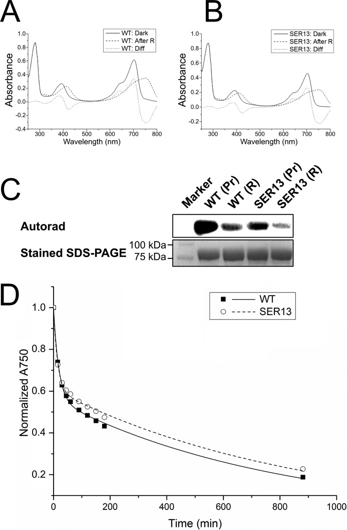
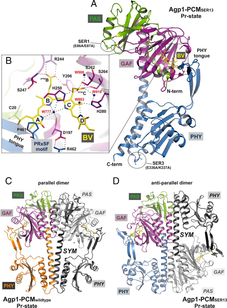
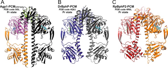
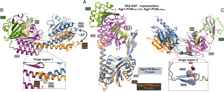
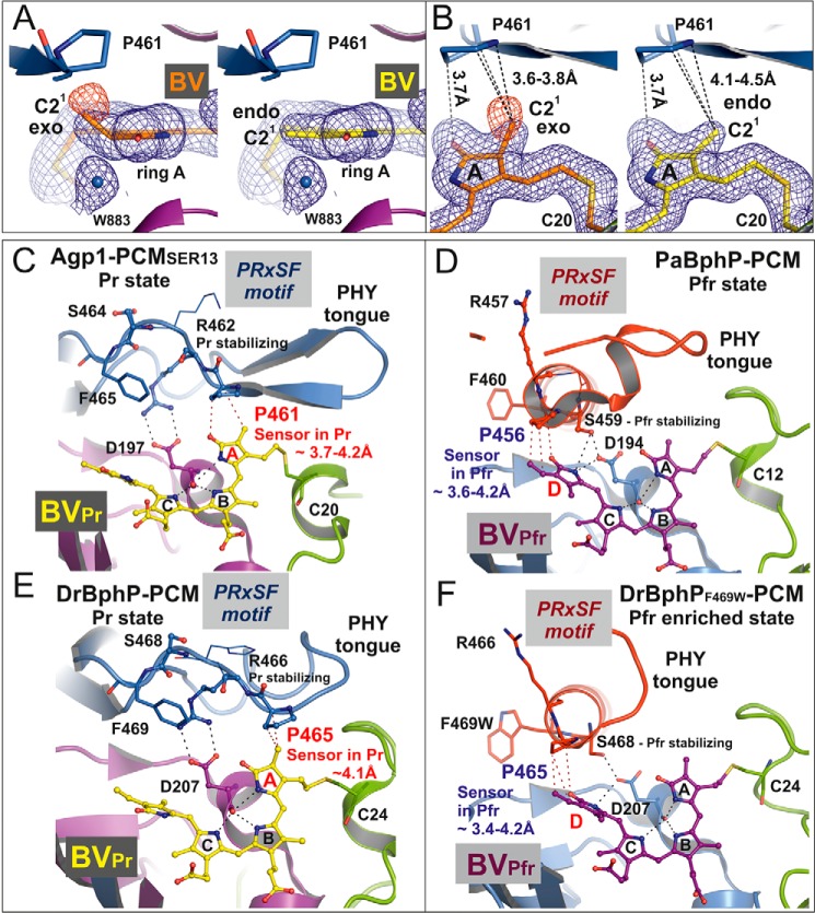
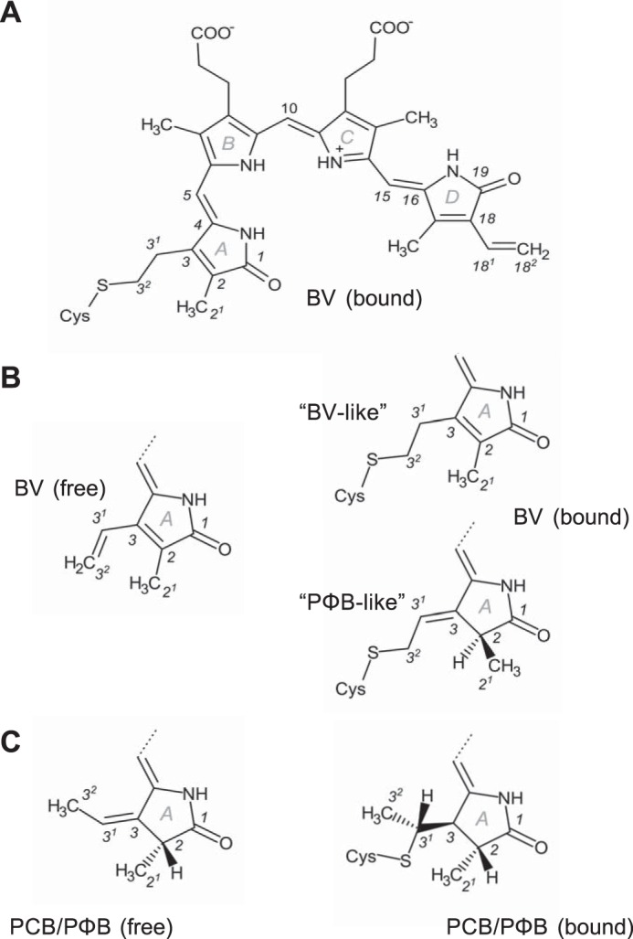
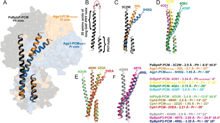
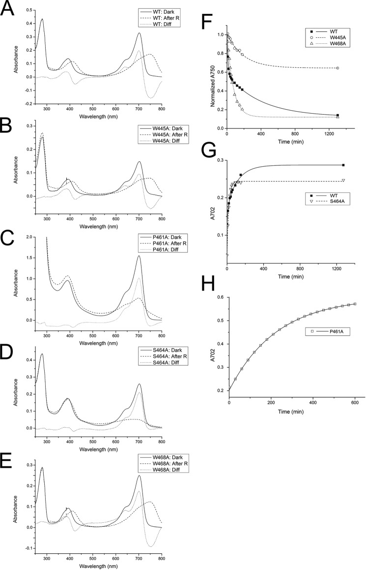
Agp1 is a canonical biliverdin-binding bacteriophytochrome from the soil bacterium Agrobacterium fabrum that acts as a light-regulated histidine kinase. Crystal structures of the photosensory core modules (PCMs) of homologous phytochromes have provided a consistent picture of the structural changes that these proteins undergo during photoconversion between the parent red light-absorbing state (Pr) and the far-red light-absorbing state (Pfr). These changes include secondary structure rearrangements in the so-called tongue of the phytochrome-specific (PHY) domain and structural rearrangements within the long α-helix that connects the cGMP-specific phosphodiesterase, adenylyl cyclase, and FhlA (GAF) and the PHY domains. We present the crystal structures of the PCM of Agp1 at 2.70 Å resolution and of a surface-engineered mutant of this PCM at 1.85 Å resolution in the dark-adapted Pr states. Whereas in the mutant structure the dimer subunits are in anti-parallel orientation, the wild-type structure contains parallel subunits. The relative orientations between the PAS-GAF bidomain and the PHY domain are different in the two structures, due to movement involving two hinge regions in the GAF-PHY connecting α-helix and the tongue, indicating pronounced structural flexibility that may give rise to a dynamic Pr state. The resolution of the mutant structure enabled us to detect a sterically strained conformation of the chromophore at ring A that we attribute to the tight interaction with Pro-461 of the conserved PRXSF motif in the tongue. Based on this observation and on data from mutants where residues in the tongue region were replaced by alanine, we discuss the crucial roles of those residues in Pr-to-Pfr photoconversion.

Keywords: Agrobacterium; conformational change; crystal structure; histidine kinase; photoreceptor; phytochrome; quaternary structure; signal transduction; tertiary structure.


Figures

FIGURE 1.
FIGURE 1.
Effect of SER mutations on spectral characteristics, histidine kinase function, and dark reversion kinetics in Agp1-PCM-SER13. A and B, UV-visible absorption spectra of full-length Agp1 wild-type (WT; A) and SER13 mutant (B) in their dark states (Dark; solid line) and after illumination with red light (After R; dashed line). The difference spectrum (Diff; dotted line) obtained by subtracting the spectrum after red light illumination from the dark spectrum shows that the wild-type and the mutant protein photoconvert into a mixture of similar proportions of Pr and Pfr. C, autoradiograph (upper panel) after SDS-PAGE of wild-type Agp1 and its SER13 mutant after incubation with [γ-32P]ATP in the dark (Pr) and illumination with red light (R). Similar to the wild-type, the mutant protein shows pronounced autophosphorylation activity in the dark that decreases after red light illumination. The lower panel shows the same section of the gel after staining with Coomassie Blue. D, dark reversion of wild-type Agp1 (■, solid line) and the SER13 mutant (○, dashed line) as measured by the time dependence of the relative absorbance at 750 nm (A750).
FIGURE 2.
FIGURE 2.
Structure of the PCM of Agp1 in the Pr state. A, ribbon representation of the Agp1-PCM-SER13 monomer. The chromophore BV and its attachment site Cys-20 are shown as balls and sticks, and carbon atoms are colored in yellow. PAS, GAF, and PHY domains are depicted in green, purple, and blue, respectively. The two SER clusters of mutated amino acid residues (SER1, E86A/E87A; SER3, E336A/K337A) are indicated by dotted circles and depicted as balls and sticks. B, rotated close-up view (same color code as in A) of a section of the chromophore binding pocket showing the potential hydrogen bond network that links biliverdin to the protein environment, including mediating water molecules. The four pyrrole rings of biliverdin are labeled A to D, and the propionate side chains of rings B and C are labeled propB and propC, respectively. Some of the potential hydrogen bonds have been omitted for clarity. The complete set of binding interactions in the chromophore binding pocket is visualized schematically in Fig. 5C. The highly conserved PRXSF motif of the tongue region from the PHY domain interacts with the chromophore via van der Waals interactions between Pro-461 and ring A of BV and stabilizes the chromophore binding pocket by a salt bridge contact between Arg-462 and Asp-197. C and D, structures of crystallographic dimers of wild-type Agp1-PCM (C) and Agp1-PCM-SER13 (D). In each dimer one monomer is colored, with PAS, GAF, and PHY domains depicted in green, purple, and orange, respectively, in C, and in D the color code is the same as in A. The second monomer (labeled SYM) related by a crystallographic symmetry axis to the first is shown in gray in C and in D. In the parallel dimer formed by the wild-type protein, the PAS, GAF, and PHY domains are juxtaposed along the symmetry axis, which is roughly parallel to the long axis of each monomer and perpendicular to the view direction in C. In the anti-parallel dimers in which the SER13 mutant protein forms in the crystals, the quaternary structure is stabilized by large interfaces comprising the GAF and PHY domains of different monomers, and the symmetry axis is perpendicular to the long axes of the monomers and parallel to the view direction in D.
FIGURE 3.
FIGURE 3.
Comparison of parallel dimer structures in prototypical phytochromes in the Pr state. A, crystallographic dimer of wild-type Agp1-PCM is shown in ribbon representation (PDB code 5I5L; same color code as in Fig. 2C). B, crystallographic dimer of DrBphP-PCM (PDB code 4Q0J) is shown, with one monomer in dark blue and the symmetry-related monomer in gray. C, non-crystallographic dimer of RpBphP2-PCM (PDB code 4R6L) is shown, with one monomer in firebrick-red and the second in orange. In all three panels the chromophore biliverdin is depicted as balls and sticks, and the structures are shown in identical views after superpositioning of the PAS-GAF bidomains. The dimer structures are similar in the regions defined by the PAS and GAF domains only. Because of the different helical spines and in particular the different bending of the long GAF-PHY helices, however, the size of the gaps between the PHY domains along the symmetry axes and the relative orientations of the C-terminal helices leading to the His kinase modules are significantly different between the three structures.
FIGURE 4.
FIGURE 4.
Structural comparison of the monomers in the crystal structures of wild-type Agp1-PCM and Agp1-PCM-SER13. A, superposition of the PAS-GAF bidomains of wild-type Agp1-PCM and Agp1-PCM-SER13. The complete PCMs are shown, with PAS and GAF domains of both structures colored green and purple, respectively, and the PHY domain is colored orange for the wild-type and blue for the SER13 mutant protein. B and C, the same superposition as in A shown in different orientations. Two potential hinge regions, hinge region 1, around Met-308 in the long GAF-PHY helix, and hinge region 2, around Trp-445 and Trp-468 in the middle of the tongue of the PHY domain, are shown in the close-up views in B and C, respectively. Met-308, Trp-445, and Trp-468 are shown in stick and sphere representations in all three panels. The different positions and orientations that the PHY domains adopt with respect to the PAS-GAF bidomains in the two structures can be described by a rotation of ∼23° about the hinges mentioned above and correlate with the different helical spines of the long GAF-PHY helix, which is almost straight in the wild-type protein but strongly bent in the SER13 mutant (B). A definition of the angles that were used to describe the relative orientations of the GAF and PHY domain is given in Fig. 8.
FIGURE 5.
FIGURE 5.
Electron density of the BV chromophore in the Agp1-PCM-SER13 crystal structure at 1. 85 Å resolution. A, BV and Asp-197 are shown as sticks with carbon atoms in yellow and purple, respectively. Water molecules (W777, W883, and W918) are depicted as blue spheres, and all other surrounding amino acid residues as thin lines. The blue mesh represents a weighted 2FoFc map (contoured at 1.5 σ) where the weights were calculated using REFMAC5 (64). B, 2FoFc simulated annealing σA-weighted omit map (green), calculated using the program PHENIX (65) for the omitted chromophore BV in the Agp1-PCM-SER13 crystal structure. C, analysis of the potential hydrogen bonds and van der Waals contacts between BV and its protein and water molecule environment. Potential hydrogen bonds were analyzed using HBPLUS (72) as implemented in the program LigPlot+ (73), which was used to draw this schematic view. Residues with closest distances less than 4 Å are considered to be in van der Waals contact.
FIGURE 6.
FIGURE 6.
Structure of the biliverdin chromophore at pyrrole ring A and the PRXSF motif as directly interacting signal transducer module in bacterial phytochromes. A, electron density of ring A of the chromophore BV after, left panel, refinement with geometric restraints for a PΦB-like structure with an exocyclic C3=C31 double bond or, right panel, with restraints for a BV-like structure with an endocyclic C2=C3 double bond in ring A. The blue mesh represents a weighted 2FoFc map contoured at 1.0 σ. That the PΦB-like chromophore structure is wrong is indicated by the negative electron density at the C21 methyl group, as shown by the red mesh that represents the weighted FoFc map at −2.0 σ. B, similar representation as in A in a different orientation. In both panels the shortest distances between Pro-461 of the PRXSF motif and the C21 atom in the PΦB-like structure with an exocyclic double bond, left panel, and in the BV-like structure with an endocyclic double bond, right panel, are indicated by dashed lines. C–F, chromophore binding pockets in the crystal structures of different bacteriophytochromes are shown with BV, the highly conserved Asp of the conserved PXXDIP motif in the GAF domain, and the amino acids of PRXSF motif as balls and sticks or thin lines (x and Phe of the PRXSF motif). The polypeptide chains are depicted in ribbon representation. C and E, in the crystal structures of Agp1-PCM-SER13 (PDB code 5I5L) and DrBphP-PCM (PDB code 4Q0J), respectively, in their Pr states, the tongue of the PHY domain is folded as a β-hairpin loop over the chromophore binding pocket. In the Pr structure of Agp1-PCM-SER13, Pro-461 of the PRXSF motif in the tongue region (as well as Pro-465 in the Pr state of DrBphP-PCM) undergoes van der Waals interactions with ring A of the chromophore BV, thereby being poised to act as sensor of structural changes that occur at ring A after photoactivation of the Pr state. Arg-462 (Arg-466 in DrBphP) stabilizes Asp-197 (Asp-207 in DrBphP) via a salt bridge. Ser-464 (Ser-468 in DrBphP) is solvent-exposed and without any interaction with the chromophore region in the Pr state crystal structures. D and F, in the crystal structures of PaBphP-PCM (PDB code 3NHQ) and DrBphP-PCM (PDB code 5C5K) in their Pfr (D) or Pfr-enriched (F) states, respectively, the tongue of the PHY domain is folded as α-helical and loop structure over the chromophore binding pocket. Pro-456 of the PRXSF motif in PaBphP-PCM (or Pro-465 in the Pfr-enriched state of DrBphP-PCM) is in van der Waals contact with ring D of the chromophore BV, in a position to sense structural changes that occur at ring D after photoactivation of the Pfr state. Ser-459 (Ser-468 in DrBphP) stabilizes Asp-194 (Asp-207 in DrBphP) of the conserved PXXDIP motif via a hydrogen bond. Arg-457 in PaBphP and the corresponding residue Arg-466 in DrBphP are solvent-exposed and without any interactions with the chromophore regions in the crystal structures of the Pfr and the Pfr-enriched state, respectively. Parts of the secondary structures in front of the chromophore binding pocket were removed for clarity in C–F.
FIGURE 7.
FIGURE 7.
Structures of the bilin chromophores referred to in this work. A, complete structure of BV resulting from covalent attachment to the conserved Cys in bacteriophytochromes via 1,2-addition. The stereochemistry of this structure is 5Zs10Zs15Za (often abbreviated ZZZssa), indicating whether the central carbon atoms of the methine bridges that link adjacent pyrrole rings participate in single bonds that are either in syn (s) or anti (a) conformation and stating that all the corresponding double bonds are in Z configuration. Moreover, the structure contains an endocyclic C2=C3 double bond in ring A. Numbering is shown for all atoms that are mentioned in the text, tables, or figures. B and C, ring A structures of the free and bound chromophores BV, PCB, and PΦB as found in phytochrome crystal structures. Covalent attachment of BV (B) via 1,4-addition results in the PΦB-like structure with an exocyclic C3=C31 double bond in the C3 side chain that was found in the CBD structure of DrBphP (24) and is different from the BV-like structure also shown in A.
FIGURE 8.
FIGURE 8.
Variation in relative orientations between GAF and PHY domains in the known PCM structures of phytochromes. A, structural comparison of the long helices that connect the GAF and the PHY domains (GAF-PHY helices) and the C-terminal helices of the PHY domains (PHY helices) of PaBphP-PCM (black), wild-type Agp1-PCM (orange), and Agp1-PCM-SER13 (blue) shown in ribbon representation. The complete PCM structures are shown in surface representation in the background. The structural alignment was obtained by superpositioning the 10 C-terminal amino acid residues of the GAF-PHY helices and the PHY helices. B, for each structure the relative orientation between the GAF and the PHY domain is characterized as indicated here for chain A of PaBphP-PCM (PDB code 3C2W) by measuring the angle between the helix axes of the segment defined by the 20 N-terminal residues of the GAF-PHY helix on the one hand and the PHY helix on the other hand. The angles between helix axes were calculated by the Chou algorithm (77) using QHELIX (55). The signs of the angles are as defined in QHELIX. C–F, superpositions of helices of phytochrome PCM structures as in A, PDB codes indicated. For each structure shown, the text inset provides information on the type of phytochrome PCM together with the corresponding PDB code, the resolution and the photochromic state of the crystal structure, and the mean angle between helices (including its standard deviation if there is more than one monomer per asymmetric unit) as defined above. The color code is the same as in A–F.
FIGURE 9.
FIGURE 9.
Photoconversion and dark reversion of Agp1 mutant proteins where selected residues of the tongue region of the PHY domain were replaced by Ala. A–E, UV-visible absorption spectra of full-length Agp1 wild-type (WT; A), as well as mutants W445A (B), P461A (C), S464A (D), and W468A (E) in their dark states (Dark; solid line) and after illumination with red light (After R; dashed line). Comparison of the difference spectra (Diff; dotted line) obtained for each sample by subtracting the spectrum after red light illumination from the dark spectrum shows that the W445A and the W468A mutant proteins photoconvert into a mixture of similar proportions of Pr- and Pfr-like states as the wild-type. Red light illumination of P461A and S464A that exhibit normal Pr-like dark spectra, however, generates bleached species that lack typical features of a Pfr state. F, dark reversion of wild-type Agp1 (■, solid line), the W445A (○, dashed line), and the W468A mutant (Δ, dotted line) as measured by the time dependence of the relative absorbance at 750 nm (A750). Dark reversion is incomplete for W445A, whereas Trp-468 exhibits complete reversion to the dark state. In contrast to dark reversion of wild-type Agp1, which can be described by bi-exponential kinetics with time constants of 13.0 ± 0.4 and 400 ± 20 min, the corresponding processes are characterized by mono-exponential kinetics with time constants of 190 ± 20 and 70 ± 4 min in W445A and W468A, respectively. G and H, dark reversion of wild-type Agp1 and the S464A and P461A mutants as measured by the time dependence of the increase in absorbance at 702 nm (A702). Data from the Agp1 wild-type (■, solid line) and the S464A mutant (▿, dashed line) are shown in G, and data from P461A (□, solid line) are shown in H. Although the data from the wild-type protein can be explained by bi-exponential kinetics with time constants of 11 ± 3 and 120 ± 10 min, the data of S464A and P461A were fitted by assuming only one kinetic phase for each mutant with time constants of 18.0 ± 0.5 and 230.0 ± 0.4 min, respectively.

References

    1. Auldridge M. E., and Forest K. T. (2011) Bacterial phytochromes: more than meets the light. Crit. Rev. Biochem. Mol. Biol. 46, 67–88 - PubMed
    1. Ulijasz A. T., and Vierstra R. D. (2011) Phytochrome structure and photochemistry: recent advances toward a complete molecular picture. Curr. Opin. Plant Biol. 14, 498–506 - PubMed
    1. Vierstra R. D., and Zhang J. (2011) Phytochrome signaling: solving the Gordian knot with microbial relatives. Trends Plant Sci. 16, 417–426 - PubMed
    1. Duanmu D., Bachy C., Sudek S., Wong C. H., Jiménez V., Rockwell N. C., Martin S. S., Ngan C. Y., Reistetter E. N., van Baren M. J., Price D. C., Wei C. L., Reyes-Prieto A., Lagarias J. C., and Worden A. Z. (2014) Marine algae and land plants share conserved phytochrome signaling systems. Proc. Natl. Acad. Sci. U.S.A. 111, 15827–15832 - PMC - PubMed
    1. Bai Y., Rottwinkel G., Feng J., Liu Y., and Lamparter T. (2016) Bacteriophytochromes control conjugation in Agrobacterium fabrum. J. Photochem. Photobiol. B 161, 192–199 - PubMed

Publication types