. 2016 Jul 28;11(7):e0160004.
doi: 10.1371/journal.pone.0160004. eCollection 2016.

Immune-Modulation by Epidermal Growth Factor Receptor Inhibitors: Implication on Anti-Tumor Immunity in Lung Cancer

Affiliations

Immune-Modulation by Epidermal Growth Factor Receptor Inhibitors: Implication on Anti-Tumor Immunity in Lung Cancer

Jin S Im et al. PLoS One. .

Abstract

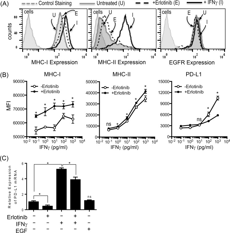
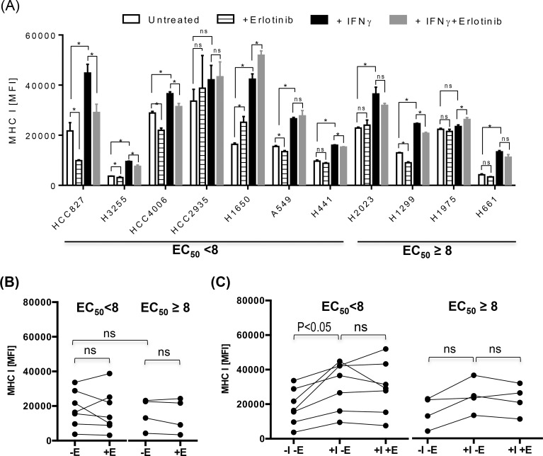
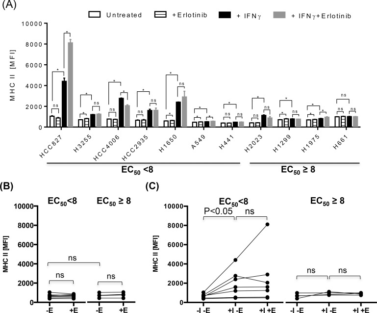
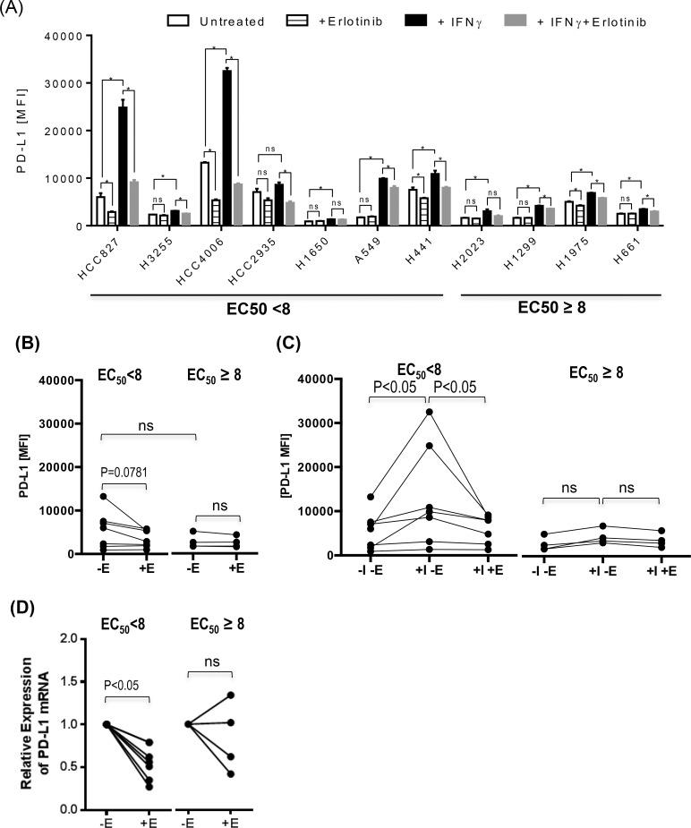
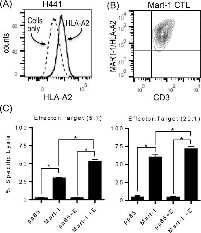
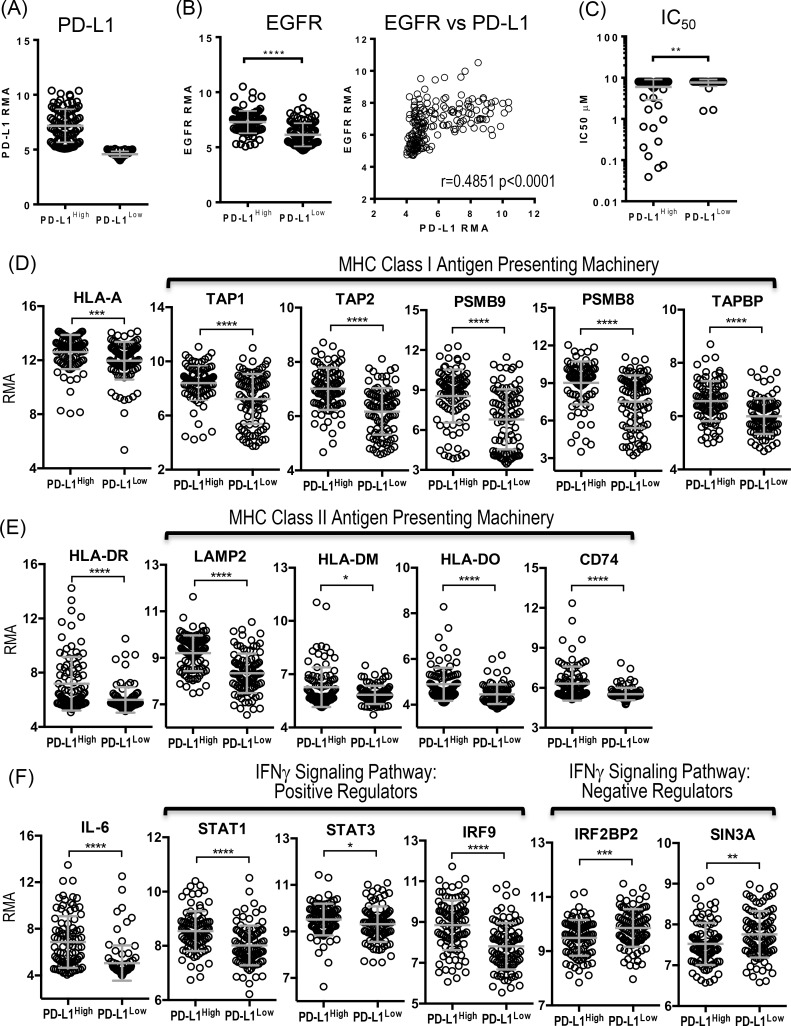
Skin toxicity is the most common toxicity caused by Epidermal Growth Factor Receptor (EGFR) inhibitors, and has been associated with clinical efficacy. As EGFR inhibitors enhance the expression of antigen presenting molecules in affected skin keratinocytes, they may concurrently facilitate neo-antigen presentation in lung cancer tumor cells contributing to anti-tumor immunity. Here, we investigated the modulatory effect of the EGFR inhibitor, erlotinib on antigen presenting molecules and PD-L1, prominent immune checkpoint protein, of skin keratinocytes and lung cancer cell lines to delineate the link between EGFR signaling pathway inhibition and potential anti-tumor immunity. Erlotinib up-regulated MHC-I and MHC-II proteins on IFNγ treated keratinocytes but abrogated IFNγ-induced expression of PD-L1, suggesting the potential role of infiltrating autoreactive T cells in the damage of keratinocytes in affected skin. Interestingly, the surface expression of MHC-I, MHC-II, and PD-L1 was up-regulated in response to IFNγ more often in lung cancer cell lines sensitive to erlotinib, but only expression of PD-L1 was inhibited by erlotinib. Further, erlotinib significantly increased T cell mediated cytotoxicity on lung cancer cells. Lastly, the analysis of gene expression dataset of 186 lung cancer cell lines from Cancer Cell Line Encyclopedia demonstrated that overexpression of PD-L1 was associated with sensitivity to erlotinib and higher expression of genes related to antigen presenting pathways and IFNγ signaling pathway. Our findings suggest that the EGFR inhibitors can facilitate anti-tumor adaptive immune responses by breaking tolerance especially in EGFR driven lung cancer that are associated with overexpression of PD-L1 and genes related to antigen presentation and inflammation.


Conflict of interest statement

Competing Interests: WH is supported by the Samsung Foundation Grant (http://www.samsungfoundation.org/html/eng/). There are no patents, products in development or marketed products to declare. This does not alter the authors' adherence to PLOS ONE policies on sharing data and materials.

Figures

Fig 1
Fig 1. EGFR inhibitor differentially modulates the expression of antigen presenting molecules and PD-L1 on HaCaT cells.
(A) HaCaT cells were treated with IFNγ at concentration of 1000 pg/ml or erlotinib at concentration of 20 μM for 24 hours, and assessed for surface expression of MHC-I, MHC-II, and EGFR. Erlotinib up-regulated the expression of MHC-I on HaCaT cells while IFNγ upregulated both MHC-I and MHC-II. Neither treatments affected EGFR expression. Gray dotted line: unstained HaCaT cells, gray solid line: untreated HaCaT cells stained for MHC-I, MHC-II, or EGFR. Black dotted line: erlotinib treated HaCaT cells stained for MHC-I, MHC-II, or EGFR. Black solid line: IFNγ treated HaCaT cells stained for MHC-I, MHC-II, or EGFR. (B) HaCaT cells were treated with IFNγ at different concentrations ranging 1 pg/ml to 1000 pg/ml in the presence or absence of erlotinib at concentration of 10 μM for 24 hours, and assessed for surface expression of MHC-I, MHC-II, and PD-L1 in Mean Fluorescence Intensity (MFI). While the expression of MHC-I and MHC-II on HaCaT cells was further up-regulated in the presence of erlotinib and IFNγ at all dose range tested for MHC-I and at a greater than 1pg/ml for MHC-II compared to IFNγ alone, IFNγ induced overexpression of PD-L1 was abrogated by the addition of erlotinib to IFNγ at a greater than 100 pg/ml. The experiment shown here was performed in triplicates, and a representative of three independent experiments.(C) A total RNA from HaCaT cells treated with or without erlotinib, IFNγ, or EGF was assessed for the relative expression of PD-L1 transcripts via Reverse Transcription quantitative Polymerase Chain Reaction (RT-qPCR) in quadruplicates. Erlotinib treatment resulted in statistically significant decrease in PD-L1 transcripts on HaCaT cells P-value less than 0.05 (*) was determined as being statistically significance. “NS” represent “statistically not-significant”.
Fig 2
Fig 2. Differential effects on MHC-I in lung cancer cell lines by IFNγ and EGFR inhibitors.
Various lung cancer cell lines were treated with IFNγ at a concentration of 1000 pg/ml in the presence or absence of erlotinib at concentration of 10 μM for 24 hours, and assessed for surface expression of MHC-I. (A) The average mean fluorescence intensities (MFIs) ± standard deviation (SD) of MHC-I staining were represented as bar graphs.(B and C) For further statistical analysis, lung cancer cell lines were divided into two groups according to the half maximum effective concentration, EC50 (EC50 <8 vs EC50 ≥ 8) of erlotinib, and average MFI of MHC I for individual cell line was represented as a single dot for further statistical analysis to assess the impact of treatment with IFNγ and/or erlotinib on MHC I expression within group of lung cancer cell lines sensitive to erlotinib (EC50 <8) or resistant to erlotinib (EC50 ≥8) There was a trend toward higher expression of MHC-I in untreated lung cancer cell lines sensitive to erlotinib (BB), and the treatment with IFNγ further up-regulated the expression of MHC-I more often on lung cancer cell lines sensitive to erlotinib compared to lung cancer cell line resistant to erlotinib (C). The addition of erlotinib to untreated or IFNγ treated lung cancer cell lines did not significantly altered the expression of MHC-I in most cell lines regardless of sensitivity to erlotinib (B and C). Abbreviations used in this experiment are as follow, E: erlotinib, I: IFNγ. The experiment shown here was performed in triplicates, and a representative of three independent experiments. P-value less than 0.05 was determined as statistically significance. “NS” represent “statistically non-significant”.
Fig 3
Fig 3. Differential effects on MHC-II in lung cancer cell lines by IFNγ and EGFR inhibitors.
Various lung cancer cell lines were treated with IFNγ at a concentration of 1000 pg/ml in the presence or absence of erlotinib at concentration of 10 μM for 24 hours, and assessed for surface expression of MHC-II. (A) The average MFI ± SD of MHC-II staining were represented as bar graphs. (B and C) Lung cancer cell lines were divided into two groups according to the half maximum effective concentration, EC50 (EC50 <8 vs EC50 ≥ 8) of erlotinib, and average MFI of MHC-II for individual cell line was represented as a single dot for further statistical analysis to assess the effect of IFNγ and/or erlotinib on the expression of MHC-II. There was only minimal baseline expression of MHC-II in untreated lung cancer cell lines regardless of sensitivity to erlotinib (B), but the treatment with IFNγ significantly up-regulated the expression of MHC-II more often on lung cancer cell lines sensitive to erlotinib compared to lung cancer cell line resistant to erlotinib (C). The addition of erlotinib to untreated or IFNγ treated lung cancer cell lines did not significantly altered the expression of MHC-II in most cell lines (B and C). Abbreviations used in this experiment are as follow, E: erlotinib, I: IFNγ. The experiment shown here was performed in triplicates, and a representative of three independent experiments. P-value less than 0.05 (*) was determined as being significant, “ns” represents “not statistically significant”.
Fig 4
Fig 4. EGFR inhibitor down-regulates the PD-L1 expression on lung cancer cell lines.
Various lung cancer cell lines were treated with IFNγ at a concentration of 1000 pg/ml in the presence or absence of erlotinib at concentration of 10 μM for 24 hours, and assessed for surface expression of PD-L1. (A) The average MFIs± SD of PD-L1 staining were represented as bar graphs in (A) (B and C) Lung cancer cell lines were divided into two groups according to EC50 (EC50 <8 vs EC50 ≥ 8) of erlotinib, and average MFI of PD-L1staining for individual cell line was represented as a single dot in dot plots for further statistical analysis to assess the influence of IFNγ and/or erlotinib on PD-L1 expression. There was a trend toward higher expression of PD-L1 in untreated lung cancer cell lines sensitive to erlotinib, and erlotinib treatment down-regulated PD-L1 expression on lung cancer cell lines sensitive to erlotinib but not statistically significant (B). The treatment with IFNγ significantly up-regulated the expression of PD-L1 proteins on lung cancer cell lines sensitive to erlotinib, and the addition of erlotinib significantly inhibited IFNγ-induced PD-L1 overexpression on lung cancer cell lines sensitive to erlotinib (C). Abbreviation used in this experiment is as follows. E: Erlotinib, I: IFNγ. The experiment shown here was performed in triplicates, and a representative of three independent experiments. (D) Various lung cancer cells were cultured in the presence or absence of erlotinib at a concentration of 10 μM for 24 hours, and the total RNA was isolated for Reverse Transcription quantitative Polymerase Chain Reaction (RT-qPCR) performed in quadruplicates to evaluate the expression of PD-L1 transcripts. The relative PD-L1 expression of individual lung cancer cell line treated with erlotinib to untreated control was represented as a single dot. Lung cancer cell lines were again divided into two groups according to EC50 (EC50 <8 vs EC50 ≥ 8) of erlotinib. Erlotinib down-regulated PD-L1 expression at the transcript level, and inhibitory effect was more pronounced in lung cancer cells sensitive to EGFR inhibitors. P-value less than 0.05 was determined as statistically significance. “NS” represent “statistically non-significant”.
Fig 5
Fig 5. EGFR inhibitor increases antigen specific T cell mediated tumor killing.
(A) The expression of HLA-A2 on HLA-A2+ lung cancer cell line, H441, was confirmed using flowcytometry. (B) Mart-1 (M27) specific cytotoxic T cell line (CTL), 1007G, was stained with anti-CD3 and Mart-1 (M27)/HLA-A2 tetramer to confirm the antigenic specificity. More than 90% of T cells expressed T cell receptors specific for Mart-1(M27)/HLA-A2 tetramers. (C) H441 cells pretreated with or without erlotinib in the presence or absence of Mart-1(M27) or pp65 control peptide were used as target cells for standard cytotoxic T cell (CTL) assay using Mart-1(M27) specific CTLs as effector T cells. Percent Specific cytotoxicity was calculated as follows: [(test release-spontaneous release)/(maximum release-spontaneous release)] × 100. Mart-1 (M27) specific CTL efficiently lysed H441 only in the presence of Mart-1 peptide but not control peptide, and the treatment of erlotinib significantly increased T cell mediated killing. The experiment shown here was performed in quadruplicates, and a representative of two independent experiments.
Fig 6
Fig 6. Overexpression of PD-L1 is associated with higher expression of EGFR and immune-related genes, and sensitivity to EGFR inhibitors.
(A) Lung cancer cell lines (N186) from Cancer Cell Line Encyclopedia (CCLE) database were divided into two groups according to median expression of PD-L1 at Log2 Robust Multi-array Average (RMA), 5.087. Mean Log2 RMAs of PD-L1 expression of PD-L1high and PD-L1low groups were 7.171 ±1.508 and 4.578 ± 0.2630, respectively. (B) EGFR expression of PD-L1high and PD-L1low lung cancer cell lines, and correlation between EGFR and PD-L1 expression (C) The half maximum inhibitory concentration (IC50) of Erlotinib for PD-L1high lung cancer cell lines (N51) and PD-L1low lung cancer cell lines (N40). (D) Expression of MHC-I and genes associated with MHC-I antigen presentation from PD-L1high and PD-L1low lung cancer cell lines. (E) Expression of MHC-II and genes associated with MHC-II antigen presentation from PD-L1high and PD-L1low lung cancer cell lines. (F) Expression of genes associated with inflammation from PD-L1high and PD-L1low lung cancer cell lines. PD-L1high lung cancer cell lines are associated with higher expression of EGFR and sensitivity to erlotinib (B and C). In addition, PD-L1high lung cancer cell lines express genes associated with MHC-I and II antigen presenting pathways higher than PD-L1low lung cancer cell lines (D and E). Significantly higher expression of IL-6 and positive regulators for IFNγ signaling pathway such as STAT1, STAT3, and IRF9 were associated with PD-L1high lung cancer cell lines, while the expression of negative regulators for IFNγ signaling pathway such as IRF2BP2 and SIN3A were significantly lower in PD-L1high lung cancer cell lines (F). P values were calculated by Wilcoxon-Man-Whitney test and summarized as follows, ****: p < 0.0001, ***: p = 0.0001 to 0.001, **: p = 0.001 to 0.01, *: p = 0.01 to 0.05, ns: p > 0.05.

References

    1. Siegel RL, Miller KD, Jemal A (2015) Cancer statistics, 2015. CA Cancer J Clin 65: 5–29. 10.3322/caac.21254 - DOI - PubMed
    1. Howlader N, Noone AM, krapcho M (2014) SEER Cancer Statistics Review, 1975–2011. Available at http://seercancergov/csr/1975_2011.
    1. Ettinger DS, Wood DE, Akerley W, Bazhenova LA, Borghaei H, Camidge DR, et al. (2015) Non-small cell lung cancer, version 6.2015. J Natl Compr Canc Netw 13: 515–524. - PubMed
    1. Reguart N, Remon J (2015) Common EGFR-mutated subgroups (Del19/L858R) in advanced non-small-cell lung cancer: chasing better outcomes with tyrosine kinase inhibitors. Future Oncol 11: 1245–1257. 10.2217/fon.15.15 - DOI - PubMed
    1. Kobayashi S, Boggon TJ, Dayaram T, Janne PA, Kocher O, Meyerson M, et al. (2005) EGFR mutation and resistance of non-small-cell lung cancer to gefitinib. N Engl J Med 352: 786–792. - PubMed

MeSH terms