

Resting-State Functional MR Imaging for Determining Language Laterality in Intractable Epilepsy
- PMID: 27467465
- PMCID: PMC5047125
- DOI: 10.1148/radiol.2016141010
Resting-State Functional MR Imaging for Determining Language Laterality in Intractable Epilepsy
Abstract
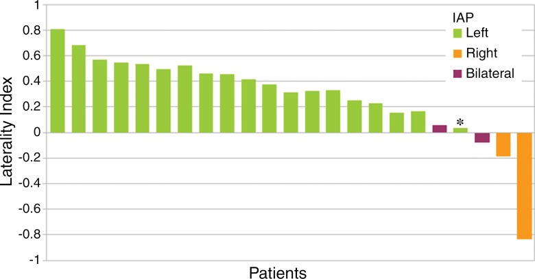
Purpose To measure the accuracy of resting-state functional magnetic resonance (MR) imaging in determining hemispheric language dominance in patients with medically intractable focal epilepsies against the results of an intracarotid amobarbital procedure (IAP). Materials and Methods This study was approved by the institutional review board, and all subjects gave signed informed consent. Data in 23 patients with medically intractable focal epilepsy were retrospectively analyzed. All 23 patients were candidates for epilepsy surgery and underwent both IAP and resting-state functional MR imaging as part of presurgical evaluation. Language dominance was determined from functional MR imaging data by calculating a laterality index (LI) after using independent component analysis. The accuracy of this method was assessed against that of IAP by using a variety of thresholds. Sensitivity and specificity were calculated by using leave-one-out cross validation. Spatial maps of language components were qualitatively compared among each hemispheric language dominance group. Results Measurement of hemispheric language dominance with resting-state functional MR imaging was highly concordant with IAP results, with up to 96% (22 of 23) accuracy, 96% (22 of 23) sensitivity, and 96% (22 of 23) specificity. Composite language component maps in patients with typical language laterality consistently included classic language areas such as the inferior frontal gyrus, the posterior superior temporal gyrus, and the inferior parietal lobule, while those of patients with atypical language laterality also included non-classical language areas such as the superior and middle frontal gyri, the insula, and the occipital cortex. Conclusion Resting-state functional MR imaging can be used to measure language laterality in patients with medically intractable focal epilepsy. (©) RSNA, 2016 Online supplemental material is available for this article.
Figures


References
Publication types
MeSH terms
Substances
Grants and funding
LinkOut - more resources
Full Text Sources
Other Literature Sources
Medical
Research Materials
