. 2016 Oct:497:125-135.
doi: 10.1016/j.virol.2016.07.016. Epub 2016 Jul 26.

Monkeypox virus induces the synthesis of less dsRNA than vaccinia virus, and is more resistant to the anti-poxvirus drug, IBT, than vaccinia virus

Affiliations

Monkeypox virus induces the synthesis of less dsRNA than vaccinia virus, and is more resistant to the anti-poxvirus drug, IBT, than vaccinia virus

William D Arndt et al. Virology. 2016 Oct.

Abstract

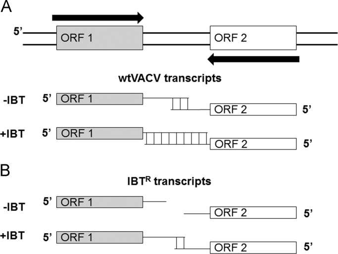
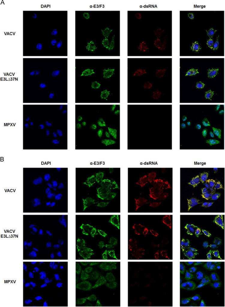
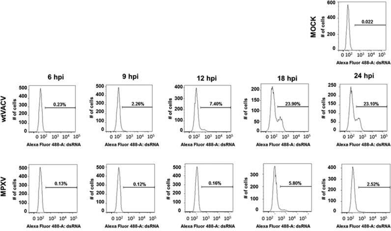
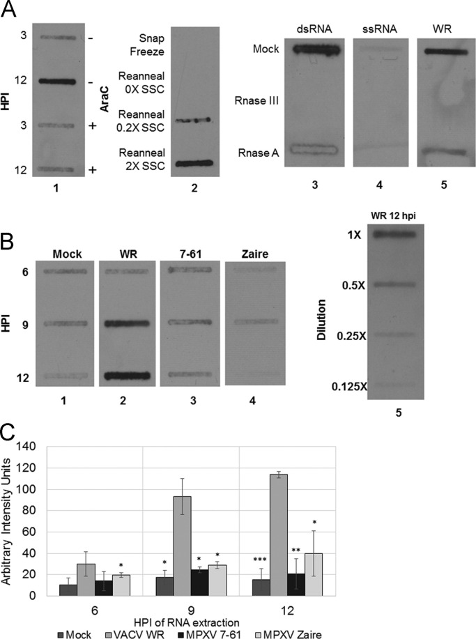
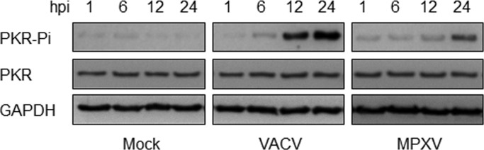
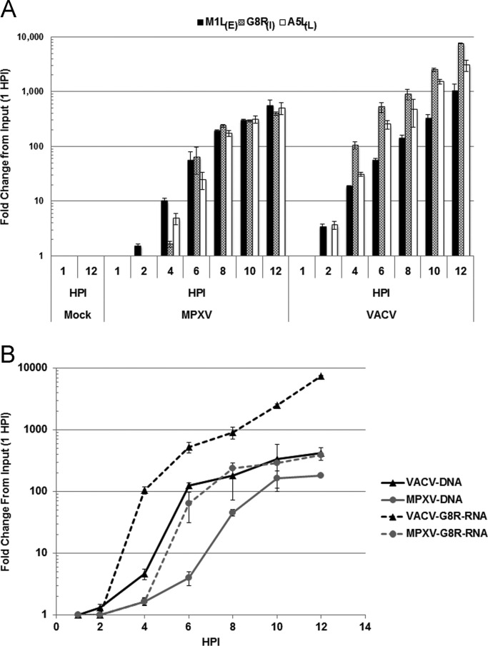
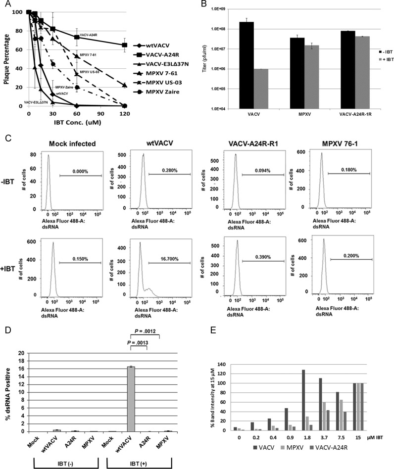
Monkeypox virus (MPXV) infection fails to activate the host anti-viral protein, PKR, despite lacking a full-length homologue of the vaccinia virus (VACV) PKR inhibitor, E3. Since PKR can be activated by dsRNA produced during a viral infection, we have analyzed the accumulation of dsRNA in MPXV-infected cells. MPXV infection led to less accumulation of dsRNA than VACV infection. Because in VACV infections accumulation of abnormally low amounts of dsRNA is associated with mutations that lead to resistance to the anti-poxvirus drug isatin beta-thiosemicarbazone (IBT), we investigated the effects of treatment of MPXV-infected cells with IBT. MPXV infection was eight-fold more resistant to IBT than wild-type vaccinia virus (wtVACV). These results demonstrate that MPXV infection leads to the accumulation of less dsRNA than wtVACV, which in turn likely leads to a decreased capacity for activation of the dsRNA-dependent host enzyme, PKR.

Keywords: E3 gene; F3 gene; IBT resistance; Innate immune evasion; Monkeypox virus; Poxvirus pathogenesis; Poxvirus transcription; Vaccinia virus; Virulence; dsRNA.


Figures

Fig. 1
Fig. 1
dsRNA accumulation in orthopoxvirus-infected cells. (A) dsRNA is thought to accumulate from convergent transcripts. At intermediate and late times post-infection, termination of transcription is imprecise, leading to long run-on transcripts. In wild-type VACV, these transcripts can base-pair with transcripts read from the opposite strand of the genome, forming dsRNA. The anti-poxvirus drug IBT increases processivity of the viral polymerase, leading to longer transcripts and accumulation of more dsRNA. (B) IBTR mutants of VACV generate shorter transcripts with less overlap, and thus accumulate less dsRNA. IBT treatment of cells infected with IBTR mutants of VACV still increases the length of viral transcripts, but the length of these transcripts is thought to be less than the length of transcripts in IBT-treated cells infected with wtVACV.
Fig. 2
Fig. 2
VACV and MPXV dsRNA levels in HeLa cells at 6 hpi and 9 hpi. HeLa cells were infected at an MOI of 5 with VACV, VACV-E3L∆37N and MPXV. At 6 hpi (A) or 9 hpi (B) the cells were fixed with methanol and stained for the presence of E3 or F3 (green) and dsRNA (Red). Nuclei (blue) were stained with DAPI. Merge panels represent an overlay of all three images. Yellow indicates the co-localization of E3 or F3 and dsRNA.
Fig. 3
Fig. 3
Flow cytometry assay of dsRNA in infected cells. HeLa cells were mock infected or infected with either wtVACV or MPXV. At 6, 9, 12, 18 or 24 hpi, cells were trypsinized and fixed with CytoPerm/Fix kit (BD). Cells were stained with antibodies against dsRNA and analyzed by flow cytometry the next day. Total cells were gated to exclude non-viable cells.
Fig. 4
Fig. 4
Slot blot detection of dsRNA in extracts from virus-infected cells. Cells were infected with virus and then total RNA was extracted at multiple time points post-infection using the RNeasy Kit (Qiagen). Equal volumes of extract were then transferred onto BrightStar®-Plus Positively Charged Nylon Membrane (Ambion) using a vacuum slot apparatus. dsRNA was visualized on the blot by probing with J2 anti-dsRNA antibodies and Goat anti-mouse HRP conjugate secondary antibody. (A) To verify the specificity of the J2 antibody in a slot blot, the following controls were performed: Lane 1: HeLa cells were mock pretreated or pretreated with 40 µg/mL AraC 1 h prior to infection with VACV WR. Total RNA was extracted at 3 and 12 hpi. Lane 2: BSC-40 cells were infected with WR and total RNA was extracted at 8 hpi. RNA was denatured by boiling at 95 °C for 5 min; samples where then either snap frozen or left at room temperature to reanneal in either no, low, or high salt conditions. Lanes 3–5: dsRNA ladder, ssRNA ladder, and WR 12 hpi-extract were either mock treated or treated with RNAse III (dsRNA specific) or RnaseA (ssRNA specific) RnaseA digestion was done in 2XSSC (300 mM NaCl and 30 mM Sodium Citrate). (B) Lanes 1–4: HeLa cells were mock infected or infected with VACV WR, MPXV 7-61, or MPXV Zaire at an MOI of 5. Total RNA was collected at 6, 9, and 12 hpi. RNA extractions were performed in triplicate; figure shows representative results. Lane 5: Serial 2-fold dilutions of the VACV WR 12 hpi-extract were included to verify that the exposure was in the linear range. (C) Band intensity of slot blots was analyzed with TotalLab Quant. The intensity of the WR 12 hpi-extraction was calibrated to 100 arbitrary units. The triplicate extraction intensities were averaged; error bars show standard deviation. Significance was calculated with unpaired 2-tailed t-test. Asterisks indicate significance of difference from corresponding VACV WR time point. *p<0.05, **p<0.01, and ***p<0.005.
Fig. 5
Fig. 5
ELISA for VACV and MPXV dsRNA levels. HeLa cells were infected with VACV and MPXV at an MOI 5. At 1, 2, 4, 6, 8, 10, and 12 hpi cells were harvested, total RNA was extracted, and 500 ng of total RNA was added to ELISA plate. Signal was quantified by Luminescent plate reader. Data presented are means with standard error of multiple experiments.
Fig. 6
Fig. 6
Use of PKR activation to detect dsRNA in extracts from the infected cells. HeLa cells were either mock infected or infected with VACV or MPXV. RNA was extracted at 1, 6, 12, and 24 hpi. RNA was added to an in vitro PKR activation assay with IFN-treated HeLa lysate. The lysates were than analyzed by Western blot, using antiserum that recognizes PKR phosphorylation at Threonine 446.
Fig. 7
Fig. 7
Accumulation of viral RNA and DNA in HeLa cells. HeLa cells were either mock infected or infected with VACV and MPXV at an MOI of 5. RNA (A) or DNA (B) was extracted at 1, 2, 4, 6, 8, 10, and 12 hpi. (A) Real-Time quantitative reverse-transcriptase PCR was performed with gene-specific primers for M1L (early), G8R (intermediate) and A5L (late) viral transcripts. (B) Real-Time quantitative PCR for viral DNA was performed with G8R-specific primers on 500 ng of total DNA.
Fig. 8
Fig. 8
MPXV sensitivity to IBT. (A) MPXV sensitivity to IBT: Plaque Reduction. BSC-40 cells were infected with ~100 pfu of virus and treated with two-fold increasing concentrations of IBT. Viruses used were VACV, VACV-A24R-R1, VACV-E3LΔ37N, two West African strains of MPXV (7-61 and US-2003), and MPXV Zaire (Central African strain). Plaques were stained with crystal violet and counted. Results appear as the percentage of the number of plaques present in the untreated cells from three replicates. (B) MPXV sensitivity to IBT: Multi-cycle Growth. Multi-cycle growth kinetics assays were performed in BSC-40 cells. The cells were infected with VACV, MPXV, and VACV-A24R-R1 at an MOI of 0.01 in the presence of absence of 60 µM IBT. Viruses were harvested at 0 and 72 hpi and titered by plaque assay in RK-E3L cells. Data presented are means with standard error of multiple experiments. (C) MPXV sensitivity to IBT: Accumulation of Free dsRNA. HeLa cells were mock infected or infected with either wt VACV, VACV A24R-R1 or MPXV. Infected cells were either untreated or treated with 45 µM IBT. After 6 hpi, cells were trypsinized and fixed. Cells were stained with antibodies against total VACV and dsRNA and were subjected to flow cytometry the next day. Total cells were gated to exclude non-viable cells. (D) Quantitation of replicate results from Panel C. Statistical difference was determined by t test using Holm-Sidak method with alpha=5%. (E) MPXV sensitivity to IBT: PKR Phosphorylation. Hela cells were infected with VACV, MPXV, or VACV-A24R-R1 at an MOI of 5 and treated one hour post infection with two-fold increasing concentrations of IBT. Western blot analyses were performed with antibodies directed against the phosphorylated form of PKR and with antibodies to the viral protein E3/F3. Western blot band intensities were measured with Image Quant, normalized to GAPDH levels, and shown as a percentage of the band intensity at the highest (15 µM) level of IBT treatment.

References

    1. Altschul S.F., Madden T.L., Schaffer A.A., Zhang J., Zhang Z., Miller W., Lipman D.J. Gapped BLAST and PSI-BLAST: a new generation of protein database search programs. Nucleic Acids Res. 1997;25:3389–3402. - PMC - PubMed
    1. Arndt W.D., Cotsmire S., Trainor K., Harrington H., Hauns K., Kibler K.V., Huynh T.P., Jacobs B.L. Evasion of the innate immune type I interferon system by monkeypox virus. J. Virol. 2015;89:10489–10499. - PMC - PubMed
    1. Bayliss C.D., Condit R.C. Temperature-sensitive mutants in the vaccinia virus A18R gene increase double-stranded RNA synthesis as a result of aberrant viral transcription. Virology. 1993;194:254–262. - PubMed
    1. Brandt T., Heck M.C., Vijaysri S., Jentarra G.M., Cameron J.M., Jacobs B.L. The N-terminal domain of the vaccinia virus E3L-protein is required for neurovirulence, but not induction of a protective immune response. Virology. 2005;333:263–270. - PubMed
    1. Brandt T.A., Jacobs B.L. Both carboxy- and amino-terminal domains of the vaccinia virus interferon resistance gene, E3L, are required for pathogenesis in a mouse model. J. Virol. 2001;75:850–856. - PMC - PubMed

Publication types