. 2016 Jul 28;11(7):e0160055.
doi: 10.1371/journal.pone.0160055. eCollection 2016.

MRI as a Novel In Vivo Approach for Assessing Structural Changes of Chlamydia Pathology in a Mouse Model

Affiliations

MRI as a Novel In Vivo Approach for Assessing Structural Changes of Chlamydia Pathology in a Mouse Model

Catherine D G Hines et al. PLoS One. .

Abstract

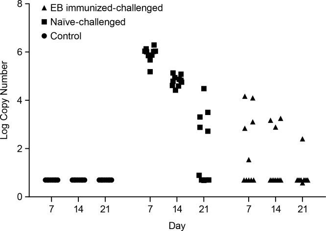
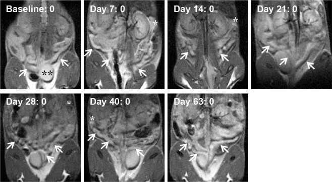
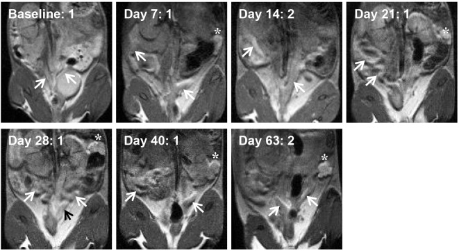
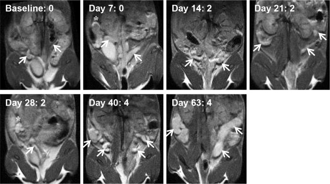
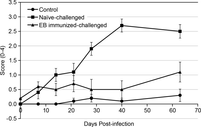
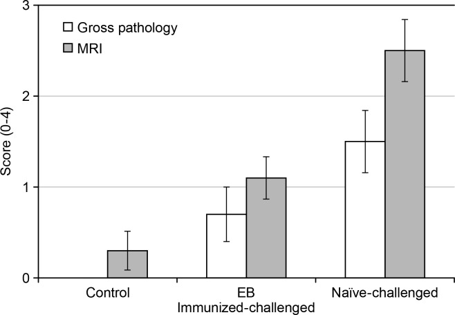
Chlamydia trachomatis is among the most prevalent of sexually transmitted diseases. While Chlamydia infection is a reportable event and screening has increased over time, enhanced surveillance has not resulted in a reduction in the rate of infections, and Chlamydia infections frequently recur. The development of a preventative vaccine for Chlamydia may be the only effective approach for reducing infection and the frequency of pathological outcomes. Current vaccine research efforts involve time consuming and/or invasive approaches for assessment of disease state, and MRI presents a clinically translatable method for assessing infection and related pathology both quickly and non-invasively. Longitudinal T2-weighted MRI was performed over 63 days on both control or Chlamydia muridarum challenged mice, either with or without elementary body (EB) immunization, and gross necropsy was performed on day 65. A scoring system was developed to assess the number of regions affected by Chlamydia pathology and was used to document pathology over time and at necropsy. The scoring system documented increasing incidence of pathology in the unimmunized and challenged mice (significantly greater compared to the control and EB immunized-challenged groups) by 21 days post-challenge. No differences between the unchallenged and EB immunized-challenged mice were observed. MRI scores at Day 63 were consistently higher than gross necropsy scores at Day 65, although two of the three groups of mice showed no significant differences between the two techniques. In this work we describe the application of MRI in mice for the potential evaluation of disease pathology and sequelae caused by C. muridarum infection and this technique's potential for evaluation of vaccines for Chlamydia.


Conflict of interest statement

Competing Interests: The authors of this manuscript have read the journal's policy and have the following competing interests: CDGH, SW, XM, JMS and MAB are currently employees of Merck & Co., Inc. JHH and JGS were employees of Merck & Co., Inc. at the initiation of this work, and are currently employed by Sanofi Pasteur and Pfizer, respectively. All work stated in the submitted manuscript was performed at Merck & Co., Inc., and all work was funded by Merck & Co., Inc. As such, the authors above have competing financial interests in the form of employment and/or ownership of stocks or shares. The spouse of author CDGH consults for W. H. Freeman, an educational company. This does not alter the authors’ adherence to PLOS ONE policies on sharing data and materials.

Figures

Fig 1
Fig 1. Bacterial counts recovered by vaginal swab.
Summary of the course of infection over the first three weeks after challenge, as measured by qPCR.
Fig 2
Fig 2. MRI images of an example control mouse at all time points during the study (n = 10 mice in control group).
The coronal images are acquired such that the animal’s head is at the top of the figures, the tail at the bottom, and as if the animal is facing the viewer such that the animal’s left side is on the right side of the figures. Arrows point to uterine horns, asterisks denote an ovary/oviduct when present in the image plane, and the double asterisk identifies the bladder at baseline. Images are shown at the approximate plate of the uterine horn branching or to show both horns. The MRI image score is shown next to the time point, and the gross pathology score at Day 65 was 0 for this animal.
Fig 3
Fig 3. MRI images of an example EB immunized-challenged mouse at all time points during the study (n = 10 mice in EB immunized-challenged group).
Arrows point to uterine horns, and asterisks denote an ovary/oviduct when present in the image. Black arrows are used only to provide contrast on hyperintense regions. Images are shown at the approximate plate of the uterine horn branching or to show horns. The presence of fluid in the animal’s left ovary/oviduct was seen from Day 21 through Day 63. The MRI image score is shown next to the time point, and the gross pathology score at Day 65 was 1 for this animal.
Fig 4
Fig 4. MRI images of an example naïve-challenged mouse at all time points of the study (n = 10 mice in naïve-challenged group).
Arrows point to uterine horns, and asterisks denote an ovary/oviduct when present in the image. Images are shown at the approximate plate of the uterine horn branching or to show horns. Excessive fluid in the uterine horns is seen at Day 63. The MRI image score is shown next to the time point, and the gross pathology score at Day 65 was 3 for this animal.
Fig 5
Fig 5. Group means of the MRI scores during the course of the study.
Error bars represent the standard error of the mean (SEM).
Fig 6
Fig 6. Comparison of the mean MRI image and gross pathology scores at the end of the study for the three groups of mice.
MRI image scores at Day 63 were consistently higher than gross pathology scores at Day 65, although no significant differences were seen between the techniques for the control and EB immunized-challenged groups. Error bars represent SEM.

Similar articles

Cited by

  • Update on Chlamydia trachomatis Vaccinology.
    de la Maza LM, Zhong G, Brunham RC. de la Maza LM, et al. Clin Vaccine Immunol. 2017 Apr 5;24(4):e00543-16. doi: 10.1128/CVI.00543-16. Print 2017 Apr. Clin Vaccine Immunol. 2017. PMID: 28228394 Free PMC article. Review.

References

    1. Centers for Disease Control and Prevention. 2011 Sexually Transmitted Disease Surveillance: Chlamydia. Division of STD Prevention, National Center for HIV/AIDS, Viral Hepatitis, STD and TB Prevention, Centers for Disease Control and Prevention. 2012;12:13 Available: http://www.cdc.gov/std/stats11/chlamydia.htm.
    1. Centers for Disease Control and Prevention. CDC Grand Rounds: Chlamydia prevention: challenges and strategies for reducing disease burden and sequelae. MMWR Morb Mortal Wkly Rep. 2011;60(12):370–3. - PubMed
    1. Gottlieb SL, Brunham RC, Byrne GI, Martin DH, Xu F, Berman SM. Introduction: The natural history and immunobiology of Chlamydia trachomatis genital infection and implications for chlamydia control. J Infect Dis. June 2010; 201 Suppl 2:S85–7. 10.1086/652392 - DOI - PubMed
    1. Brunham RC, Rey-Ladino J. Immunology of Chlamydia infection: implications for a Chlamydia trachomatis vaccine. Nat Rev Immunol. February 2005; 5(2): 149–61. - PubMed
    1. Hafner L, Beagley K, Timms P. Chlamydia trachomatis infection: host immune responses and potential vaccines. Mucosal Immunology. 2008. March; 1(2):116–30. 10.1038/mi.2007.19 - DOI - PubMed