

Review
doi: 10.1016/j.sbi.2016.07.010.
Epub 2016 Jul 25.
Structure-guided wavelength tuning in far-red fluorescent proteins
Affiliations
- PMID: 27468111
- PMCID: PMC5548387
- DOI: 10.1016/j.sbi.2016.07.010
Item in Clipboard
Review
Structure-guided wavelength tuning in far-red fluorescent proteins
Curr Opin Struct Biol.
2016 Aug.
Abstract
In recent years, protein engineers have succeeded in tuning the excitation spectra of natural fluorescent proteins from green wavelengths into orange and red wavelengths, resulting in the creation of a series of fluorescent proteins with emission in the far-red portions of the optical spectrum. These results have arisen from the synergistic combination of structural knowledge of fluorescent proteins, chemical intuition, and high-throughput screening methods. Here we review structural features found in autocatalytic far-red fluorescent proteins, and discuss how they add to our understanding of the biophysical mechanisms of wavelength tuning in biological chromophores.
Copyright © 2016. Published by Elsevier Ltd.
Figures
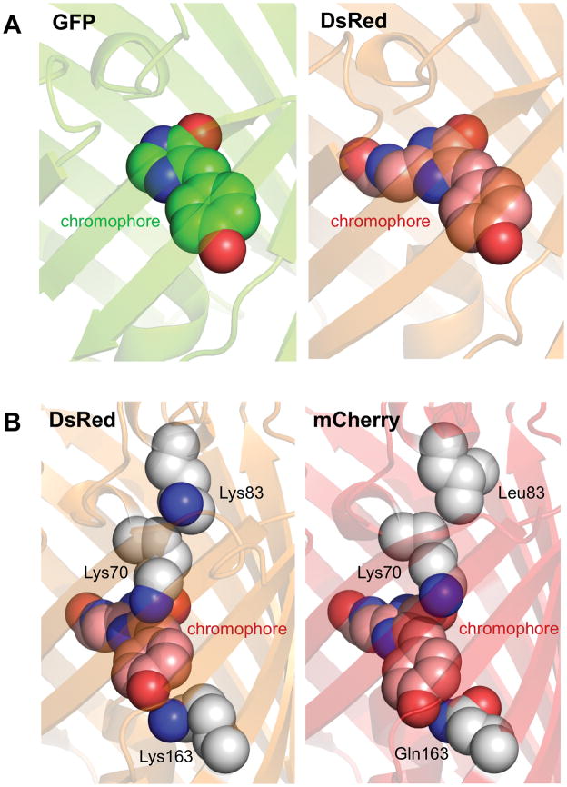
Basic structural biology of RFPs. (A) Structures of GFP and DsRed chromophores. The GFP chromophore (left), shown with carbon in green, nitrogen blue, and oxygen red, is a π-conjugated system consisting of a phenolate ring (front) connected to a imidazolinone ring (rear) by a methylene bridge. The DsRed chromophore (right), shown with carbon in pink, consists of the same groups formed from cyclization of a Gln-Tyr-Gly sequence, but with conjugation extended through an acylimine group formed by additional oxidation of the backbone N-Cα bond of the Gln residue. (B) Altered electronic interactions of the chromophore with its environment in mCherry. In DsRed (left), the positive charges of Lys70 and Lys163 stabilize the ground state, where electron density preferentially resides in the phenol ring. In mCherry (right), movement of Lys70 away from the chromophore and mutation of Lys163 to Gln reduces this effect. Movement of Lys70 in mCherry is due to loss of electrostatic repulsion by Leu83. Side chains and alpha carbons of these important amino acids are shown a spheres, with carbon in white, nitrogen blue, and oxygen red.
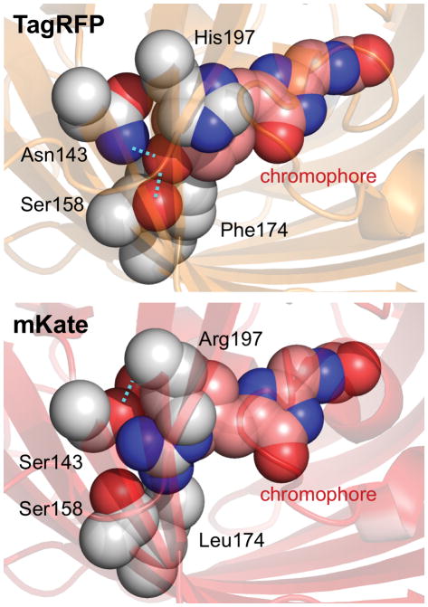
Chromophore isomerization, reduced hydrogen bonding, and altered electrostatic interactions in mKate. The chromophore rings in TagRFP are in a trans conformation, where the phenolate oxygen makes two hydrogen bonds (cyan dotted lines) and the His197 engages in a cation-π interaction with the phenolate ring. In mKate, mutations Asn143Ser, Phe174Leu, and His197Arg cause the chromophore to assume a cis conformation, losing one hydrogen bond and the cation-π interaction.
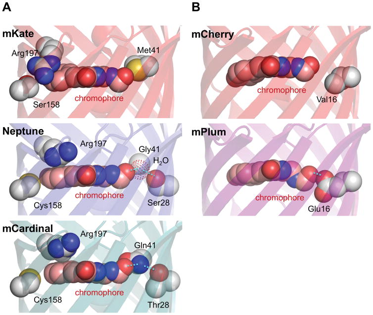
Structural basis of red-shifted excitation in far-RFPs. (A) Increased chromophore planarity and a new hydrogen bond in Neptune and mCardinal. In mKate, the Arg197 side chain extends across and to the side of the phenolate ring of the chromophore, which is not coplanar with the imidazolinone ring. In Neptune, Cys158 pushes Arg197 to a new position above the chromophore, allowing for a more coplanar arrangement of the two chromophore rings. In addition, mutation Met41Gly creates a new cavity for a water molecule (red dotted sphere) to reside in near the acylimine. The white sphere labeled Gly41 is the Gly41 alpha carbon. Hydrogen bonding between the water molecule and the acylimine oxygen stabilizes the excited state and leads to an excitation red-shift. In mCardinal, the role of the water molecule is replaced by Gln41, which is held in position by Thr28. (B) Excited-state energetic relaxations near the acylimine in the excited state of mPlum. mPlum contains a new hydrogen bond from Glu16 to the acylimine oxygen of the chromophore. Conformational changes to the chromophore or to Glu16 occur in the excited state, causing a large Stokes shift.
Similar articles
-
Structure-guided rational design of red fluorescent proteins: towards designer genetically-encoded fluorophores.Curr Opin Struct Biol. 2017 Aug;45:91-99. doi: 10.1016/j.sbi.2016.12.001. Epub 2016 Dec 27. Curr Opin Struct Biol. 2017. PMID: 28038355 Review.
-
Monomeric Garnet, a far-red fluorescent protein for live-cell STED imaging.Sci Rep. 2015 Dec 9;5:18006. doi: 10.1038/srep18006. Sci Rep. 2015. PMID: 26648024 Free PMC article.
-
The structure of a far-red fluorescent protein, AQ143, shows evidence in support of reported red-shifting chromophore interactions.Protein Sci. 2014 Aug;23(8):1148-53. doi: 10.1002/pro.2498. Epub 2014 Jun 14. Protein Sci. 2014. PMID: 24888769 Free PMC article.
-
Optimized and far-red-emitting variants of fluorescent protein eqFP611.Chem Biol. 2008 Mar;15(3):224-33. doi: 10.1016/j.chembiol.2008.02.008. Chem Biol. 2008. PMID: 18355722
-
Red fluorescent proteins: chromophore formation and cellular applications.Curr Opin Struct Biol. 2012 Oct;22(5):679-88. doi: 10.1016/j.sbi.2012.09.002. Epub 2012 Sep 20. Curr Opin Struct Biol. 2012. PMID: 23000031 Free PMC article. Review.
Cited by
-
Far-Red Fluorescent Proteins: Tools for Advancing In Vivo Imaging.Biosensors (Basel). 2024 Jul 24;14(8):359. doi: 10.3390/bios14080359. Biosensors (Basel). 2024. PMID: 39194588 Free PMC article. Review.
-
Tissue clearing may alter emission and absorption properties of common fluorophores.Sci Rep. 2022 Apr 1;12(1):5551. doi: 10.1038/s41598-022-09303-9. Sci Rep. 2022. PMID: 35365729 Free PMC article.
-
A Bright, Nontoxic, and Non-aggregating red Fluorescent Protein for Long-Term Labeling of Fine Structures in Neurons.Front Cell Dev Biol. 2022 Jun 29;10:893468. doi: 10.3389/fcell.2022.893468. eCollection 2022. Front Cell Dev Biol. 2022. PMID: 35846353 Free PMC article.
-
Recent Advances in Gene Therapy for Cancer Theranostics.Curr Opin Biomed Eng. 2021 Dec;20:100300. doi: 10.1016/j.cobme.2021.100300. Epub 2021 Jul 15. Curr Opin Biomed Eng. 2021. PMID: 34738046 Free PMC article.
-
Fluorescent indicators for simultaneous reporting of all four cell cycle phases.Nat Methods. 2016 Dec;13(12):993-996. doi: 10.1038/nmeth.4045. Epub 2016 Oct 31. Nat Methods. 2016. PMID: 27798610 Free PMC article.
References
-
- Shimomura O, Johnson FH, Saiga Y. Extraction, purification and properties of aequorin, a bioluminescent protein from the luminous hydromedusan, Aequorea. J Cell Comp Physiol. 1962;59:223–239. - PubMed
-
- Matz MV, Fradkov AF, Labas YA, Savitsky AP, Zaraisky AG, Markelov ML, Lukyanov SA. Fluorescent proteins from nonbioluminescent Anthozoa species. Nat Biotechnol. 1999;17:969–973. - PubMed
Publication types
MeSH terms
Substances
Grants and funding
LinkOut - more resources
Full Text Sources
Other Literature Sources
