

Blood Biomarkers for Evaluation of Perinatal Encephalopathy
- PMID: 27468268
- PMCID: PMC4942457
- DOI: 10.3389/fphar.2016.00196
Blood Biomarkers for Evaluation of Perinatal Encephalopathy
Abstract
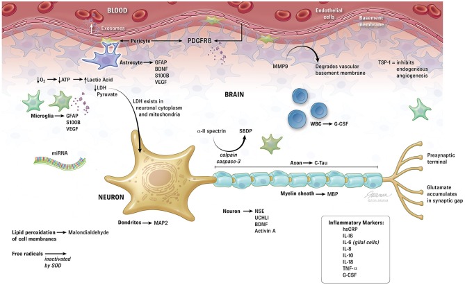
Recent research in identification of brain injury after trauma shows many possible blood biomarkers that may help identify the fetus and neonate with encephalopathy. Traumatic brain injury shares many common features with perinatal hypoxic-ischemic encephalopathy. Trauma has a hypoxic component, and one of the 1st physiologic consequences of moderate-severe traumatic brain injury is apnea. Trauma and hypoxia-ischemia initiate an excitotoxic cascade and free radical injury followed by the inflammatory cascade, producing injury in neurons, glial cells and white matter. Increased excitatory amino acids, lipid peroxidation products, and alteration in microRNAs and inflammatory markers are common to both traumatic brain injury and perinatal encephalopathy. The blood-brain barrier is disrupted in both leading to egress of substances normally only found in the central nervous system. Brain exosomes may represent ideal biomarker containers, as RNA and protein transported within the vesicles are protected from enzymatic degradation. Evaluation of fetal or neonatal brain derived exosomes that cross the blood-brain barrier and circulate peripherally has been referred to as the "liquid brain biopsy." A multiplex of serum biomarkers could improve upon the current imprecise methods of identifying fetal and neonatal brain injury such as fetal heart rate abnormalities, meconium, cord gases at delivery, and Apgar scores. Quantitative biomarker measurements of perinatal brain injury and recovery could lead to operative delivery only in the presence of significant fetal risk, triage to appropriate therapy after birth and measure the effectiveness of treatment.
Keywords: Glial injury; biomarkers; hypoxic-ischemic encephalopathy; neonatal encephalopathy; neuronal injury.
Figures
Similar articles
-
Focal Brain Injury Associated with a Model of Severe Hypoxic-Ischemic Encephalopathy in Nonhuman Primates.Dev Neurosci. 2017;39(1-4):107-123. doi: 10.1159/000456658. Epub 2017 Mar 25. Dev Neurosci. 2017. PMID: 28343228 Free PMC article.
-
Cesarean section on request at 39 weeks: impact on shoulder dystocia, fetal trauma, neonatal encephalopathy, and intrauterine fetal demise.Semin Perinatol. 2006 Oct;30(5):276-87. doi: 10.1053/j.semperi.2006.07.009. Semin Perinatol. 2006. PMID: 17011400 Review.
-
[Perinatal differences in asphyxic full-term newborns in relation to the presence of hypoxic-ischemic encephalopathy].Rev Neurol. 1997 Aug;25(144):1187-94. Rev Neurol. 1997. PMID: 9340143 Spanish.
-
[Perinatal asphyxia, hypoxic-ischemic encephalopathy and neurological sequelae in full-term newborns. II. Description and interrelation].Rev Neurol. 1996 Aug;24(132):969-76. Rev Neurol. 1996. PMID: 8755359 Spanish.
-
Neonatal hypoxic ischemic encephalopathy-related biomarkers in serum and cerebrospinal fluid.Clin Chim Acta. 2015 Oct 23;450:282-97. doi: 10.1016/j.cca.2015.08.021. Epub 2015 Aug 28. Clin Chim Acta. 2015. PMID: 26320853 Review.
Cited by
-
Comparability of the small RNA secretome across human biofluids concomitantly collected from healthy adults.PLoS One. 2020 Apr 10;15(4):e0229976. doi: 10.1371/journal.pone.0229976. eCollection 2020. PLoS One. 2020. PMID: 32275679 Free PMC article.
-
Blood biomarkers for evaluation of perinatal encephalopathy: state of the art.Curr Opin Pediatr. 2018 Apr;30(2):199-203. doi: 10.1097/MOP.0000000000000591. Curr Opin Pediatr. 2018. PMID: 29346139 Free PMC article. Review.
-
Window of opportunity for human amnion epithelial stem cells to attenuate astrogliosis after umbilical cord occlusion in preterm fetal sheep.Stem Cells Transl Med. 2021 Mar;10(3):427-440. doi: 10.1002/sctm.20-0314. Epub 2020 Oct 26. Stem Cells Transl Med. 2021. PMID: 33103374 Free PMC article.
-
Promise of extracellular vesicles for diagnosis and treatment of epilepsy.Epilepsy Behav. 2021 Aug;121(Pt B):106499. doi: 10.1016/j.yebeh.2019.106499. Epub 2019 Oct 18. Epilepsy Behav. 2021. PMID: 31636006 Free PMC article. Review.
-
Seizures May Worsen Outcomes of Neonatal Hypoxic-Ischemic Encephalopathy: A Longitudinal Serum Biomarkers Study.Pediatr Neurol. 2025 May;166:55-64. doi: 10.1016/j.pediatrneurol.2025.02.008. Epub 2025 Feb 21. Pediatr Neurol. 2025. PMID: 40101305
References
-
- American College of Obstetricians Gynecologists American Academy of Pediatrics. (2014). Neonatal Encephalopathy and Neurologic Outcome, 2nd Edn. Washington DC: American College of Obstetricians and Gynecologists.
-
- Banks W. A., Gray A. M., Erickson M. A., Salameh T. S., Damodarasamy M., Sheibani N., et al. . (2015). Lipopolysaccharide-induced blood-brain barrier disruption: roles of cyclooxygenase, oxidative stress, neuroinflammation, and elements of the neurovascular unit. J. Neuroinflammation 12:223. 10.1186/s12974-015-0434-1 - DOI - PMC - PubMed
Publication types
Grants and funding
LinkOut - more resources
Full Text Sources
Other Literature Sources