. 2016 Jul 29:6:30754.
doi: 10.1038/srep30754.

Effect of leptin combined with CoCl2 on healing in Sprague Dawley Rat fracture model

Affiliations

Effect of leptin combined with CoCl2 on healing in Sprague Dawley Rat fracture model

Pengcheng Liu et al. Sci Rep. .

Abstract

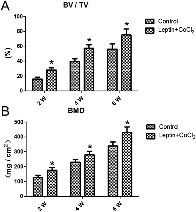
To evaluate the effect of leptin combined with CoCl2 on rat femur fracture healing. 48 male Sprague Dawley rats were randomly divided into two main groups. Then standardized femur fractures were created to all rats. Control group rats were treated with 0.5 mL physiological saline, and experimental group rats were treated with 5 μg/Kg.d leptin and 15 mg/Kg.d CoCl2 along with 0.5 mL physiological saline for 42 days intraperitoneally. Each main group was divided into three subgroups for each evaluation at second, fourth and sixth weeks, each subgroup included eight rats. The radiological evaluation showed that the fracture healing progress of experimental group was superior to control group from second week. At fourth week, experimental group had better fracture healing progress than control group significantly. Results of biomechanics show the ultimate load (N) and deflection ultimate load (mm) of experimental group was significantly increased than that in control group from fourth week. The present result demonstrated that leptin combined with CoCl2 significantly increased the mRNA expression levels of HIF1A, Vegfa, Runx2, Bmp2, Bglap and Alpl. It suggested that leptin combined with CoCl2 have a positive effect on rat femur fracture healing by activating the HIF1A pathway.


Figures

Figure 1
Figure 1
(A) Different degrees of union radiological images of two groups. (B) Callus diameter of X-ray (mm) of experimental group were larger than control group. (C) Volume of fracture callus (mm3) of experimental group were larger than control group. *P < 0.05; **P < 0.01 vs. Control group at indicated time.
Figure 2
Figure 2
(A) Bone volume fraction (BV/TV) of experimental group were larger than that of control group from fourth week. (B) Bone mineral density (BMD) of the callus in the experimental group were significantly increased compared with control group from fourth week. *P < 0.05; **P < 0.01 vs. Control group at indicated time.
Figure 3
Figure 3
(A) Microscopy of callus with tetracycline bone double labeling fluorescence showed the growth distance of new bone between groups had significantly difference at sixth weeks. (B) Histomorphology showed the mineral apposition rate (MAR) between groups had significantly difference from fourth week. *P < 0.05; **P < 0.01 vs. Control group at indicated time.
Figure 4
Figure 4
(A)Ultimate load (N) of experimental group was significantly increased than that of control group at fourth and sixth weeks. (B) Deflection ultimate load (mm) had significantly difference at fourth and sixth weeks. *P < 0.05; **P < 0.01 vs. Control group at indicated time.
Figure 5
Figure 5. Relative mRNA expression levels of Alpl, Bglap, Bmp2, Runx2, Vegfa and HIF1A were determined by quantitative polymerase chain reaction.
*P < 0.05; **P < 0.01 vs. Control group at indicated time. Alpl, Alkaline phosphatase, liver/bone/kidney; Bglap, Bone gamma-carboxyglutamate (gla) protein; Bmp2, Bone morphogenetic protein 2; Runx2, Runt-related transcription factor 2; Vegfa, Vascular endothelial growth factor A; HIF1A, hypoxia-inducible factor-1α.

References

    1. Dimitri P. et al. Leptin May Play a Role in Bone Microstructural Alterations in Obese Children. J Clin Endocrinol Metab 100, 594–602, 10.1210/jc.2014-3199 (2015). - DOI - PMC - PubMed
    1. Chen X. X. & Yang T. Roles of Leptin in Bone Metabolism and Bone Diseases. J Bone Miner Metab 33, 474–485, 10.1007/s00774-014-0569-7 (2015). - DOI - PubMed
    1. Liu K., Liu P., Liu R., Wu X. & Cai M. Relationship Between Serum Leptin Levels and Bone Mineral Density: A Systematic Review and Meta-Analysis. Clin Chim Acta 444, 260–263, 10.1016/j.cca.2015.02.040 (2015). - DOI - PubMed
    1. Yamauchi M. et al. Plasma Leptin Concentrations are Associated with Bone Mineral Density and the Presence of Vertebral Fractures in Postmenopausal Women. Clin Endocrinol (Oxf) 55, 341–347 (2001). - PubMed
    1. Cornish J. et al. Leptin Directly Regulates Bone Cell Function in Vitro and Reduces Bone Fragility in Vivo. J Endocrinol 175, 405–415 (2002). - PubMed

Publication types

MeSH terms