. 2016 Oct;284(Pt A):50-62.
doi: 10.1016/j.expneurol.2016.07.018. Epub 2016 Jul 25.

Deficiency in matrix metalloproteinase-2 results in long-term vascular instability and regression in the injured mouse spinal cord

Affiliations

Deficiency in matrix metalloproteinase-2 results in long-term vascular instability and regression in the injured mouse spinal cord

Alpa Trivedi et al. Exp Neurol. 2016 Oct.

Abstract

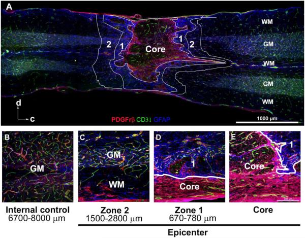
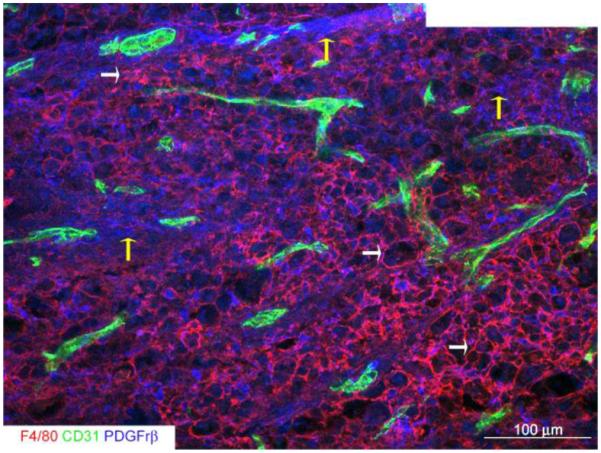
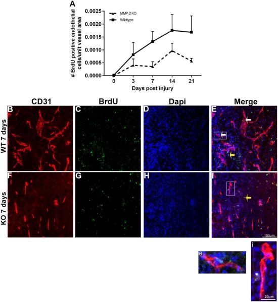
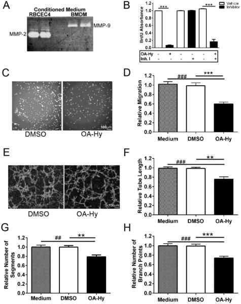
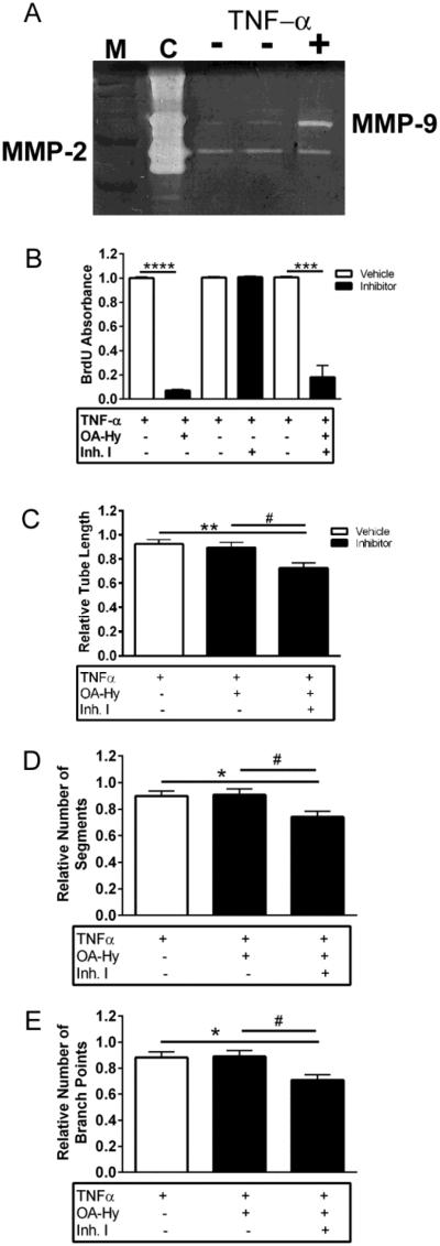
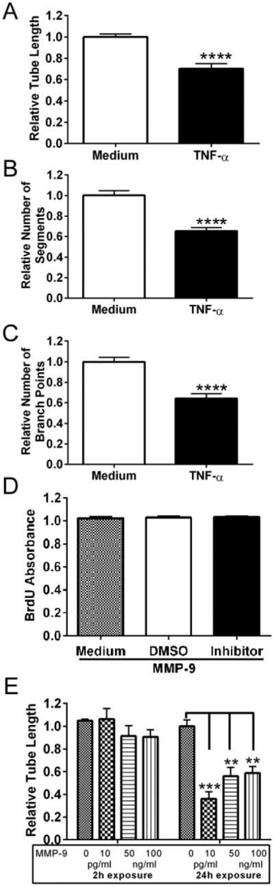
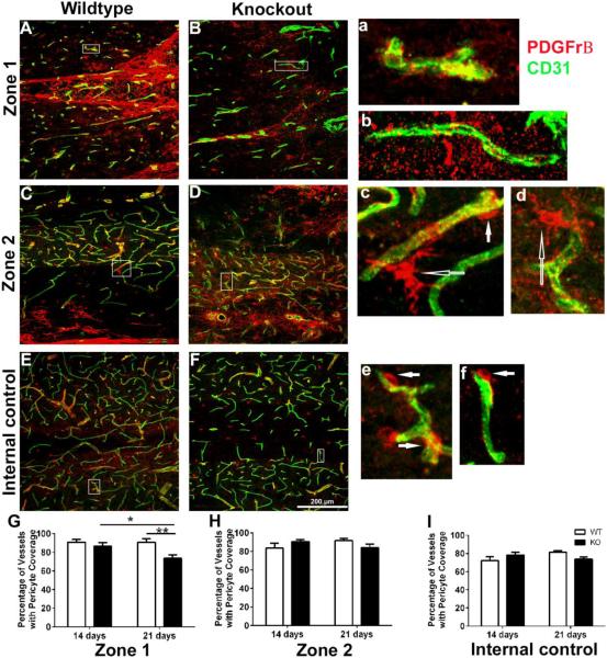
Angiogenesis plays a critical role in wound healing after spinal cord injury. Therefore, understanding the events that regulate angiogenesis has considerable relevance from a therapeutic standpoint. We evaluated the contribution of matrix metalloproteinase (MMP)-2 to angiogenesis and vascular stability in spinal cord injured MMP-2 knockout and wildtype (WT) littermates. While MMP-2 deficiency resulted in reduced endothelial cell division within the lesioned epicenter, there were no genotypic differences in vascularity (vascular density, vascular area, and endothelial cell number) over the first two weeks post-injury. However, by 21days post-injury MMP-2 deficiency resulted in a sharp decline in vascularity, indicative of vascular regression. Complementary in vitro studies of brain capillary endothelial cells confirmed MMP-2 dependent proliferation and tube formation. As deficiency in MMP-2 led to prolonged MMP-9 expression in the injured spinal cord, we examined both short-term and long-term exposure to MMP-9 in vitro. While MMP-9 supported endothelial tube formation and proliferation, prolonged exposure resulted in loss of tubes, findings consistent with vascular regression. Vascular instability is frequently associated with pericyte dissociation and precedes vascular regression. Quantification of PDGFrβ+ pericyte coverage of mature vessels within the glial scar (the reactive gliosis zone), a known source of MMP-9, revealed reduced coverage in MMP-2 deficient animals. These findings suggest that acting in the absence of MMP-2, MMP-9 transiently supports angiogenesis during the early phase of wound healing while its prolonged expression leads to vascular instability and regression. These findings should be considered while developing therapeutic interventions that block MMPs.

Keywords: Angiogenesis; Contusion injury; Matrix metalloproteinases; PDGFrβ positive pericytes; Proliferation; Vascular regression; Vascularity.


Figures

Figure 1
Figure 1. The epicenter of the contused spinal cord at 3 weeks post-injury is characterized by distinct regions, defined by glial and pericyte scarring
A, This montage illustrates the typical appearance of the core and Zones 1 and 2, demarcated by white lines. The core typically consists of scattered CD31+ vessels and PBGFrβ+ pericytes, enclosed within a prominent pericyte scar. This pericyte scar is surrounded by Zone 1, consisting of dense GFAP+ glial scarring. Zone 2 represents the region immediately peripheral to the astrocyte scar and consists of both grey and white matter structures. Arrows indicate orientation of the cord as dorsal (d) and caudal (c). B, An internal control, located approximately 6700-8000 μm from core, represents baseline expression and distribution of blood vessels, pericytes, and astrocytes. C-E, Examples of Zones 1 and 2, noting distance from the core, with white lines delineating the boundary between Zone 1 and the core. GM: gray matter, WM: white matter
Figure 2
Figure 2. The epicenter core of the contused spinal cord at 3 weeks post-injury is primarily comprised of macrophages and thick pericyte scarring
This montage illustrates the core of the epicenter where there is a characteristic dense accumulation of F4/80+ macrophages (white arrows), CD31+ vascular structures and pronounced pericyte scarring (yellow arrows).
Figure 3
Figure 3. Endothelial cell proliferation is reduced in the spinal cord injured MMP-2 KO
A, Endothelial proliferation is significantly lower in MMP-2 KO as compared to the WT group [(two-way ANOVA, effect of genotype, p=0.0083, Values are means + SEM, (n = 5 animals/genotype/time-point)]. B-E, A representative image from the core of the epicenter of a WT at 7 days post-injury. Note the presence of both proliferating endothelial cells (BrdU+, Dapi+, and CD31+ vessels) (white arrows, E) and non-endothelial cells (yellow arrow). Enclosed box in E is shown at higher magnification in e, illustrating the co-localization of BrdU+ Dapi+ CD31+ in a vascular segment. F-I, A representative image from the core of the epicenter of a KO at 7 days post-injury. While there are prominent CD31+ vessels, none appear to co-localize with BrdU and there is evidence for BrdU+ CD31-cells (yellow arrow, I). Enclosed box in I is shown at higher magnification in i.
Figure 4
Figure 4. Loss of MMP-2 results in long-term vascular regression and increased expression of MMP-9
Vascularity was analyzed by densitometric (A) and morphometric (B, C) analyses within the core of the epicenter. Change in vascularity, expressed relative to uninjured mice, was analyzed over time across genotypes (two-way ANOVA followed Sidak’s multiple comparisons test). A, While vascular density is increased in the MMP-2 KO at 7 (**p<0.01) and 14 (*p<0.05) days post injury relative to the WT group, there is a loss of vascular density by 21 days (**p<0.01). B-C, There is a significant loss of vascular area (B, *p<0.05), and endothelial cells (C, ****p<0.0001) at 21 days post-injury in the MMP-2 KO mice. Values are mean + SEM, (n=5 animals/time point/genotype). D, Gelatin zymography confirms greater expression of MMP-9 proforms (105kDa) in the injured (inj) MMP-2 KO at 21 days post-injury as compared to injured WT mice and sham (sh) controls. Note that only the proenzyme form of MMP-9 protein is detected by zymography, an in vivo finding consistent with other studies (Hsu et al., 2008; Hsu et al., 2006; Wang et al., 2000). This is not surprising as the ability to detect active MMP-9 may be compromised by its rapid degradation after binding to the cell surface (Yu and Stamenkovic, 1999). Purified human MMP-2 and MMP-9 served as MMP standards (MMP STD).
Figure 5
Figure 5. MMP-2 induces endothelial cell proliferation and migration and facilitates tube formation in vitro
A, While RBCEC-4 cells endogenously express MMP-2 at high levels by gelatin zymography, MMP-9 is not detected (RBCEC4, lanes 1 and 2). In contrast, bone marrow derived macrophages (BMDM, lanes 3 and 4) express high levels of MMP-9 but no detectable levels of MMP-2. B, RBCEC-4 cell proliferation, based upon BrdU ELISAs, is reduced in the presence of an MMP-2 inhibitor (OA-Hy), whereas an MMP-9 inhibitor (Inh.I) has no detectable effect. (unpaired two-tailed t-test, ***p<0.001). C, Representative images of migrating RBCEC-4 cells on a transwell membrane in the presence of the MMP-2 inhibitor, OA-Hy, or vehicle (DMSO). D, Migrating RBCEC4 cells, expressed relative to medium alone, are reduced in the presence of the MMP-2 inhibitor, OA-Hy, relative to vehicle (DMSO), [(one-way ANOVA followed by Bonferroni’s multiple comparisons test, ###p<0.001 vs. medium, ***p<0.001 vs. vehicle)]. Bars represent mean + SEM averaged over 5 fields/membrane, 3 transwells/condition, and 3 independent experiments. E, Representative images of RBCEC-4 cells plated on Matrigel and treated with the MMP-2 inhibitor, OA-Hy, or vehicle (DMSO). F-H, Tube formation is attenuated in the presence of the MMP-2 inhibitor OA-Hy relative to the vehicle, DMSO (one-way ANOVA followed by Bonferroni’s multiple comparisons test), as assessed by F, measures of tube length (###p<0.001 vs. medium, **p<0.01 vs. vehicle), G, number of segments (##p<0.01 vs. medium, **p<0.01 vs. vehicle) and H, number of branch points (###p<0.001 vs. medium, ***p<0.001 vs. vehicle). Values were normalized to medium alone. Bars represent mean + SEM averaged over 4 wells/condition and 3 independent experiments.
Figure 6
Figure 6. MMP-9, in the absence of MMP-2, does not modulate endothelial cell proliferation but does support endothelial tube formation
A, By gelatin zymography, MMP-9 is upregulated in RBCEC-4 cells in the presence of TNF-α. Molecular weight markers (M) and purified MMP-2 and MMP-9 served as controls (C), and medium was prepared from RBCEC-4 cells, treated with (+) and without (-) TNF-α. B, Whereas inhibition of MMP-9 (Inh. I) has no effect on proliferation of activated (TNF-α treated) RBCEC-4 cells, OA-Hy inhibits cell proliferation in both the presence and absence of Inh I. (Unpaired two-tailed t-test, ***p<0.001, ****p<0.0001). Absorbance was normalized to medium + TNF-α wells. C-E, Inhibition of MMP-2 with OA-Hy in activated (MMP-9-expressing) RBCEC-4 cells has no effect on tube formation (p>0.05), whereas inhibition of both MMP-2 and MMP-9 resulted in a decrease (one-way ANOVA followed by Tukey’s multiple comparisons test) in C, tube length (**p<0.01 vs. medium, #p<0.05 vs. OA-Hy), D, number of segments (*p<0.05 vs. medium, #p<0.05 vs. OA-Hy) and E, number of branch points (*p<0.05 vs. medium, #p<0.05 vs. OA-Hy). All measures were normalized to medium + TNF-α wells. Bars represent mean + SEM averaged over 4 wells/condition and 3 independent experiments.
Figure 7
Figure 7. Prolonged exposure to TNF-α or exogenously added MMP-9 leads to a decrease in tube formation but does not alter endothelial cell proliferation
A-C, There is a reduction in tube length, number of segments and number of branch points in response to prolonged MMP-9 expression (unpaired two-tailed t-test, ****p<0.0001). All measures were normalized to medium alone. Bars represent mean + SEM averaged over 4 wells/condition and 3 independent experiments. D, Proliferation of RBCEC-4 cells, exposed to exogenously added MMP-9 for 24 hours, was analyzed by BrdU ELISA. Long-term exposure to MMP-9 did not alter cell proliferation (one-way ANOVA, p>0.05). Absorbance was normalized to medium. Bars represent mean + SEM averaged over 8 replicates. E, Formation of tubes by RBCEC-4 cells was analyzed by a Matrigel assay after either short-term (2h) or prolonged (24h) exposure to MMP-9. Prolonged exposure to MMP-9 leads to a decrease in tube length (one-way ANOVA followed by Tukey’s multiple comparisons test, ***p<0.001; **p<0.01). Measures of tube length were normalized to medium alone. Bars represent mean + SEM, averaged over 8 replicates.
Figure 8
Figure 8. Blood vessels associated with pericytes within the glial scar of the MMP-2 KO are reduced in numbers by 21 days post-injury
A-F, Immunolocalization of CD31+ endothelial cells and PBGFrβ+ pericytes within Zone 1, Zone 2, and in an internal control in representative sections, prepared from spinal cord injured WT and KO mice. Enclosed boxes are shown at higher magnification in a, c, and e (WT) and b, d, and f (KO) showing CD31+ vessels and PBGFrβ+ pericytes. Filled arrows (c, e and f) show pericytes in close proximity to blood vessels. Open arrows (c, d) delineate ramified pericytes. G, Blood vessels in the MMP-2 KO mice show reduced pericyte coverage of vessels in Zone 1 as compared to WT mice at 21 days post-injury (**p<0.01) and over time (*p<0.05, 21 days post-injury as compared to 14 days post-injury) [(two-way ANOVA followed by Sidak’s multiple comparisons test)]. H-I, No differences in pericyte coverage was observed in both genotypes over time (14 and 21 days post-injury) or between WT and MMP-2 KO mice at each of the time points in zone 2 or the internal control. Bars represent mean + SEM, (n=5 mice/genotype/time-point).

Similar articles

Cited by

References

    1. Alameddine RS, Hamieh L, Shamseddine A. From sprouting angiogenesis to erythrocytes generation by cancer stem cells: evolving concepts in tumor microcirculation. BioMed research international. 2014;2014:986768. - PMC - PubMed
    1. Ambili M, Sudhakaran PR. 60K gelatinase in involuting rat mammary gland is produced as a 90K proenzyme. Biochemistry and molecular biology international. 1998;45:389–399. - PubMed
    1. Armulik A, Abramsson A, Betsholtz C. Endothelial/pericyte interactions. Circulation research. 2005;97:512–523. - PubMed
    1. Armulik A, Genove G, Betsholtz C. Pericytes: developmental, physiological, and pathological perspectives, problems, and promises. Developmental cell. 2011;21:193–215. - PubMed
    1. Baluk P, Raymond WW, Ator E, Coussens LM, McDonald DM, Caughey GH. Matrix metalloproteinase-2 and -9 expression increases in Mycoplasma-infected airways but is not required for microvascular remodeling. Am J Physiol Lung Cell Mol Physiol. 2004;287:L307–317. - PubMed