. 2016 Jul 28;14(1):226.
doi: 10.1186/s12967-016-0976-8.

In vivo monitoring of lung inflammation in CFTR-deficient mice

Affiliations

In vivo monitoring of lung inflammation in CFTR-deficient mice

Fabio Stellari et al. J Transl Med. .

Abstract

Background: Experimentally, lung inflammation in laboratory animals is usually detected by the presence of inflammatory markers, such as immune cells and cytokines, in the bronchoalveolar lavage fluid (BALF) of sacrificed animals. This method, although extensively used, is time, money and animal life consuming, especially when applied to genetically modified animals. Thus a new and more convenient approach, based on in vivo imaging analysis, has been set up to evaluate the inflammatory response in the lung of CFTR-deficient (CF) mice, a murine model of cystic fibrosis.

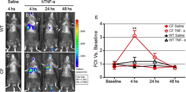
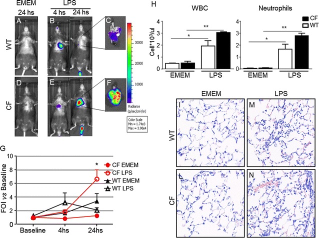
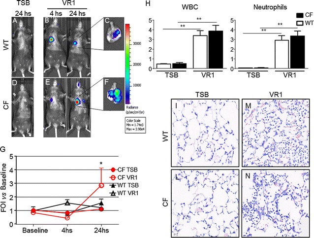
Methods: Wild type (WT) and CF mice were stimulated with P. aeruginosa LPS, TNF-alpha and culture supernatant derived from P. aeruginosa (strain VR1). Lung inflammation was detected by measuring bioluminescence in vivo in mice transiently transgenized with a luciferase reporter gene under the control of a bovine IL-8 gene promoter.

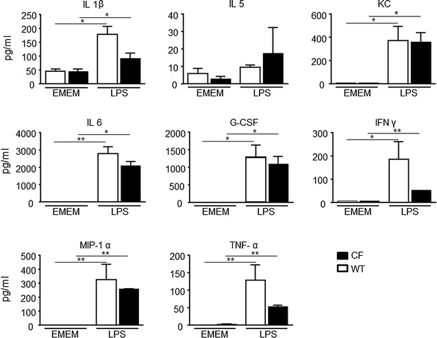
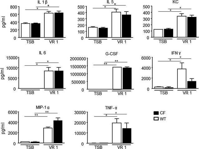
Results: Differences in bioluminescence (BLI) signal were revealed by comparing the two types of mice after intratracheal challenge with pro-inflammatory stimuli. BLI increased at 4 h after stimulation with TNF-alpha and at 24 h after administration of LPS and VR1 supernatant in CF mice with respect to untreated animals. The BLI signal was significantly more intense and lasted for longer times in CF animals when compared to WT mice. Analysis of BALF markers: leukocytes, cytokines and histology revealed no significant differences between CF and WT mice.

Conclusions: In vivo gene delivery technology and non-invasive bioluminescent imaging has been successfully adapted to CFTR-deficient mice. Activation of bIL-8 transgene promoter can be monitored by non-invasive BLI imaging in the lung of the same animal and compared longitudinally in both CF or WT mice, after challenge with pro-inflammatory stimuli. The combination of these technologies and the use of CF mice offer the unique opportunity of evaluating the impact of therapies aimed to control inflammation in a CF background.

Keywords: CFTR-deficient mice; In vivo bioluminescence imaging; Lung inflammation mouse model; Pseudomonas aeruginosa.


Figures

Fig. 1
Fig. 1
Representative in vivo images of WT and CF mice transiently transgenized with bIL-8-Luc and intratracheally challenged with hTNF-alpha (1 µg/mouse) and monitoring of bIL-8 activation by BLI. a, c: WT and CF mice treated with saline solution (control). b, d: WT and CF mice after intratracheal instillation with hTNF-alpha and monitoring of IL-8 activation at 4, 24 and 48 h by BLI. e: Time-course of IL-8-Luc activation in WT and CF mice. Results are reported as folds of increase (FOI) over baseline as mean ± SEM, n = 9 each group. Statistical differences were tested by one-way ANOVA followed by Dunnett’s t post hoc test for group comparisons. *p < 0.05 and **p < 0.01
Fig. 2
Fig. 2
Representative in vivo images of WT and CF mice transiently transgenized with bIL-8-Luc and intratracheally treated with Pseudomonas LPS (6.25 µg/mouse). a, d: WT and CF mice treated with EMEM (control). b, e: WT and CF mice after intratracheal instillation with LPS and monitored IL-8 activation at 4 and 24 h by BLI. c, f: ex vivo images of IL-8 activation in lungs excised from WT and CF mice at 24 hs after LPS challenge. Panel G: Time-course of IL-8-luc activation in WT and CF mice. Results are reported as folds of increase (FOI) over baseline as mean ± SEM, n = 6 at each group. h: Cellular infiltration into the lung of WT and CF mice after intratracheal instillation with LPS at 24 h. Three mice were used for every time point and the experiment was replicated three times. Values are expressed as mean ± SEM of the three different experiments. Statistical differences were tested by one-way ANOVA followed by Dunnett’s t post hoc test for group comparisons. *p < 0.05 and **p < 0.01 WT and CF TSB vs WT and CF LPS group. (In): Representative photomicrographs of Giemsa stained lung of WT and CF mice after intratracheal instillation with EMEM (I, L) and LPS (M, N) at 24 h. Magnification 40×
Fig. 3
Fig. 3
Cytokines quantification in BALFs. WT and CF mice, treated with either EMEM or LPS and sacrificed at 24 h. A panel of 23 cytokines was analysed using a Bio-Plex™ Cytokine Assay Kit (Bio-Rad Laboratories). Eight resulted up-regulated compared to the control animals: IL-1β, IL-5, KC, IL-6, G-CSF, INFγ, MIP-1α, and TNF-α Three mice were used for every time point and the experiment was replicated three times. Values are expressed as mean ± SEM. Statistical differences were tested by one-way ANOVA followed by Dunnett’s t post hoc test for group comparisons. *p < 0.05 and **p < 0.01
Fig. 4
Fig. 4
Representative in vivo images of WT and CF mice transiently transgenized with bIL-8-Luc intratracheally challenged with VR1 culture supernatant (10X/mouse), and monitoring of bIL-8 activation by BLI. a, d: WT and CF mice after intratracheal instillation with TSB (control). Panel B, E: WT and CF mice after intratracheal instillation with VR1Sn and monitoring of IL-8 activation at 4 and 24 h by BLI. c, f: ex vivo images of IL-8 activation in lungs excised from WT and CF mice at 24 h. g: Time-course of IL-8-luc activation in WT and CF mice after challenge with intratracheal VR1Sn. Results are reported as FOI over baseline as mean ± SEM, n = 6 for each group. Panel H: Cellular infiltration into the lung of WT and CF mice after intratracheal instillation with VR1Sn. VR1Sn-induced neutrophils and white blood cells (WBC) recruitment in the airways at 24 h. Three mice were used for every time point and the experiment was replicated three times. Values are expressed as mean ± SEM of the three different experiments. Statistical differences were tested by one-way ANOVA followed by Dunnett’s t post hoc test for group comparisons. *p < 0.05 and **p < 0.01 WT and CF TSB vs WT and CF VR1 group. (in) : Representative micro-photographs Giemsa staining of lung of WT and CF mice after intratracheal instillation with TSB(I, L), and VR1Sn (M, N) at 24 h. Magnification 40×
Fig. 5
Fig. 5
Cytokines quantification in BALFs. WT and CF mice, treated with either TSB or VR1Sn and sacrificed at 24 h. A panel of 23 cytokines was analysed using a Bio-Plex™ Cytokine Assay Kit (Bio-Rad Laboratories), and eight resulted up-regulated compared to the control animals: IL-1β, IL-5, KC, IL-6, G-CSF, INFγ, MIP-1α, and TNF-α Three mice were used for every time point and the experiment was replicated three times. Values are expressed as mean ± SEM. Statistical differences were tested by one-way ANOVA followed by Dunnett’s t post hoc test for group comparisons. *p < 0.05 and **p < 0.01

References

    1. Galluzzo M, Ciraolo E, Lucattelli M, Hoxha E, Ulrich M, Campa CC, et al. Genetic deletion and pharmacological inhibition of PI3 K γ reduces neutrophilic airway inflammation and lung damage in mice with cystic fibrosis-like lung disease. Mediators Inflamm. 2015;2015:545417. doi: 10.1155/2015/545417. - DOI - PMC - PubMed
    1. Cohen-Cymberknoh M, Kerem E, Ferkol T, Elizur A. Airway inflammation in cystic fibrosis: molecular mechanisms and clinical implications. Thorax. 2013;68(12):1157–1162. doi: 10.1136/thoraxjnl-2013-203204. - DOI - PubMed
    1. Hauser AR, Jain M, Bar-Meir M, McColley SA. Clinical significance of microbial infection and adaptation in cystic fibrosis. Clin Microbiol Rev. 2011;24(1):29–70. doi: 10.1128/CMR.00036-10. - DOI - PMC - PubMed
    1. Sorio C, Montresor A, Bolomini-Vittori M, Caldrer S, Rossi B, Dusi S, et al. Mutations of cystic fibrosis transmembrane conductance regulator (CFTR) gene cause a monocyte-selective adhesion deficiency. Am J Respir Crit Care Med. 2015. - PubMed
    1. Belessis Y, Dixon B, Hawkins G, Pereira J, Peat J, MacDonald R, et al. Early cystic fibrosis lung disease detected by bronchoalveolar lavage and lung clearance index. Am J Respir Crit Care Med. 2012;185(8):862–873. doi: 10.1164/rccm.201109-1631OC. - DOI - PubMed

Publication types

MeSH terms

Substances

LinkOut - more resources