. 2016 Sep;26(9):995-1006.
doi: 10.1038/cr.2016.89. Epub 2016 Jul 29.

The Central domain of RyR1 is the transducer for long-range allosteric gating of channel opening

Affiliations

The Central domain of RyR1 is the transducer for long-range allosteric gating of channel opening

Xiao-Chen Bai et al. Cell Res. 2016 Sep.

Abstract

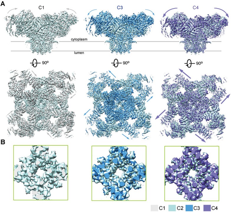
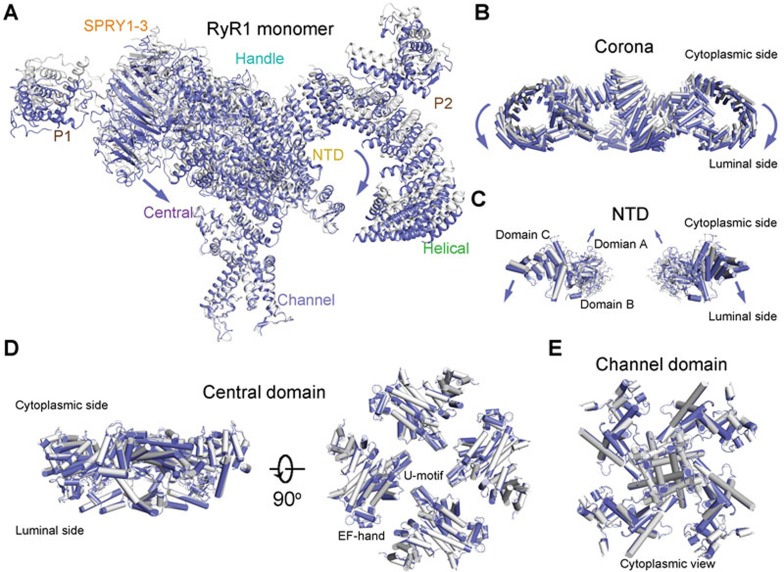
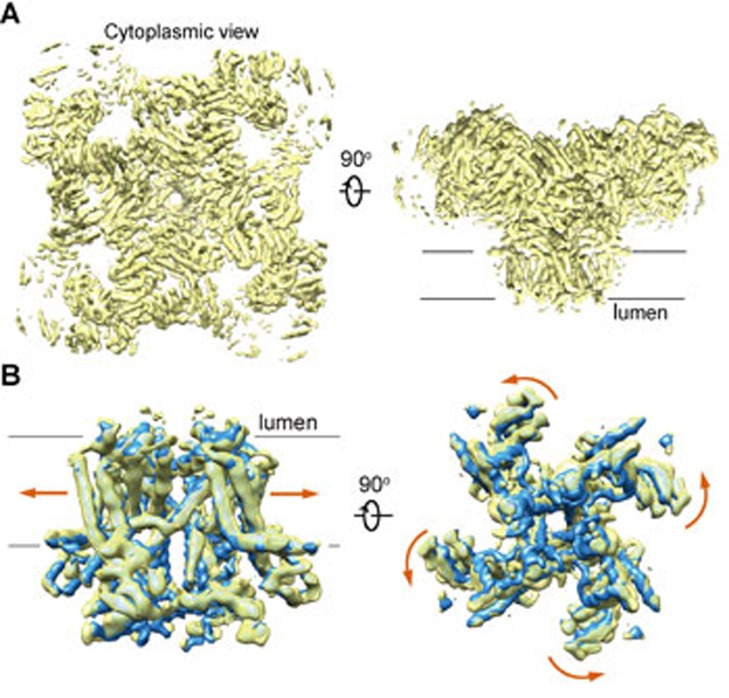
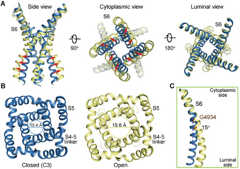
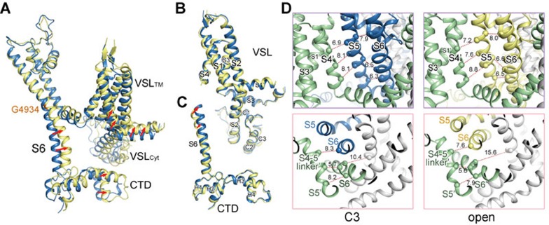
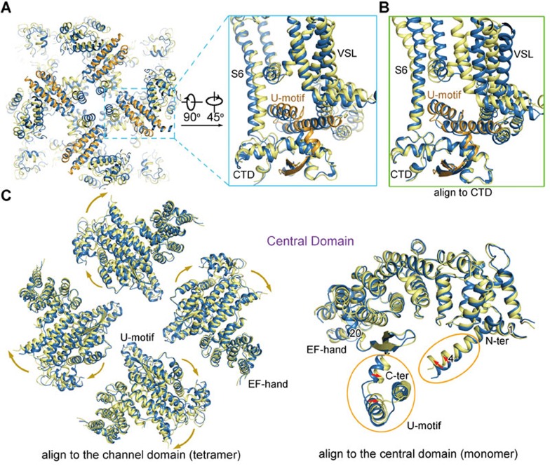
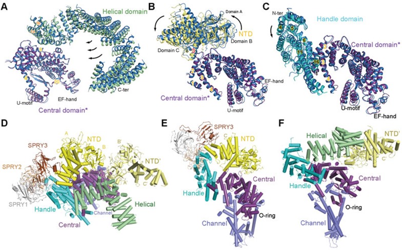
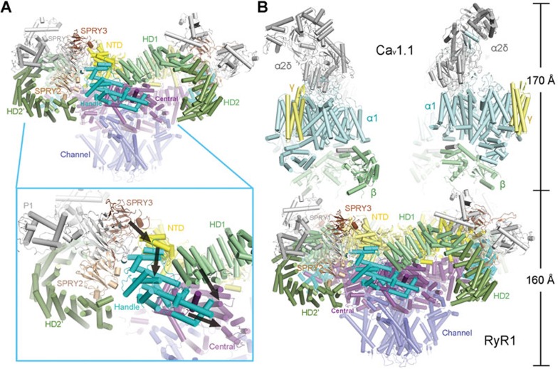
The ryanodine receptors (RyRs) are intracellular calcium channels responsible for rapid release of Ca(2+) from the sarcoplasmic/endoplasmic reticulum (SR/ER) to the cytoplasm, which is essential for the excitation-contraction (E-C) coupling of cardiac and skeletal muscles. The near-atomic resolution structure of closed RyR1 revealed the molecular details of this colossal channel, while the long-range allosteric gating mechanism awaits elucidation. Here, we report the cryo-EM structures of rabbit RyR1 in three closed conformations at about 4 Å resolution and an open state at 5.7 Å. Comparison of the closed RyR1 structures shows a breathing motion of the cytoplasmic platform, while the channel domain and its contiguous Central domain remain nearly unchanged. Comparison of the open and closed structures shows a dilation of the S6 tetrahelical bundle at the cytoplasmic gate that leads to channel opening. During the pore opening, the cytoplasmic "O-ring" motif of the channel domain and the U-motif of the Central domain exhibit coupled motion, while the Central domain undergoes domain-wise displacement. These structural analyses provide important insight into the E-C coupling in skeletal muscles and identify the Central domain as the transducer that couples the conformational changes of the cytoplasmic platform to the gating of the central pore.


Figures

Figure 1
Figure 1
Cryo-EM structures of RyR1 in three additional closed conformations. (A) Three additional RyR1 structures were obtained. Compared with the previously reported RyR1 structure, the cytoplasmic region of the three new classes undergoes pronounced shifts. The four classes of closed RyR1 are designated C1 through C4, among which C2 is the previously reported one. Shown here are the superimpositions of the three new structures with the C2 conformer. The arrows indicate the structural shifts from C2 to the indicated conformer. Please refer to Supplementary information, Movie S1 for the conformational transition from C1 to C4. (B) The channel domain remains nearly identical in the four classes. Shown here are the cytoplasmic views of the pore-forming segments. The EM maps were generated in Chimera.
Figure 2
Figure 2
Conformational changes of the individual domains between C1 and C4 conformers. (A) Structural comparison between C1 (gray) and C4 (violet) protomers. The two tetrameric structures are superimposed relative to the channel domain. The violet arrows indicate the conformational changes from C1 to C4. The same is applied to the other panels. (B) Structural shifts of the corona between C1 and C4 tetramers. The outskirt of the corona, composed of the Handle and Helical domains, moves towards the SR lumen from C1 to C4. Shown here is a side view. (C) The NTD undergoes a rocking motion with the central domains A and B upwards and the outer domain C downwards. For visual clarity, only two diagonal protomers are shown in side view. (D, E) The Central (D) and channel (E) domains remain nearly unchanged between C1 and C4 states. All structure figures were prepared with PyMol.
Figure 3
Figure 3
The cryo-EM reconstruction of an open RyR1. (A) The overall cryo-EM map of the open RyR1 at 5.7 Å resolution. Two perpendicular views are shown. (B) Comparison of the channel domain between the open and the C3 conformers. Shown on the right is the cytoplasmic view. The arrows indicate the conformational change from the C3 (blue) to the open (yellow) state.
Figure 4
Figure 4
Dilation of the inner gate leads to channel opening. (A) The cytoplasmic gate of the S6 bundle undergoes a dilation that leads to pore opening, while the luminal segments have little change. (B) Dilation of the cytoplasmic gate. The indicated distances are measured between the Cα atoms of the gating residue Ile4937 on the S6 segments in the diagonal protomers. Please refer to Supplementary information, Movie S2 for the conformational changes of the channel domain. (C) Comparison of the S6 segment relative to the luminal halves of the pore-forming segments (residues 4 836-4 935) shows that the structural deviation between the open (yellow) and closed (blue) states occurs at Gly4934.
Figure 5
Figure 5
Coupled conformational shifts of the segments within the channel domain during pore opening. (A) Structural comparison of the channel domain in one protomer between the open and C3 structures relative to the luminal halves of the pore-forming segments (residues 4 836-4 935). The cytoplasmic “O-ring” composed of the cytoplasmic segments of S6 (S6Cyt), the VSLCyt, and the CTD appears to undergo concordant shifts. (B) The VSL appears rigid during pore opening. There is no intra-domain rearrangement observed between the VSL domain in the open and closed structures. Therefore, the VSL domain undergoes a rigid-body shift during pore opening. (C) There is no relative motion between CTD and the S6Cyt segment during the pore dilation. (D) The concerted motions between the S5 and S6 segments in the same protomer, and between the S5 segment and the S4 and S4-5 segments in the neighbouring protomers. The distances between the Cα atoms of the indicated residues are presented in Å. For visual clarity, the specified protomer is coloured blue and yellow in the closed and open states, respectively, while the neighbouring protomer is coloured green in both states. The other two protomers that are not discussed are coloured gray. Please refer to Supplementary information, Movie S3 for the concordant shifts of the channel segments during pore dilation.
Figure 6
Figure 6
Coupled conformational changes between the channel domain and the Central domain. (A) Concordant conformational changes between the cytoplasmic “O-ring” of the channel domain and the U-motif of the Central domain. Shown here are the luminal and side views of the U-motif and the O-ring. The U-motif in the open RyR1 is coloured orange. Please refer to Supplementary information, Movie S4 for the concerted conformational changes. (B) Structural comparison of the O-ring and the U-motif between the C3 conformer and the open structure relative to CTD. There is little shift of the U-motif relative to CTD. (C) Comparison of the Central domain between the open and the C3 structures relative to the tetrameric channel domain (left panel) and relative to the individual Central domain (right panel). The tetrameric Central domain is shown in cytoplasmic view. The yellow and green arrows indicate the conformational transition from C3 to the open state.
Figure 7
Figure 7
The extensive interaction network among the cytoplasmic domains provide the molecular basis for long-range allosteric gating. (A-C) The motions of the Helical domain, NTD, and Handle domain relative to the Central domain between the closed and open structures. The comparison is made relative to the Central domain (labeled with asterisk). In all panels, domains in C3 are blue, while those in the open structure are domain-coloured. (D) Extensive interfaces among the armadillo repeats-containing cytoplasmic domains, including the NTD, Helical, Handle, and Central domains. The NTD from the neighbouring protomer is also shown, coloured pale yellow and labeled NTD'. (E) The extensive internal interactions within one cytoplasmic superhelical assembly consisting of the armadillo repeats in NTD, the Handle domain, and the Central domain. Note that the Central domain also interacts with the NTD. (F) The extensive interaction network involving the Helical domain, the Handle domain, the Central domain in one protomer and the NTD in the adjacent protomer. Please refer to our previous publication for detailed analysis of the cytoplasmic interaction network.
Figure 8
Figure 8
Speculative mechanism of the excitation-contraction coupling. (A) Conformational changes to any cytoplasmic domain may be propagated to the Central domain along the interaction network described in Figure 7. Shown here are side views of tetrameric RyR1. Inset: an example of a speculative route (black arrows) for the propagation of conformational changes that can be triggered by motion of the SPRY3 domain. (B) Speculative model of the complex between RyR1 and the Cav1.1 complex. Structural determination of both RyR1 and the Cav1.1 complex provides the foundation for elucidating the molecular mechanism of RyR1 opening induced by depolarization of the plasma membrane. Structures of the Cav1.1 complex (PDB code: 3JBR) and RyR1 were manually docked in COOT.

References

    1. Pessah IN, Waterhouse AL, Casida JE. The calcium-ryanodine receptor complex of skeletal and cardiac muscle. Biochem Biophys Res Commun 1985; 128:449–456. - PubMed
    1. Inui M, Saito A, Fleischer S. Purification of the ryanodine receptor and identity with feet structures of junctional terminal cisternae of sarcoplasmic reticulum from fast skeletal muscle. J Biol Chem 1987; 262:1740–1747. - PubMed
    1. Lai FA, Erickson HP, Rousseau E, Liu QY, Meissner G. Purification and reconstitution of the calcium release channel from skeletal muscle. Nature 1988; 331:315–319. - PubMed
    1. Lanner JT, Georgiou DK, Joshi AD, Hamilton SL. Ryanodine receptors: structure, expression, molecular details, and function in calcium release. Cold Spring Harb Perspect Biol 2010; 2:a003996. - PMC - PubMed
    1. Takeshima H, Nishimura S, Matsumoto T, et al. Primary structure and expression from complementary DNA of skeletal muscle ryanodine receptor. Nature 1989; 339:439–445. - PubMed

Publication types

MeSH terms

Substances