Core Mechanisms Regulating Developmentally Timed and Environmentally Triggered Abscission
- PMID: 27468996
- PMCID: PMC5074626
- DOI: 10.1104/pp.16.01004
Core Mechanisms Regulating Developmentally Timed and Environmentally Triggered Abscission
Abstract
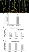
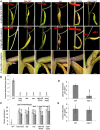
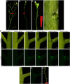
Drought-triggered abscission is a strategy used by plants to avoid the full consequences of drought; however, it is poorly understood at the molecular genetic level. Here, we show that Arabidopsis (Arabidopsis thaliana) can be used to elucidate the pathway controlling drought-triggered leaf shedding. We further show that much of the pathway regulating developmentally timed floral organ abscission is conserved in regulating drought-triggered leaf abscission. Gene expression of HAESA (HAE) and INFLORESCENCE DEFICIENT IN ABSCISSION (IDA) is induced in cauline leaf abscission zones when the leaves become wilted in response to limited water and HAE continues to accumulate in the leaf abscission zones through the abscission process. The genes that encode HAE/HAESA-LIKE2, IDA, NEVERSHED, and MAPK KINASE4 and 5 are all necessary for drought-induced leaf abscission. Our findings offer a molecular mechanism explaining drought-triggered leaf abscission. Furthermore, the ability to study leaf abscission in Arabidopsis opens up a new avenue to tease apart mechanisms involved in abscission that have been difficult to separate from flower development as well as for understanding the mechanistic role of water and turgor pressure in abscission.
© 2016 American Society of Plant Biologists. All rights reserved.
Figures


Similar articles
-
Leaf shedding as an anti-bacterial defense in Arabidopsis cauline leaves.PLoS Genet. 2017 Dec 18;13(12):e1007132. doi: 10.1371/journal.pgen.1007132. eCollection 2017 Dec. PLoS Genet. 2017. PMID: 29253890 Free PMC article.
-
NEVERSHED and INFLORESCENCE DEFICIENT IN ABSCISSION are differentially required for cell expansion and cell separation during floral organ abscission in Arabidopsis thaliana.J Exp Bot. 2013 Dec;64(17):5345-57. doi: 10.1093/jxb/ert232. Epub 2013 Aug 20. J Exp Bot. 2013. PMID: 23963677
-
The EPIP peptide of INFLORESCENCE DEFICIENT IN ABSCISSION is sufficient to induce abscission in arabidopsis through the receptor-like kinases HAESA and HAESA-LIKE2.Plant Cell. 2008 Jul;20(7):1805-17. doi: 10.1105/tpc.108.059139. Epub 2008 Jul 25. Plant Cell. 2008. PMID: 18660431 Free PMC article.
-
IDA: a peptide ligand regulating cell separation processes in Arabidopsis.J Exp Bot. 2013 Dec;64(17):5253-61. doi: 10.1093/jxb/ert338. Epub 2013 Oct 22. J Exp Bot. 2013. PMID: 24151306 Review.
-
Re-evaluation of the ethylene-dependent and -independent pathways in the regulation of floral and organ abscission.J Exp Bot. 2019 Mar 11;70(5):1461-1467. doi: 10.1093/jxb/erz038. J Exp Bot. 2019. PMID: 30726930 Review.
Cited by
-
A Non-Shedding Fruit Elaeis oleifera Palm Reveals Perturbations to Hormone Signaling, ROS Homeostasis, and Hemicellulose Metabolism.Genes (Basel). 2021 Oct 28;12(11):1724. doi: 10.3390/genes12111724. Genes (Basel). 2021. PMID: 34828330 Free PMC article.
-
Advances in Rice Seed Shattering.Int J Mol Sci. 2023 May 17;24(10):8889. doi: 10.3390/ijms24108889. Int J Mol Sci. 2023. PMID: 37240235 Free PMC article. Review.
-
Abscission in plants: from mechanism to applications.Adv Biotechnol (Singap). 2024 Aug 9;2(3):27. doi: 10.1007/s44307-024-00033-9. Adv Biotechnol (Singap). 2024. PMID: 39883313 Free PMC article. Review.
-
Arabidopsis uses a molecular grounding mechanism and a biophysical circuit breaker to limit floral abscission signaling.Proc Natl Acad Sci U S A. 2024 Oct 29;121(44):e2405806121. doi: 10.1073/pnas.2405806121. Epub 2024 Oct 25. Proc Natl Acad Sci U S A. 2024. PMID: 39453742 Free PMC article.
-
Receptor-Like Protein Kinases Function Upstream of MAPKs in Regulating Plant Development.Int J Mol Sci. 2020 Oct 15;21(20):7638. doi: 10.3390/ijms21207638. Int J Mol Sci. 2020. PMID: 33076465 Free PMC article. Review.
References
-
- Addicott FT. (1982) Abscission. University of California Press, Oakland, CA
-
- Alvarez-Buylla ER, Benítez M, Corvera-Poiré A, Chaos Cador A, de Folter S, Gamboa de Buen A, Garay-Arroyo A, García-Ponce B, Jaimes-Miranda F, Pérez-Ruiz RV, et al. (2010) Flower development. Arabidopsis Book 8: e0127, doi/10.1199/tab.0127 - DOI - PMC - PubMed
-
- Anthony MF, Coggins CW Jr (1999) The efficacy of five forms of 2,4-D in controlling preharvest fruit drop in citrus. Sci Hortic (Amsterdam) 81: 267–277
MeSH terms
Substances
LinkOut - more resources
Full Text Sources
Other Literature Sources
Molecular Biology Databases

