

Pediatric Patients Demonstrate Progressive T1-Weighted Hyperintensity in the Dentate Nucleus following Multiple Doses of Gadolinium-Based Contrast Agent
- PMID: 27469211
- PMCID: PMC5161565
- DOI: 10.3174/ajnr.A4891
Pediatric Patients Demonstrate Progressive T1-Weighted Hyperintensity in the Dentate Nucleus following Multiple Doses of Gadolinium-Based Contrast Agent
Abstract
Background and purpose: While there have been recent reports of brain retention of gadolinium following gadolinium-based contrast agent administration in adults, a retrospective series of pediatric patients has not previously been reported, to our knowledge. We investigated the relationship between the number of prior gadolinium-based contrast agent doses and increasing T1 signal in the dentate nucleus on unenhanced T1-weighted MR imaging. We hypothesized that despite differences in pediatric physiology and the smaller gadolinium-based contrast agent doses that pediatric patients are typically administered based on weighted-adjusted dosing, the pediatric brain would also demonstrate dose-dependent increasing T1 signal in the dentate nucleus.
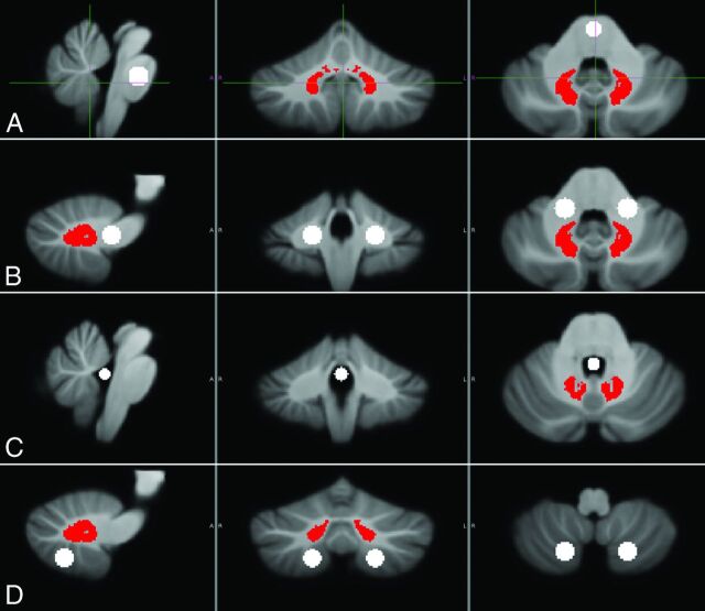
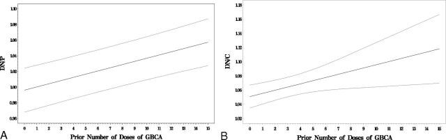
Materials and methods: We included children with multiple gadolinium-based contrast agent administrations at our institution. A blinded reader placed ROIs within the dentate nucleus and adjacent cerebellar white matter. To eliminate reader bias, we also performed automated ROI delineation of the dentate nucleus, cerebellar white matter, and pons. Dentate-to-cerebellar white matter and dentate-to pons ratios were compared with the number of gadolinium-based contrast agent administrations.
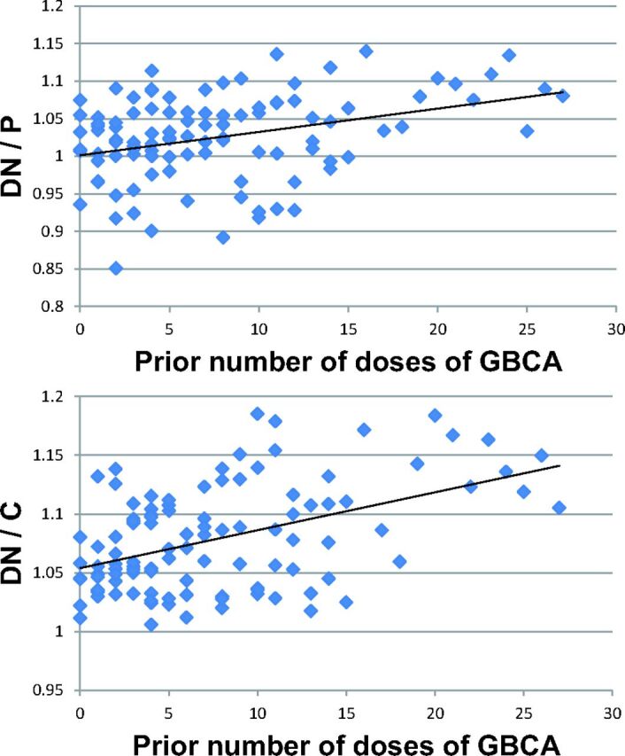
Results: During 20 years at our institution, 280 patients received at least 5 gadolinium-based contrast agent doses, with 1 patient receiving 38 doses. Sixteen patients met the inclusion/exclusion criteria for ROI analysis. Blinded reader dentate-to-cerebellar white matter ratios were significantly associated with gadolinium-based contrast agent doses (rs = 0.77, P = .001). The dentate-to-pons ratio and dentate-to-cerebellar white matter ratios based on automated ROI placement were also significantly correlated with gadolinium-based contrast agent doses (t = 4.98, P < .0001 and t = 2.73, P < .02, respectively).
Conclusions: In pediatric patients, the number of prior gadolinium-based contrast agent doses is significantly correlated with progressive T1-weighted dentate hyperintensity. Definitive confirmation of gadolinium deposition requires tissue analysis. Any potential clinical sequelae of gadolinium retention in the developing brain are unknown. Given this uncertainty, we suggest taking a cautious stance, including the use, in pediatric patients, of higher stability, macrocyclic agents, which in both human and animal studies have been shown to be associated with lower levels of gadolinium deposition, and detailed documentation of dosing. Most important, a patient should not be deprived of a well-indicated contrasted MR examination.
© 2016 by American Journal of Neuroradiology.
Figures
Similar articles
-
Progressive T1 Shortening of the Dentate Nucleus in Patients With Multiple Sclerosis: Result of Multiple Administrations of Linear Gadolinium Contrast Agents Versus Intrinsic Disease.AJR Am J Roentgenol. 2018 Nov;211(5):1099-1105. doi: 10.2214/AJR.17.19155. Epub 2018 Aug 30. AJR Am J Roentgenol. 2018. PMID: 30160975
-
Gadolinium deposition within the paediatric brain: no increased intrinsic T1-weighted signal intensity within the dentate nucleus following the administration of a minimum of four doses of the macrocyclic agent gadobutrol.Eur Radiol. 2018 Nov;28(11):4882-4889. doi: 10.1007/s00330-018-5464-5. Epub 2018 May 9. Eur Radiol. 2018. PMID: 29744642 Free PMC article.
-
Gadolinium Deposition within the Pediatric Brain: No Increased Intrinsic T1-Weighted Signal Intensity within the Dentate Nucleus following the Administration of a Minimum of 4 Doses of the Macrocyclic Agent Gadoteridol.AJNR Am J Neuroradiol. 2018 Sep;39(9):1604-1608. doi: 10.3174/ajnr.A5748. Epub 2018 Aug 9. AJNR Am J Neuroradiol. 2018. PMID: 30093477 Free PMC article.
-
The presence of the gadolinium-based contrast agent depositions in the brain and symptoms of gadolinium neurotoxicity - A systematic review.PLoS One. 2017 Feb 10;12(2):e0171704. doi: 10.1371/journal.pone.0171704. eCollection 2017. PLoS One. 2017. PMID: 28187173 Free PMC article.
-
Gadolinium deposition within the dentate nucleus and globus pallidus after repeated administrations of gadolinium-based contrast agents-current status.Neuroradiology. 2016 May;58(5):433-41. doi: 10.1007/s00234-016-1658-1. Epub 2016 Feb 12. Neuroradiology. 2016. PMID: 26873830 Review.
Cited by
-
Gadolinium: pharmacokinetics and toxicity in humans and laboratory animals following contrast agent administration.Arch Toxicol. 2022 Feb;96(2):403-429. doi: 10.1007/s00204-021-03189-8. Epub 2022 Jan 8. Arch Toxicol. 2022. PMID: 34997254 Free PMC article. Review.
-
Brain tissue gadolinium retention in pediatric patients after contrast-enhanced magnetic resonance exams: pathological confirmation.Pediatr Radiol. 2020 Mar;50(3):388-396. doi: 10.1007/s00247-019-04535-w. Epub 2020 Jan 27. Pediatr Radiol. 2020. PMID: 31989188
-
Imaging Assessment of Radiation Therapy-Related Normal Tissue Injury in Children: A PENTEC Visionary Statement.Int J Radiat Oncol Biol Phys. 2024 Jun 1;119(2):669-680. doi: 10.1016/j.ijrobp.2024.03.006. Int J Radiat Oncol Biol Phys. 2024. PMID: 38760116 Free PMC article. Review.
-
Evaluation of the effect of multiple administrations of gadopentetate dimeglumine or gadoterate meglumine on brain T1-weighted hyperintensity in pediatric patients.Pediatr Radiol. 2021 Dec;51(13):2568-2580. doi: 10.1007/s00247-021-05134-4. Epub 2021 Jul 20. Pediatr Radiol. 2021. PMID: 34286351
-
Signal intensity at unenhanced T1-weighted magnetic resonance in the globus pallidus and dentate nucleus after serial administrations of a macrocyclic gadolinium-based contrast agent in children.Pediatr Radiol. 2017 Sep;47(10):1345-1352. doi: 10.1007/s00247-017-3874-1. Epub 2017 May 19. Pediatr Radiol. 2017. PMID: 28526896
References
-
- American College of Radiology. ACR Appropriateness Criteria. http://www.acr.org/Quality-Safety/Appropriateness-Criteria. Accessed March 14, 2016.
MeSH terms
Substances
Grants and funding
LinkOut - more resources
Full Text Sources
Other Literature Sources
Medical
Miscellaneous