. 2016 Nov:177:127-142.
doi: 10.1016/j.trsl.2016.06.011. Epub 2016 Jul 9.

Stanniocalcin-1 attenuates ischemic cardiac injury and response of differentiating monocytes/macrophages to inflammatory stimuli

Affiliations

Stanniocalcin-1 attenuates ischemic cardiac injury and response of differentiating monocytes/macrophages to inflammatory stimuli

Arezoo Mohammadipoor et al. Transl Res. 2016 Nov.

Abstract

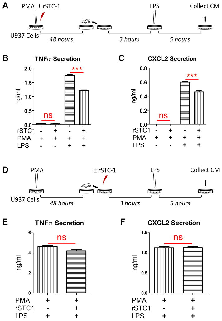
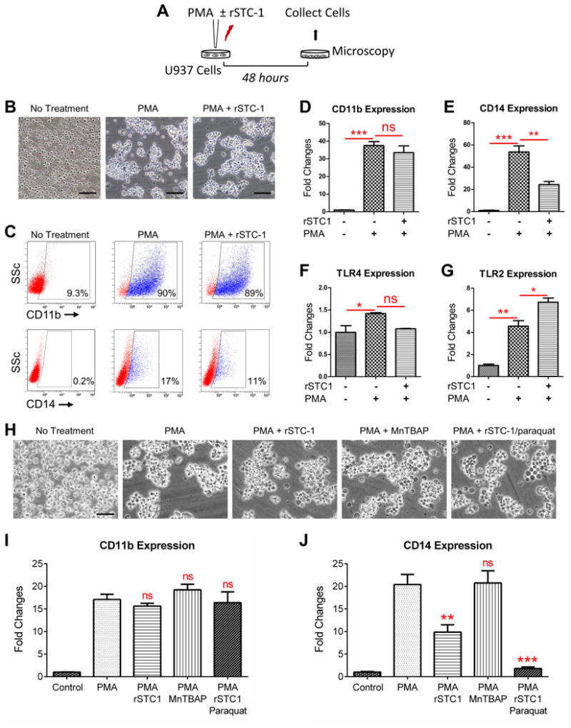
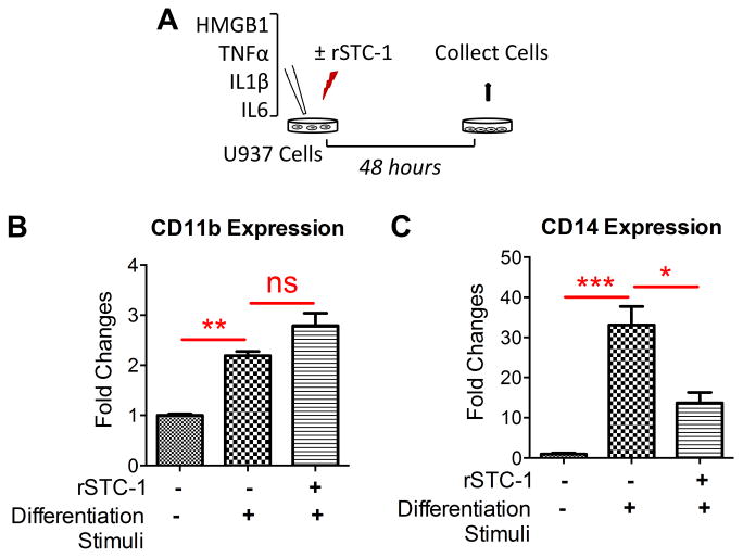
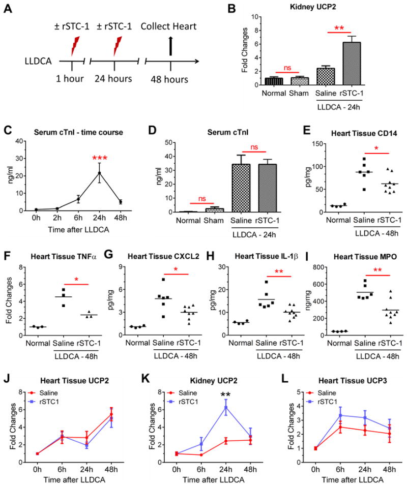
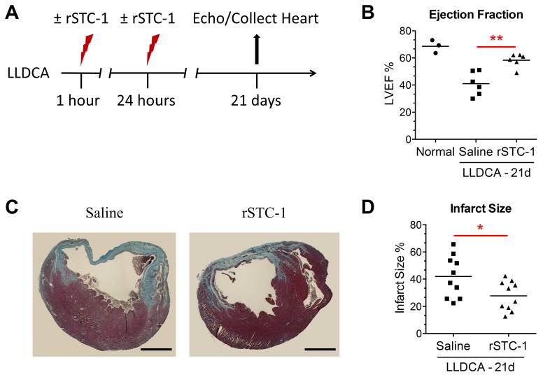
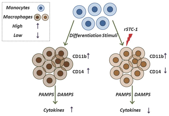
Stanniocalcin-1 (STC-1) is a multifunctional glycoprotein with antioxidant and anti-inflammatory properties. Ischemic myocardial necrosis generates "danger" signals that perpetuate detrimental inflammatory reactions often involving monocyte recruitment and their subsequent differentiation into proinflammatory macrophages. Therefore, we evaluated the effects of recombinant STC-1 (rSTC-1) on monocyte phenotype and in a mouse model of myocardial infarction. Using an established protocol to differentiate human monocytes into macrophages, we demonstrated that rSTC-1 did not alter morphology of the differentiated cells, toll-like receptor (TLR) 4 expression, or expression of the myeloid cell marker CD11b. However, rSTC-1 treatment before differentiation attenuated the rise in the expression of CD14, a TLR4 coreceptor and pathogen sensor that propagates innate immune responses, and suppressed levels of inflammatory cytokines produced by the differentiated cells in response to the CD14-TLR4 ligand lipopolysaccharide. Moreover, rSTC-1 treatment reduced CD14 expression in monocytes stimulated with endogenous danger signals. Interestingly, the effects of rSTC-1 on CD14 expression were not reproduced by a superoxide dismutase mimetic. In mice with induced myocardial infarcts, intravenous administration of rSTC-1 decreased CD14 expression in the heart as well as levels of tumor necrosis factor alpha, C-X-C motif ligand 2, interleukin 1 beta, and myeloperoxidase. It also suppressed the formation of scar tissue while enhancing cardiac function. The data suggests that one of the beneficial effects of STC-1 might be attributed to suppression of CD14 on recruited monocytes and macrophages that limits their inflammatory response. STC-1 may be a promising therapy to protect the heart and other tissues from ischemic injury.


Conflict of interest statement

All authors are in agreement with the content of the manuscript and have read the journal's authorship agreement as well as the policy on disclosure of potential conflicts of interest.

Figures

Figure 1
Figure 1. Recombinant stanniocalcin-1 (rSTC-1) treatment, prior to monocyte differentiation but not after, suppressed the inflammatory response of macrophages to danger signals
(A) Schematic illustrating the strategy used to evaluate the effects of rSTC-1 pre-treatment on macrophage response to lipopolysaccharide (LPS). Human U937 monocytes were induced to differentiate into macrophages by treatment with 100 ng/ml of phorbol 12-myristate 13-acetate (PMA) in the presence or absence of 1 μg/ml of rSTC-1. After 48 hours, differentiated macrophages were harvested, plated, and incubated for another 3 hours. Then, macrophages were stimulated with 50 ng/ml of LPS. After 5 hours, media conditioned (CM) by unstimulated or LPS-stimulated macrophages were collected and used to evaluate changes in levels of secreted TNFα (B) and CXCL2 (C) by ELISA. (D) Schematic illustrating the strategy used to evaluate the effects of rSTC-1 on the response of differentiated macrophages to LPS. U937 monocytes were plated in macrophage medium supplemented with 100 ng/ml of PMA for 48 hours. Differentiated macrophages were harvested, re-plated, and incubated with or without 1 μg/ml of rSTC-1. After 3 hours, macrophages were stimulated with LPS for an additional 5 hours. The CM was collected, clarified by centrifugation and used to determine levels of TNFα (E) and CXCL2 (F) by ELISA. Values are presented as mean + SEM (n=3). Statistical significance was determined using ANOVA (B-C) or Student's t-test (E-F) (not significant, ns p≥ 0.05; ***p< 0.001).
Figure 2
Figure 2. Expression of CD14 was reduced in differentiating monocyte/macrophages by treatment with recombinant stanniocalcin-1 (rSTC-1)
Human U937 monocytes were incubated in macrophage medium supplemented with 100 ng/ml of phorbol 12-myristate 13-acetate (PMA) in the presence or absence of 1.0 μg/ml of rSTC-1. After 48 hours, morphology of differentiated macrophages was evaluated by light microscopy. Then, macrophages were harvested and processed for real-time RT-PCR or flow cytometry analysis. (A) Schematic showing the workflow. (B) Representative images of undifferentiated U937 monocytes (No Treatment), PMA-differentiated U937 macrophages (PMA), and U937 cells stimulated with PMA and rSTC-1 (PMA+ rSTC-1). Scale bar, 100 μm. (C) Cell surface expression of CD11b and CD14 was assessed by flow cytometry. Real-time RT-PCR for CD11b (D), CD14 (E), TLR4 (F), and TLR2 (G) in undifferentiated U937 monocytes and cells stimulated with PMA for 48 hours (with and without rSTC-1 treatment). (H) Representative images of U937 cells treated for 48 hours with PMA in the presence and absence of rSTC-1, MnTBAP, and paraquat. Scale bar, 50 μm. Real-time RT-PCR for CD11b (I) and CD14 (J) in U937 cells stimulated for 48 hours with the compounds described in panel H. Values are expressed as mean + SEM (n=3-5). Data was analyzed using ANOVA (not significant, ns p 0.05, *p< 0.05, **p< 0.01, ***p< 0.001).
Figure 3
Figure 3. Recombinant stanniocalcin-1 (rSTC-1) reduced CD14 expression by monocyte/macrophages stimulated with danger signals
Human U937 monocytes were stimulated with 5 μg/ml human high mobility group box1 (HMGB1) and 10 ng/ml each of recombinant human tumor necrosis factor alpha (TNFa), interleukin-1 beta (IL-1β), and IL-6 in the presence or absence of 1.0 μg/ml rSTC-1 treatment. After 48 hours, cells were lysed for real-time RT-PCR. (A) Schematic showing the workflow. The fold changes of mRNA levels for CD11b (B) and CD14 (C) were measured in unstimulated U937 monocytes, stimulated U937 monocytes, and stimulated cells treated simultaneously with rSTC-1. Values are expressed as mean + SEM (n=3). Data were analyzed with ANOVA (not significant, ns p 0.05, *p< 0.05, **p< 0.01, ***p< 0.001).
Figure 4
Figure 4. Intravenous administration of recombinant stanniocalcin-1 (rSTC-1) reduced the expression of CD14 in cardiac tissue and attenuated inflammation following myocardial infarction
NOD/SCID mice were subjected to ischemic cardiac injury by permanent ligation of the left descending coronary artery (LLDCA). At 1 hour and 24 hours after ligation, 2.0 mg/kg of rSTC-1 or equal volume of 0.9% sodium chloride (saline) was administered intravenously. Mice were euthanized up to 48 hours after LLDCA to collect heart tissue and assess inflammatory response. Blood was collected to measure serum levels of cardiac troponin (cTnI). (A) Diagram showing the workflow of the cardiac injury model. (B) Changes in expression of kidney UCP2 were determined by real-time RT-PCR. (C) Time-dependent change in serum level of cTnI was determined by ELISA. (D) Assessment of serum cTnI, 24 hours after permanent ligation and rSTC-1 treatment. Intravenous rSTC-1 suppressed levels of (E) CD14, inflammatory cytokines TNFα (F), CXCL2 (G), and IL-1β (H), and (I) myeloperoxidase (MPO) in heart tissue lysate prepared 48 hours after LLDCA. Mean value is illustrated by the black lines. Real-time RT-PCR of UCP2 in cardiac (J) and kidney (K) tissue, and (L) UCP3 in cardiac tissue. Statistical significance was determined using Student's t-test when 2 groups were compared, or ANOVA for analysis of more than 2 groups (not significant, ns p> 0.05, * p< 0.05, ** p< 0.01).
Figure 5
Figure 5. Recombinant stanniocalcin-1 (rSTC-1) administered intravenously improved heart function and reduced infarct size
NOD/SCID mice were subjected to ischemic cardiac injury by permanent ligation of the left descending coronary artery (LLDCA). At 1 hour and 24 hours after ligation, 2.0 mg/kg of rSTC-1 or equal volume of 0.9% sodium chloride (saline) was administered intravenously. Twenty one days after ligation, the mice were anesthetized and their left ventricular ejection fraction (LVEF) was evaluated. Then, mice were euthanized to collect heart tissue and assess infarct size. (A) Diagram showing the workflow of the cardiac injury model. (B) Each data point represents the average LVEF of 2 independent recordings with 3 consecutive cardiac cycles (n=3-6). (C) Representative images showing fibrosis in heart sections stained with Masson Trichrome (Scale bar, 1 mm.). (D) Infarct size was measured at the ventricular midline of each section and expressed as the percent length of the fibrotic region relative to the ventricular midline length. Each data point represents the average of ten infarct size measurements per heart (n=10-12). Data was analyzed using Student's t-test (*p< 0.05, **p< 0.01).
Figure 6
Figure 6. Summary of recombinant stanniocalcin-1 (rSTC-1) effects on monocyte/macrophage differentiation and function
Tissue injury induces release of differentiation stimuli [such as pathogen-associated molecular pattern molecules (PAMPs), damage-associated molecular pattern molecules (DAMPs), cytokines, and chemokines] that promote monocyte differentiation and recruitment to the site of injury. Differentiation stimuli cause an increase in expression of surface molecules such as CD11b and CD14 in monocyte/macrophage and enhance secretion of inflammatory cytokines and chemokines (for example TNFα, IL-1β, and CXCL2) in these cells. Administration of rSTC-1 during the differentiation process reduces expression of CD14 by monocytes/macrophages, which subsequently decreases their response to inflammatory stimuli including DAMPS and PAMPS.

References

    1. Sheikh-Hamad D. Mammalian stanniocalcin-1 activates mitochondrial antioxidant pathways: new paradigms for regulation of macrophages and endothelium. Am J Physiol Renal Physiol. 2010;298(2):F248–54. - PMC - PubMed
    1. Re RN, Cook JL. The mitochondrial component of intracrine action. Am J Physiol Heart Circ Physiol. 2010;299(3):H577–83. - PMC - PubMed
    1. Yeung BH, Law AY, Wong CK. Evolution and roles of stanniocalcin. Mol Cell Endocrinol. 2012;349(2):272–280. - PubMed
    1. Sazonova O, James KA, McCudden CR, Segal D, Talebian A, Wagner GF. Stanniocalcin-1 secretion and receptor regulation in kidney cells. Am J Physiol Renal Physiol. 2008;294(4):F788–94. - PubMed
    1. Sheikh-Hamad D, Bick R, Wu GY, et al. Stanniocalcin-1 is a naturally occurring L-channel inhibitor in cardiomyocytes: relevance to human heart failure. Am J Physiol Heart Circ Physiol. 2003;285(1):H442–8. - PubMed

Publication types

MeSH terms