. 2016 Aug 21;61(16):6132-53.
doi: 10.1088/0031-9155/61/16/6132. Epub 2016 Jul 29.

A neural network-based method for spectral distortion correction in photon counting x-ray CT

Affiliations

A neural network-based method for spectral distortion correction in photon counting x-ray CT

Mengheng Touch et al. Phys Med Biol. .

Abstract

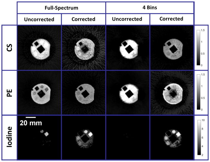
Spectral CT using a photon counting x-ray detector (PCXD) shows great potential for measuring material composition based on energy dependent x-ray attenuation. Spectral CT is especially suited for imaging with K-edge contrast agents to address the otherwise limited contrast in soft tissues. We have developed a micro-CT system based on a PCXD. This system enables both 4 energy bins acquisition, as well as full-spectrum mode in which the energy thresholds of the PCXD are swept to sample the full energy spectrum for each detector element and projection angle. Measurements provided by the PCXD, however, are distorted due to undesirable physical effects in the detector and can be very noisy due to photon starvation in narrow energy bins. To address spectral distortions, we propose and demonstrate a novel artificial neural network (ANN)-based spectral distortion correction mechanism, which learns to undo the distortion in spectral CT, resulting in improved material decomposition accuracy. To address noise, post-reconstruction denoising based on bilateral filtration, which jointly enforces intensity gradient sparsity between spectral samples, is used to further improve the robustness of ANN training and material decomposition accuracy. Our ANN-based distortion correction method is calibrated using 3D-printed phantoms and a model of our spectral CT system. To enable realistic simulations and validation of our method, we first modeled the spectral distortions using experimental data acquired from (109)Cd and (133)Ba radioactive sources measured with our PCXD. Next, we trained an ANN to learn the relationship between the distorted spectral CT projections and the ideal, distortion-free projections in a calibration step. This required knowledge of the ground truth, distortion-free spectral CT projections, which were obtained by simulating a spectral CT scan of the digital version of a 3D-printed phantom. Once the training was completed, the trained ANN was used to perform distortion correction on any subsequent scans of the same system with the same parameters. We used joint bilateral filtration to perform noise reduction by jointly enforcing intensity gradient sparsity between the reconstructed images for each energy bin. Following reconstruction and denoising, the CT data was spectrally decomposed using the photoelectric effect, Compton scattering, and a K-edge material (i.e. iodine). The ANN-based distortion correction approach was tested using both simulations and experimental data acquired in phantoms and a mouse with our PCXD-based micro-CT system for 4 bins and full-spectrum acquisition modes. The iodine detectability and decomposition accuracy were assessed using the contrast-to-noise ratio and relative error in iodine concentration estimation metrics in images with and without distortion correction. In simulation, the material decomposition accuracy in the reconstructed data was vastly improved following distortion correction and denoising, with 50% and 20% reductions in material concentration measurement error in full-spectrum and 4 energy bins cases, respectively. Overall, experimental data confirms that full-spectrum mode provides superior results to 4-energy mode when the distortion corrections are applied. The material decomposition accuracy in the reconstructed data was vastly improved following distortion correction and denoising, with as much as a 41% reduction in material concentration measurement error for full-spectrum mode, while also bringing the iodine detectability to 4-6 mg ml(-1). Distortion correction also improved the 4 bins mode data, but to a lesser extent. The results demonstrate the experimental feasibility and potential advantages of ANN-based distortion correction and joint bilateral filtration-based denoising for accurate K-edge imaging with a PCXD. Given the computational efficiency with which the ANN can be applied to projection data, the proposed scheme can be readily integrated into existing CT reconstruction pipelines.


Figures

Fig. 1
Fig. 1
Spectral CT data acquired for a physical 3D-printed phantom in full-spectrum and 4 bins mode using the same photon flux. (Top) System-calibrated and log-transformed sinograms. (Bottom) FBP reconstructions of the sinogram data. Note the noise in the sinograms and reconstructions in full-spectrum mode. The units of the calibration bars are in cm−1.
Fig. 2
Fig. 2
A flowchart showing (a) neural network training as a calibration process using a distorted tomographic projection scan of a calibration phantom and its synthesized, digital, ideal projection. (b) Application of the neural network to perform distortion correction before the projections are reconstructed, filtered, and decomposed into basis materials.
Fig. 3
Fig. 3
Phantoms used in training (first row) and testing (second row) of our neural network. The first column shows phantoms used in simulation containing iodine solution from 0 to 14 mg/ml in water. The second and third columns show the digital models and their corresponding 3D printed physical phantoms used in our experiment for training and applying the neural network, respectively.
Fig. 4
Fig. 4
Spectral basis functions including the photoelectric effect (PE), Compton scattering (CS), and iodine. The PE and CS functions are scaled to sum to the attenuation of water.
Fig. 5
Fig. 5
Distortion modeling [1]. In (A) and (B) we plot the fitting model and the corresponding measured spectra for Cd-109 and Ba-133, respectively. Plot (C) shows the expected, undistorted 75-kVp polychromatic spectrum (red), as well as the same spectrum after applying the spectral distortion model (solid, blue line). A corresponding measured spectrum acquired with our PCXD at 75 kVp is also shown (blue circles). The 2D display in (D) represents the DRF, showing the low energy tailing effect (black arrow) and the K escape peak for CdTe for incident photons with an energy higher than 25 keV (white arrow).
Fig. 6
Fig. 6
Comparison of spectral images for full-spectrum (left) and 4 energy bins (right) before and after applying ANN distortion correction. With correction, the iodine K-edge is clearly visible at 34 keV for full-spectrum mode (red rectangle). The expected results are also shown.
Fig. 7
Fig. 7
The decomposed material images using Compton scattering (CS) and photoelectric (PE) physical basis functions (normalized to water) and iodine from uncorrected and distortion corrected spectral CT data. When uncorrected, the spectral distortion resulted in tremendous iodine contamination in the CS image. Once the distortion was corrected for, the misclassification was reduced, resulting in more uniform images of the vials containing water and the PLA material of the phantom container itself.
Fig. 8
Fig. 8
Reconstructed images for full-spectrum (left) and 4 bins (right) using FBP from experimental data of the 3D-printed training phantom before and after joint bilateral filtration (BF). The absolute residual images show no visible structure or bias from the filtration operation. For full-spectrum mode, 24 energy bins (from 27 keV to 50 keV) were used to provide the distortion correction.
Fig. 9
Fig. 9
Spectral CT images acquired by our PCXD in full-spectrum and 4 bins mode and reconstructed with FBP before and after applying the distortion correction. After the distortion correction, bilateral filtration was applied to further reduce noise. Without correction, the iodine K-edge was smeared out over multiple energy bins. Once distortion was corrected for, the K-edge was notably recovered at ~34 keV (red rectangles).
Fig. 10
Fig. 10
Measured attenuation in a vial containing 8 mg/ml of iodine and a vial containing water before (circles) and after (lines) application of ANN distortion correction for (a) full-spectrum mode and (b) 4 bins mode. The K-edge enhancement from iodine at ~34 keV was notably recovered with the distortion correction. However, there is bias in the water attenuation spectrum at the K-edge of iodine after the correction.
Fig. 11
Fig. 11
Decomposed material images corresponding to full-spectrum and 4 bins mode produced from uncorrected and distortion corrected projections. Without distortion correction, the iodine is greatly underestimated and misclassified into the CS and PE maps. The results are improved with distortion correction.
Fig. 12
Fig. 12
Comparison of measured iodine concentrations in (a) full-spectrum and (b) 4 bins mode, with (blue) and without (red) distortion correction. The material decomposition was greatly improved with distortion correction resulting in more accurate iodine concentration estimation.
Fig. 13
Fig. 13
(a) Summary of spectral CT results in a mouse scan. 2D cross sectional slices across the heart of the mouse are shown corresponding to energy bins from 30 keV to 35 keV for full-spectrum mode and all bins in 4 bins mode. The images were reconstructed using FBP from distorted projections, distortion corrected projections, and distortion corrected projections with bilateral filtration. The enhancement in the heart corresponds to the presence of iodine in the blood. The loss of K-edge contrast in the spectrally distorted images was recovered by the ANN distortion correction, as highlighted in the jump of enhancement between the 33 and 34 keV energy bins (b). A high-resolution scan is shown in (c) to provide an anatomical reference. The reference slice across the heart of the mouse is averaged along the axial dimension to match the resolution of the lower resolution full-spectrum image.
Fig. 14
Fig. 14
Material decomposition of the mouse data for full-spectrum and 4 bins mode. The decomposition was improved with the ANN distortion correction, as shown in the relatively homogeneous CS and PE images. The iodine map appears to be more accurately identified at locations corresponding to the heart (red arrow). The composite images show the iodine map (green) superimposed on the CS and PE images (black and white) without and with distortion correction.

References

    1. Clark DP, et al. Simultaneous imaging of multiple contrast agents using full-spectrum micro-CT. Medical Imaging 2015: Physics of Medical Imaging. 2015:9412.
    1. Taguchi K, Iwanczyk JS. Vision 20/20: Single photon counting x-ray detectors in medical imaging. Med Phys. 2013;40(10):100901. - PMC - PubMed
    1. Ballabriga R, et al. Review of hybrid pixel detector readout ASICs for spectroscopic X-ray imaging. Journal of Instrumentation. 2016:11.
    1. Bhavane R, et al. Dual-energy computed tomography imaging of atherosclerotic plaques in a mouse model using a liposomal-iodine nanoparticle contrast agent. Circ Cardiovasc Imaging. 2013;6(2):285–94. - PMC - PubMed
    1. Badea CT, et al. Dual-energy micro-CT of the rodent lung. American Journal of Physiology-Lung Cellular and Molecular Physiology. 2012;302(10):L1088–L1097. - PMC - PubMed