. 2016 Jul 29:6:30473.
doi: 10.1038/srep30473.

A small-angle X-ray scattering study of alpha-synuclein from human red blood cells

Affiliations

A small-angle X-ray scattering study of alpha-synuclein from human red blood cells

Katsuya Araki et al. Sci Rep. .

Abstract

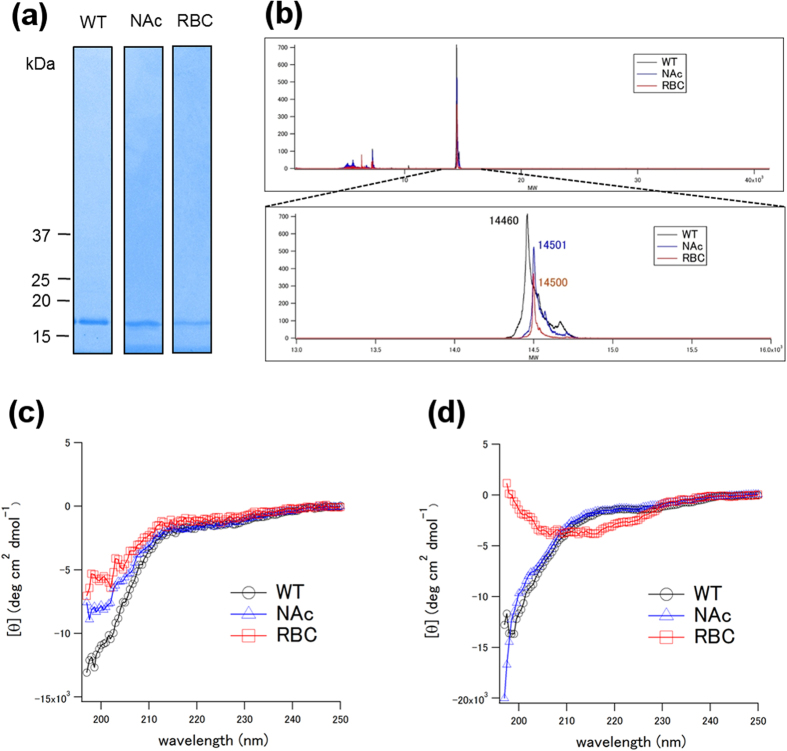
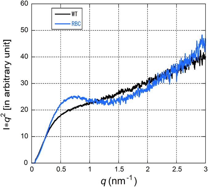
α-synuclein (α-syn) is the main component of Lewy bodies, which are neuropathological hallmarks of patients with Parkinson's disease. As it has been controversial whether human α-syn from erythrocytes exists as a tetramer under physiological conditions, we tried solving this issue by the small-angle X-ray solution scattering method. Under two different conditions (high ionic strength with a Tris buffer and low ionic strength with an ammonium acetate buffer), no evidence was found for the presence of tetramer. When comparing erythrocyte and recombinant α-syn molecules, we found no significant difference of the molecular weight and the secondary structure although the buffer conditions strongly affect the radius of gyration of the protein. The results indicate that, even though a stable tetramer may not be formed, conformation of α-syn depends much on its environment, which may be the reason for its tendency to aggregate in cells.


Figures

Figure 1
Figure 1
(a) SDS-PAGE/Coomassie Brilliant Blue (or silver) staining. WT, NAc and RBC represents wildtype α-syn, N-terminally acetylated α-syn and α-syn purified from human RBCs, respectively. (b) MALDI spectra of α-syn. Theoretical molecular weights (MWs) of Wild type (WT) and N-terminally acetylated (NAc) are 14460 and 14502, respectively. RBC represents α-syn purified from human RBCs. (c) Circular dichroism spectra of WT, NAc and RBC in the AA buffer (10 mM ammonium acetate (pH 7.4)). (d) Circular dichroism spectra of WT, NAc and RBC in the Tris buffer (50 mM Tris-HCl, 150 mM NaCl (pH 7.4))
Figure 2
Figure 2
(a) A typical set of SAXS profiles from human RBC α-syn at four different concentrations. (b) Guinier plots of the data in (a). (c) A typical set of SAXS profiles from WT α-syn in the Tris buffer at four different concentrations. (d) Guinier plots of the data in (d). (e) A typical set of SAXS profiles from WT α-syn in the AA buffer at three different concentrations. (f) Guinier plots of the data in (e).
Figure 3
Figure 3
(a) Concentration dependence of the Rg values in three types of α-syn. The value at the abscissa was used as the Rg value at infinite dilution. (b) Concentration dependence of the I(0)/c value.
Figure 4
Figure 4
(a) An elusion profile of recombinant α-syn from a size-exclusion column. The flow rate was changed from 0.5 to 0.2 ml/min at 12.7 ml. (b) An X-ray scattering profile of α-syn pooled around the elusion peak. The data in 30 frames (5 min) across the elusion peak, corresponding to 1.0 ml of elusion volume, were summed for the analysis. The linear region of the Guinier plot was q = 0.145 to 0.359 nm−1. The data were truncated at around q = 1.5 nm−1 because of the small size of the X-ray detector but plotted so as to be compared with Fig. 1c.
Figure 5
Figure 5. Kratky plots of recombinant α-syn (WT) and RBC α-syn (RBC) in the Tris buffer.

References

    1. Spillantini M. G., Crowther R. A., Jakes R., Hasegawa M. & Goedert M. α-Synuclein in filamentous inclusions of Lewy bodies from Parkinson disease and dementia with Lewy bodies. Proc. Natl. Acad. Sci. 95, 6469–6473 (1998). - PMC - PubMed
    1. Baba M. et al. Aggregation of α-synuclein in Lewy bodies of sporadic Parkinson disease and dementia with Lewy bodies. Am. J. Pathol. 152, 879–884 (1998). - PMC - PubMed
    1. Araki K. et al. Synchrotron FTIR micro-spectroscopy for structural analysis of Lewy bodies in the brain of Parkinson’s disease patients. Sci Rep. 5, 17625 (2015). - PMC - PubMed
    1. Tuttel M. D. et al. Solid-state NMR structure of a pathogenic fibril of full-length human α-synuclein. Nat Struct Mol Biol. 23, 409–415 (2016). - PMC - PubMed
    1. Maroteaux L., Campanelli J. T. & Scheller R. H. Synuclein – a neuron-specific protein localized to the nucleus and presynaptic nerve terminal. J. Neurosci. 8, 2804–2815 (1988). - PMC - PubMed

Publication types