. 2016 Oct 1;473(19):3371-83.
doi: 10.1042/BCJ20160243. Epub 2016 Jul 28.

Engineering tyrosine electron transfer pathways decreases oxidative toxicity in hemoglobin: implications for blood substitute design

Affiliations

Engineering tyrosine electron transfer pathways decreases oxidative toxicity in hemoglobin: implications for blood substitute design

Gary G A Silkstone et al. Biochem J. .

Abstract

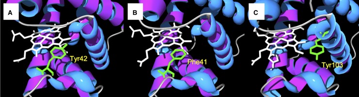
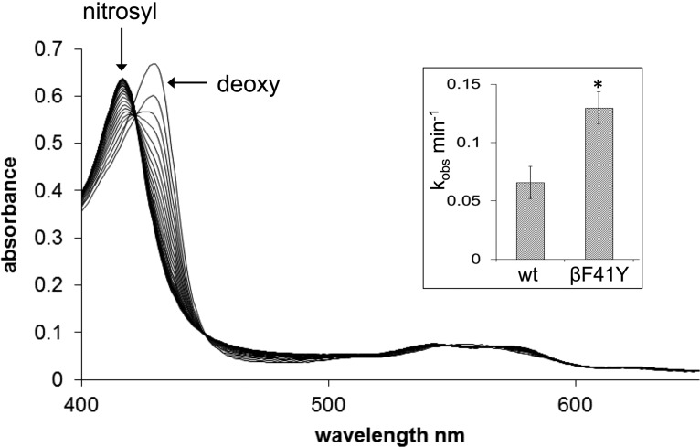
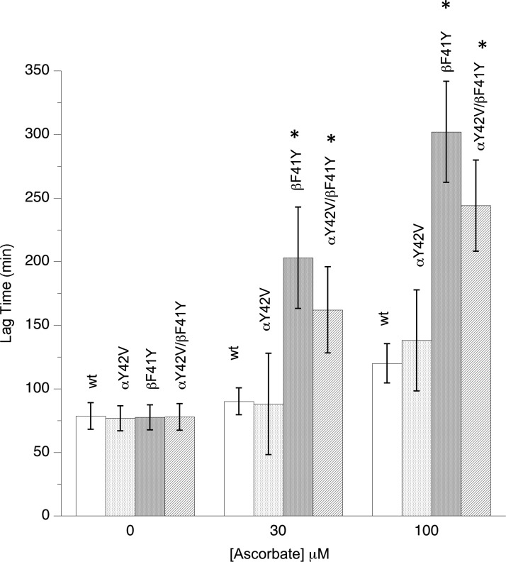
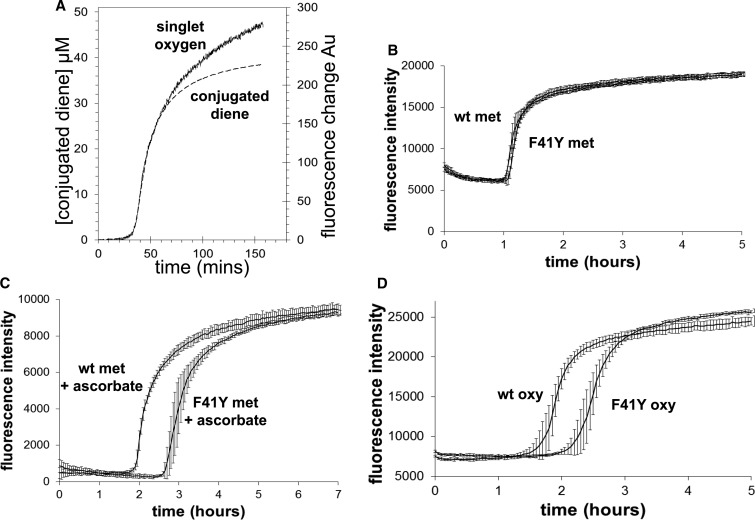
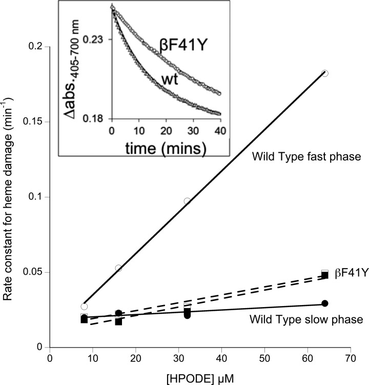
Hemoglobin (Hb)-based oxygen carriers (HBOC) have been engineered to replace or augment the oxygen-carrying capacity of erythrocytes. However, clinical results have generally been disappointing due to adverse side effects linked to intrinsic heme-mediated oxidative toxicity and nitric oxide (NO) scavenging. Redox-active tyrosine residues can facilitate electron transfer between endogenous antioxidants and oxidative ferryl heme species. A suitable residue is present in the α-subunit (Y42) of Hb, but absent from the homologous position in the β-subunit (F41). We therefore replaced this residue with a tyrosine (βF41Y, Hb Mequon). The βF41Y mutation had no effect on the intrinsic rate of lipid peroxidation as measured by conjugated diene and singlet oxygen formation following the addition of ferric(met) Hb to liposomes. However, βF41Y significantly decreased these rates in the presence of physiological levels of ascorbate. Additionally, heme damage in the β-subunit following the addition of the lipid peroxide hydroperoxyoctadecadieoic acid was five-fold slower in βF41Y. NO bioavailability was enhanced in βF41Y by a combination of a 20% decrease in NO dioxygenase activity and a doubling of the rate of nitrite reductase activity. The intrinsic rate of heme loss from methemoglobin was doubled in the β-subunit, but unchanged in the α-subunit. We conclude that the addition of a redox-active tyrosine mutation in Hb able to transfer electrons from plasma antioxidants decreases heme-mediated oxidative reactivity and enhances NO bioavailability. This class of mutations has the potential to decrease adverse side effects as one component of a HBOC product.

Keywords: hemoglobin; oxidative stress; reactive oxygen species.


Figures

Figure 1.
Figure 1.. Tyrosine electron transfer pathways in globins.
Active site structure of Hb α-subunit (A), Hb β-subunit (B), and myoglobin (C); sites of redox-active tyrosine residues are indicated. Note that in the Hb β-subunit, the tyrosine is substituted with a phenylalanine. Mutating this to a tyrosine (βF41Y) creates a new ascorbate reducible site. This diagram was adapted from a figure originally published in The Journal of Biological Chemistry. Reeder, B. J. et al. Tyrosine residues as redox cofactors in human hemoglobin: Implications for engineering non toxic blood substitutes. J. Biol. Chem. 2008; 283:30780–30787 © the American Society for Biochemistry and Molecular Biology.
Figure 2.
Figure 2.. HPLC comparison of recombinant and native Hb.
Reversed-phase HPLC separation of heme cofactor and protein subunits from native Hb, wt recombinant Hb and the βF41Y mutant.
Figure 3.
Figure 3.. Heme loss from recombinant Hb.
Heme loss from metHb and binding to the apo H64Y/V68F mutant. The spectral changes show the reaction following mixing metHb with the apo H64Y/V68F mutant for wt (bottom) and βF41Y (top). The main figure shows the spectral changes every 13 min from t = 0 to t = 4 h. An expanded region (×7) of the visible spectra in the 600 nm region is also shown. The inset shows the time course for the reaction in the visible region used for further kinetic analysis. Conditions: 100 mM NaPi, pH 7.2, containing 0.15 M sucrose; T = 37°C; [Hb] = 2.5 μM; [apo mbH64Y/V68F] = 30 μM.
Figure 4.
Figure 4.. Hb nitrite reductase activity.
Formation of nitrosyl Hb from deoxy Hb, wt, and βF41Y recombinant forms, in the presence of 0.2 mM nitrite. Conditions: sodium phosphate buffer (20 mM, pH 7.4, 25°C); [Hb] = 5 μM. Inset: the rate (mean ± SD, n = 6) of conversion from deoxy to nitrosyl. Asterisk indicates significant difference from wt (P < 0.0001).
Figure 5.
Figure 5.. Effect of tyrosine mutations on Hb-catalyzed lipid peroxidation.
Liposome oxidation monitored by conjugate diene formation following the addition of recombinant metHb in the presence or absence of ascorbate. Assay conditions: sodium phosphate (20 mM, pH 7.4, 30°C); [heme] = 2 µM; data presented as mean ± SD, n = 9. Asterisks indicate significant difference from wt (P < 0.05).
Figure 6.
Figure 6.. Singlet oxygen production catalyzed by recombinant Hb.
Changes in singlet oxygen (fluorescence) and conjugated diene (absorbance) formation following the addition of metHb to liposomes (A). Comparison of wt and βF41Y metHb addition in the absence (B) and presence (C) of 50 μM ascorbate. Addition of wt and βF41Y oxyHb to liposomes (D). Assay conditions: sodium phosphate (20 mM, pH 7.4, 30°C); [heme] = 2 µM; [SOSG] = 0.25 µM. Data presented as mean ± SD, n = 8.
Figure 7.
Figure 7.. Reaction of the lipid peroxide HPODE with recombinant Hb.
Addition of 32 μM HPODE to metHb (wt and βF41Y). Time courses of heme damage (inset) were fit to double exponentials and plotted against [HPODE]. Conditions: sodium phosphate (20 mM, pH 7.4); [heme] = 2 μM after mixing; T = 25°C.

References

    1. Alayash A.I. (2014) Blood substitutes: why haven't we been more successful? Trends Biotechnol. 32, 177–185 doi:10.1016/j.tibtech.2014.02.006 - DOI - PMC - PubMed
    1. Varnado C.L., Mollan T.L., Birukou I., Smith B.J.Z., Henderson D.P. and Olson J.S. (2013) Development of recombinant hemoglobin-based oxygen carriers. Antioxid. Redox Signal. 18, 2314–2328 doi:10.1089/ars.2012.4917 - DOI - PMC - PubMed
    1. Dou Y., Maillett D.H., Eich R.F. and Olson J.S. (2002) Myoglobin as a model system for designing heme protein based blood substitutes. Biophys. Chem. 98, 127–148 doi:10.1016/S0301-4622(02)00090-X - DOI - PubMed
    1. Looker D., Abbott-Brown D., Cozart P., Durfee S., Hoffman S., Mathews A.J. et al. (1992) A human recombinant haemoglobin designed for use as a blood substitute. Nature 356, 258–260 doi:10.1038/356258a0 - DOI - PubMed
    1. Komiyama N.H., Miyazaki G., Tame J. and Nagai K. (1995) Transplanting a unique allosteric effect from crocodile into human haemoglobin. Nature 373, 244–246 doi:10.1038/373244a0 - DOI - PubMed

Publication types