. 2016 Sep;36(9):1900-9.
doi: 10.1161/ATVBAHA.116.307869. Epub 2016 Jul 28.

Essential Role of Smooth Muscle STIM1 in Hypertension and Cardiovascular Dysfunction

Affiliations

Essential Role of Smooth Muscle STIM1 in Hypertension and Cardiovascular Dysfunction

Modar Kassan et al. Arterioscler Thromb Vasc Biol. 2016 Sep.

Abstract

Objectives: Chronic hypertension is the most critical risk factor for cardiovascular disease, heart failure, and stroke.

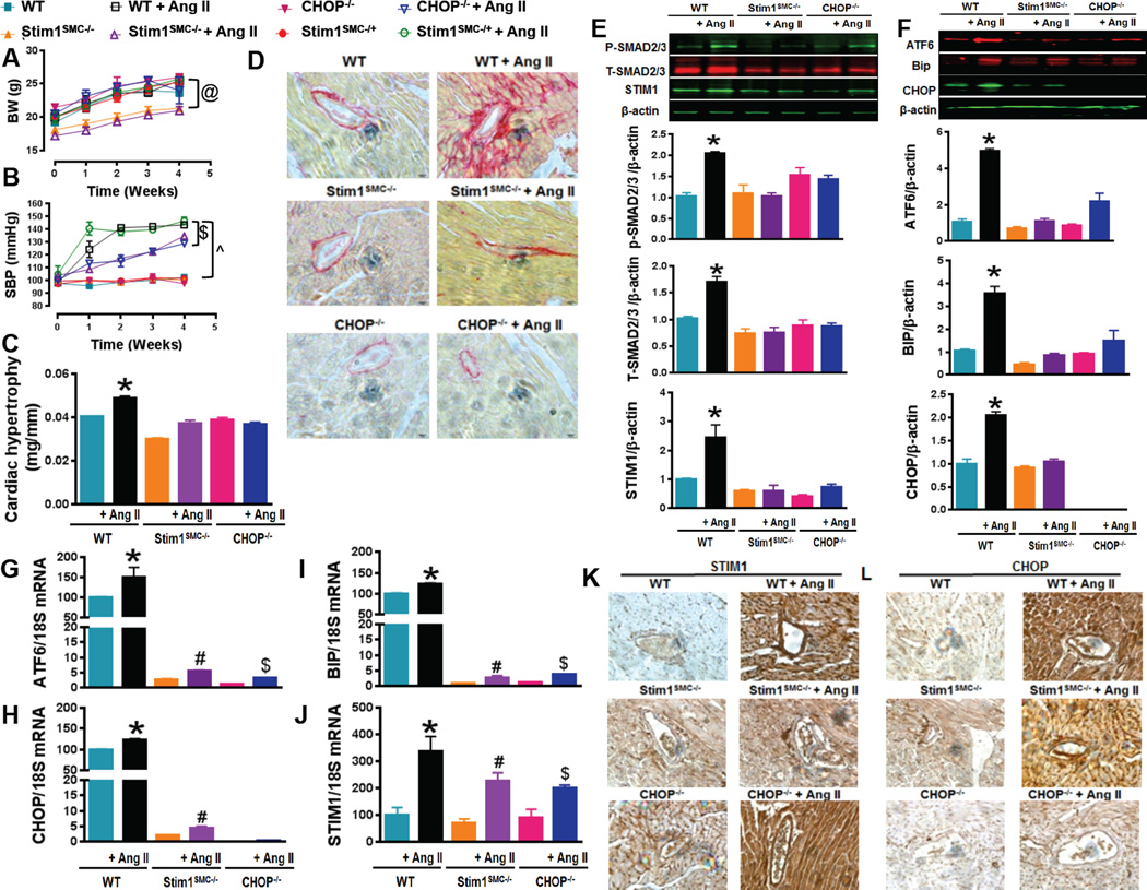
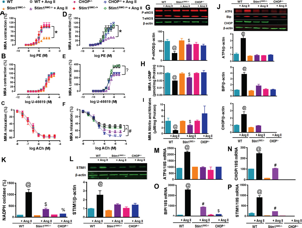
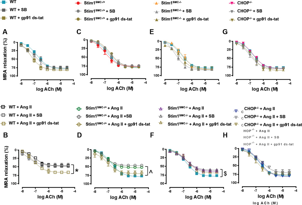
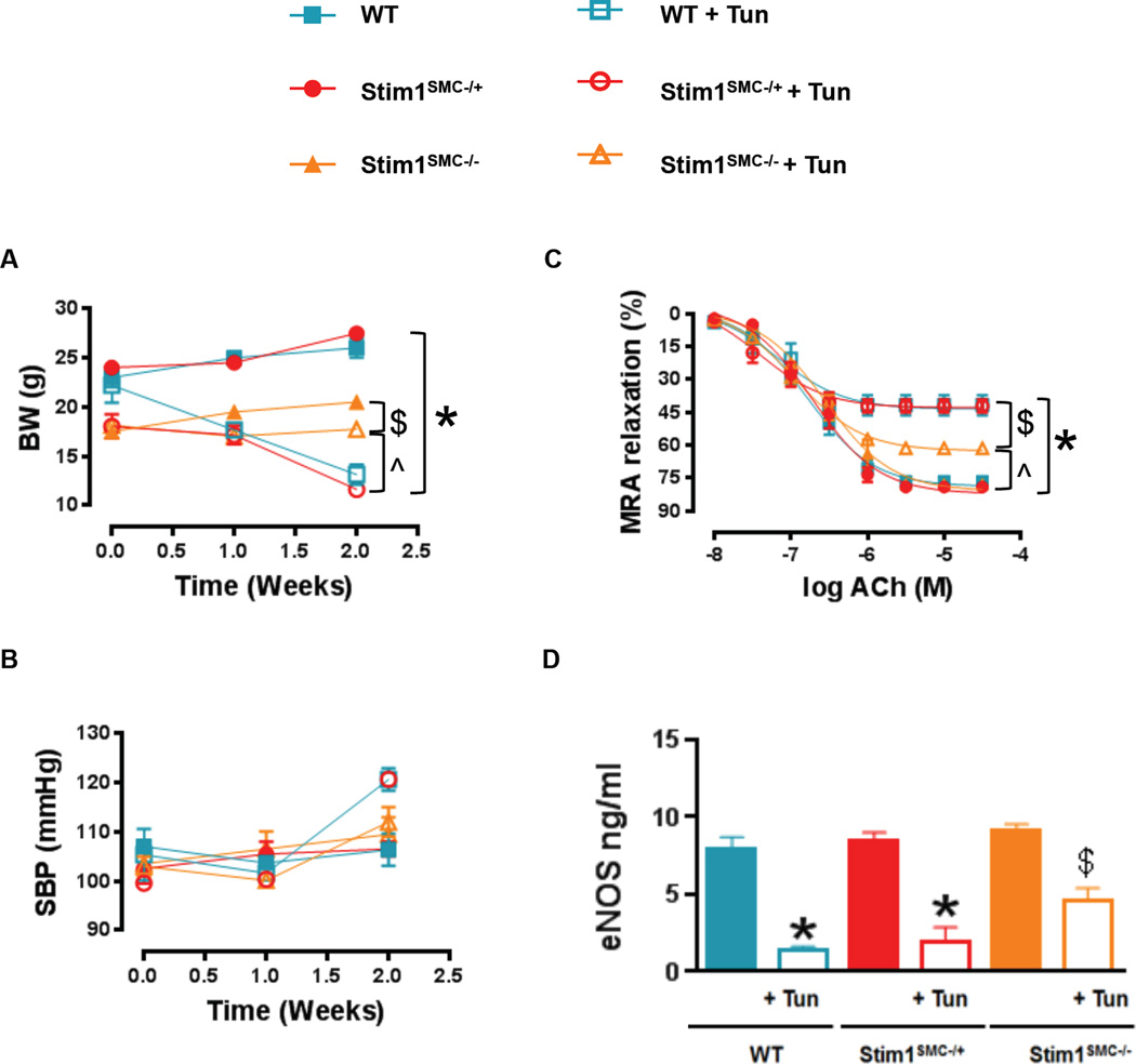
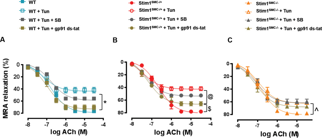
Approach and results: Here we show that wild-type mice infused with angiotensin II develop hypertension, cardiac hypertrophy, perivascular fibrosis, and endothelial dysfunction with enhanced stromal interaction molecule 1 (STIM1) expression in heart and vessels. All these pathologies were significantly blunted in mice lacking STIM1 specifically in smooth muscle (Stim1(SMC-/-)). Mechanistically, STIM1 upregulation during angiotensin II-induced hypertension was associated with enhanced endoplasmic reticulum stress, and smooth muscle STIM1 was required for endoplasmic reticulum stress-induced vascular dysfunction through transforming growth factor-β and nicotinamide adenine dinucleotide phosphate oxidase-dependent pathways. Accordingly, knockout mice for the endoplasmic reticulum stress proapoptotic transcriptional factor, CCAAT-enhancer-binding protein homologous protein (CHOP(-/-)), were resistant to hypertension-induced cardiovascular pathologies. Wild-type mice infused with angiotensin II, but not Stim1(SMC-/-) or CHOP(-/-) mice showed elevated vascular nicotinamide adenine dinucleotide phosphate oxidase activity and reduced phosphorylated endothelial nitric oxide synthase, cGMP, and nitrite levels.

Conclusions: Thus, smooth muscle STIM1 plays a crucial role in the development of hypertension and associated cardiovascular pathologies and represents a promising target for cardiovascular therapy.

Keywords: ER stress; cardiac hypertrophy; endothelial nitric oxide synthase; hypertension; nicotinamide adenine dinucleotide phosphate; stromal interaction molecule 1; vascular reactivity.


Figures

Figure 1
Figure 1. STIM1 and CHOP mediate hypertension-induced cardiac damage and fibrosis
Body weight (A, n=6) and Systolic blood pressure measured by tail cuff machine (B, n=6) in WT, heterozygous (Stim1SMC−/+) and homozygous Stim1 knockout specifically in SMC (Stim1SMC−/−), and CHOP knockout (CHOP−/−) mice infused with or without Ang II. Cardiac hypertrophy determined by evaluating heart weight/tibia length ratios (C, n=6), Cardiac fibrosis stained with the collagen-specific Sirius-red (D, n=3), Western blot and quantification showing phosphorylated and total SMAD 2/3, STIM1 (E, n=3) and ER stress markers (ATF6, BIP CHOP) (F, n=3), mRNA levels of ER stress marker (ATF6 CHOP and BIP) (G, H, I, n=3) and STIM1 (J, n=3) and, Immunohistochemistry showing STIM1 (K, n=3) and CHOP (L, n=3) in heart from WT, Stim1SMC−/− and CHOP−/− mice infused with saline or Ang II. Two-way repeated measured ANOVA followed by Tukey's Post-Hoc test were applied for figures (A, B). One-way ANOVA followed by Bonferroni Post-Hoc test were applied for figures (G, H, I, J, E, F). @p<0.05 between STIMSMC−/−, STIMSMC−/− + AngII VS WT, WT + Ang II, STIMSMC−/+, STIMSMC−/+ + Ang II, CHOP−/−, CHOP−/− + Ang II. $p<0.05 between STIMSMC−/− + AngII, CHOP−/− + Ang II vs. STIMSMC−/+ + Ang II, WT + Ang II. ^p<0.05 between WT, STIMSMC−/−, CHOP−/− vs WT + Ang II, STIMSMC−/− + AngII, CHOP−/− + Ang II. *p<0.05 between WT + Ang II vs WT, STIMSMC−/−, STIMSMC−/− + AngII, CHOP−/−, CHOP−/− + Ang II. #p<0.05 between STIMSMC−/− vs STIMSMC−/− + AngII. &p<0.05 between CHOP−/− vs CHOP−/− + Ang II.
Figure 2
Figure 2. STIM1 and CHOP regulate hypertension-induced vascular damage
Wire Myograph vascular reactivity showing vessel contraction response to phenylephrine (A, D, n=5–6) and ThromboxaneA2 analogue U-46619 (B, E, n=5), and endothelial-dependent relaxation in response to acetylcholine (C, F, n=5) in mesenteric resistance arteries (MRA) from WT, heterozygous (Stim1SMC−/+) and homozygous Stim1 knockout specifically in SMC (Stim1SMC/−), and CHOP knockout (CHOP−/−) mice infused with saline or Ang II. Western blot showing phosphorylated and total eNOS (G, n=3), Elisa showing cGMP levels using a sandwich enzymelinked immunosorbent assay (H, n=3) and nitrites/nitrate levels using the Griess reaction (I, n=3), protein levels and quantification of ER stress markers (ATF6, BIP and CHOP) (J, n=3), NADPH oxidase activity using lucigenin chemiluminescence (K, n=5), Western blot showing STIM1 (L, n=3), mRNA of ER stress markers (ATF6, BIP and CHOP) (M, N, O, n=3) and STIM1 (P, n=3) in mesenteric resistance arteries from WT, Stim1SMC−/− and CHOP−/− mice infused with or without Ang II. Two-way repeated measured ANOVA followed by Tukey's Post-Hoc test were applied for figures (A, B, C, D, E, F). One-way ANOVA followed by Bonferroni Post-Hoc test were applied for figures (K, L, H, I, J, M, N, O, P). *p<0.05 between STIMSMC−/− vs. WT, STIMSMC−/+, and CHOP−/−. &p<0.05 between STIMSMC−/− + AngII vs WT + Ang II, STIMSMC−/+ + Ang II, CHOP−/− + Ang II. ?p<0.05 between STIMSMC−/− + AngII, CHOP−/− + Ang II VS WT + Ang II, STIMSMC−/+ + Ang II. ^p<0.05 between WT + Ang II, STIMSMC−/+ + Ang II vs. STIMSMC−/− + AngII. #p<0.05 between WT + Ang II, STIMSMC−/+ + Ang II Vs. STIMSMC−/− + AngII vs CHOP−/− + Ang II. @p<0.05 between WT + Ang II vs WT, STIMSMC−/−, STIMSMC−/− + AngII, CHOP−/−, CHOP−/− + Ang II. $p<0.05 between STIMSMC−/− vs STIMSMC−/− + AngII. %p<0.05 between CHOP−/− vs. CHOP−/− + Ang II.
Figure 3
Figure 3. Effect of inhibition of NADPH oxidase and TGF-β signaling on Vascular reactivity in STIM1 and CHOP knockout mice
Wire Myograph vascular reactivity showing endothelial-dependent relaxation in response to acetylcholine before and after incubation with TGF-β inhibitors (SB431542) and NADPH oxidase inhibitor (gp91 ds-tat) in mesenteric resistance arteries from: - WT mice infused with saline or Ang II (A, B, n=5) - Heterozygous Stim1 knockout specifically in SMC (Stim1SMC−/+) mice infused with saline or Ang II (C, D, n=5) - Homozygous Stim1 knockout specifically in SMC (Stim1SMC−/−) mice infused with saline or Ang II (E, F, n=5) - CHOP knockout (CHOP−/−) mice infused with saline or Ang II (G, H, n=5) Two-way repeated measured ANOVA followed by Tukey's Post-Hoc test were applied for figures (A, B, C, D, E, F, G H). *p<0.05 between WT + Ang II + gp91 ds-tat vs WT + Ang II, WT + Ang II + SB. ^p<0.05 between WT, STIMSMC−/+ + Ang II + gp91 ds-tat vs STIMSMC−/+ + Ang II, STIMSMC−/+ + Ang II + SB. $p<0.05 between WT vs. STIMSMC−/− + Ang II + gp91 ds-tat, STIMSMC−/− + Ang II, STIMSMC−/− + Ang II + SB.
Figure 4
Figure 4. Effect of tunicamycin-induced ER stress on vascular reactivity in STIM1 knockout mice
Body weight (A, n=5) and systolic blood pressure measured by tail cuff machine (B, n=5), Wire Myograph vascular reactivity showing endothelial-dependent relaxation in response to acetylcholine (C, n=5) and eNOS levels determined by an ELISA Kit (D, n=3) in MRA from WT, heterozygous (Stim1SMC−/+) and homozygous Stim1 knockout specifically in SMC (Stim1SMC−/−) mice treated with saline or Tunicamycin. Two-way repeated measured ANOVA followed by Tukey's Post-Hoc test were applied for figures (A, B, C). One-way ANOVA followed by Bonferroni Post-Hoc test were applied for figures (D). $p<0.05 between STIMSMC−/+ + Tunica vs. WT, STIMSMC−/−, STIMSMC−/+. *p<0.05 between STIMSMC−/+ + Tunica, WT + Tunica vs. WT, STIMSMC−/−, STIMSMC−/+. &p<0.05 between STIMSMC−/− vs. STIMSMC−/− + Tunica. ^p<0.05 between STIMSMC−/−, STIMSMC−/− + Tunica vs. STIMSMC−/+ + Tunica, WT + Tunica. %p<0.05 between STIMSMC−/− + Tunica vs. STIMSMC−/+ + Tunica and WT + Tunica.
Figure 5
Figure 5. Effect of inhibition of NADPH oxidase and TGF-β signaling on Vascular reactivity in tunicamycin-treated STIM1 knockout mice
Wire Myograph vascular reactivity showing endothelial-dependent relaxation in response to acetylcholine before and after incubation with TGF-β inhibitor (SB431542) and NADPH oxidase inhibitor (gp91 ds-tat) in mesenteric resistance arteries from: - WT mice treated with saline or Tunicamycin (A, n=5) - Heterozygous Stim1 knockout specifically in SMC (Stim1SMC−/+) mice treated with saline or Tunicamycin (B, n=5) - Homozygous Stim1 knockout specifically in SMC (Stim1SMC−/−) mice treated with saline or Tunicamycin (C, n=5) Two-way repeated measured ANOVA followed by Tukey's Post-Hoc test were applied for figures (A, B, C). *p<0.05 between WT, WT + Tunica + gp91 ds-tat vs Sham + Tunica, WT + Tunica + SB. $p<0.05 between STIMSMC−/+ vs STIMSMC−/+ + Tunica + gp91 ds-tat. @p<0.05 between STIMSMC−/+ + Tunica + gp91 ds-tat vs STIMSMC−/+ + Tunica, STIMSMC−/+ + Tunica + SB. $p<0.05 between STIMSMC−/− vs STIMSMC−/− + Tunica, STIMSMC−/− + Tunica + gp91 ds-tat, STIMSMC−/− + Ang II, STIMSMC−/− + Tunica + SB.

References

    1. Leong XF, Ng CY, Jaarin K. Animal models in cardiovascular research: Hypertension and atherosclerosis. Biomed Res Int. 2015;2015:528757. - PMC - PubMed
    1. Efrati S, Zaidenstein R, Dishy V, Beberashvili I, Sharist M, Averbukh Z, Golik A, Weissgarten J. Ace inhibitors and survival of hemodialysis patients. American journal of kidney diseases. 2002;40:1023–1029. - PubMed
    1. Kassan M, Galán M, Partyka M, Saifudeen Z, Henrion D, Trebak M, Matrougui K. Endoplasmic reticulum stress is involved in cardiac damage and vascular endothelial dysfunction in hypertensive mice. Arteriosclerosis, thrombosis, and vascular biology. 2012;32:1652–1661. - PMC - PubMed
    1. Galán M, Kassan M, Kadowitz PJ, Trebak M, Belmadani S, Matrougui K. Mechanism of endoplasmic reticulum stress-induced vascular endothelial dysfunction. Biochimica et Biophysica Acta (BBA)-Molecular Cell Research. 2014;1843:1063–1075. - PMC - PubMed
    1. Dharmashankar K, Widlansky ME. Vascular endothelial function and hypertension: Insights and directions. Current hypertension reports. 2010;12:448–455. - PMC - PubMed

Publication types

MeSH terms