. 2016 Sep;186(9):2337-52.
doi: 10.1016/j.ajpath.2016.07.001. Epub 2016 Jul 26.

Collaborative Interferon-γ and Interleukin-17 Signaling Protects the Oral Mucosa from Staphylococcus aureus

Affiliations

Collaborative Interferon-γ and Interleukin-17 Signaling Protects the Oral Mucosa from Staphylococcus aureus

Jobert G Barin et al. Am J Pathol. 2016 Sep.

Abstract

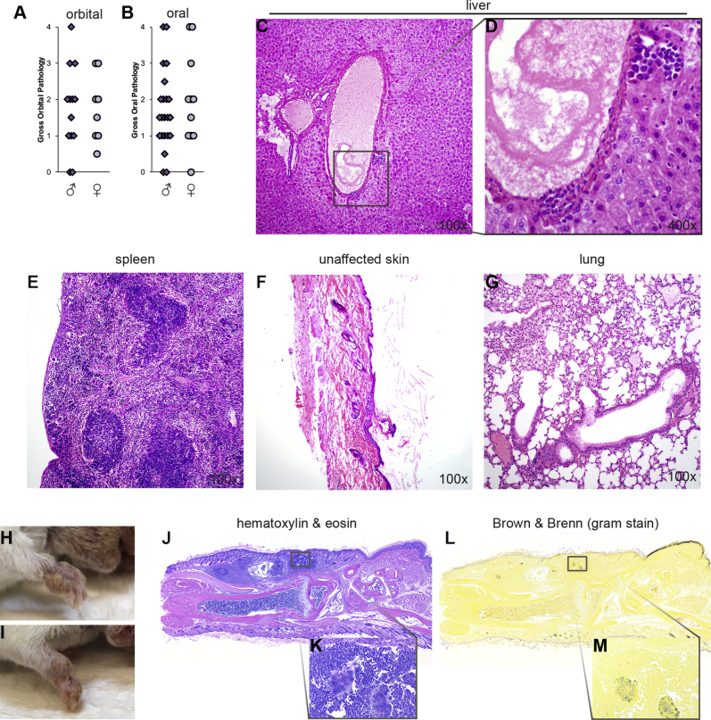
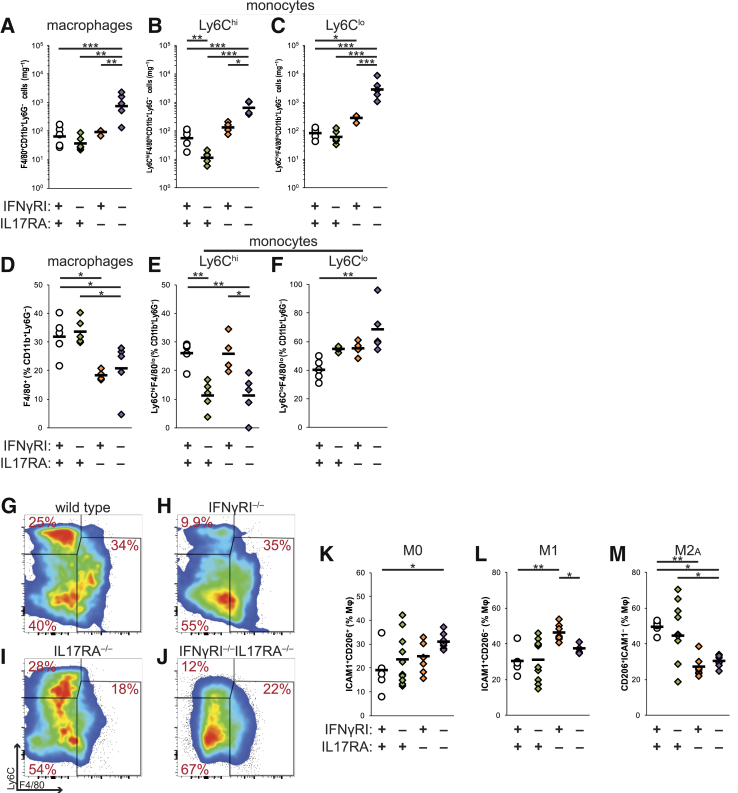
Infections with Staphylococcus aureus are a continuing and growing problem in community and hospital settings. Preclinical animal modeling of S. aureus relies on experimental infection, which carries some limitations. We describe here a novel, spontaneous model of oral staphylococcal infection in double knockout mice, deficient in the receptors for IL-17 (IL-17RA) and interferon (IFN)-γ (IFNγRI), beginning at 6 to 8 weeks of age. IFNγRI(-/-)IL17RA(-/-) (GRAKO) mice developed progressive oral abscesses. Cytometric methods revealed extensive neutrophilic infiltration of oral tissues in GRAKO mice; further investigation evidenced that IL-17 predominated neutrophil defects in these mice. To investigate the contribution of IFN-γ signaling to this native host defense to S. aureus, we observed perturbations of monocyte recruitment and macrophage differentiation in the oral tissues of GRAKO mice, and CXCL9/chemokine ligand receptor (CXCR)3-driven recruitment of T-cell oral tissues and draining lymph nodes. To address the former finding, we depleted macrophages and monocytes in vivo from IL17RA(-/-) mice using liposomes loaded with clodronate. This treatment elicited oral abscesses, recapitulating the phenotype of GRAKO mice. From these findings, we propose novel collaborative functions of IL-17 and IFN-γ, acting through neutrophils and macrophages, respectively, in native mucocutaneous host defenses to S. aureus.


Figures

Supplemental Figure S1
A and B: Comparison of gross pathologic lesions of male and female IFNγRI−/−IL17RA−/− mice. CG: Histopathologic characterization of additional disease phenotype in highly affected IFNγRI−/−IL17RA−/− mice. Subject was a 15-week-old IFNγRI−/−IL17RA−/−male mouse, with a grade 3 oral lesion. Note the limited periportal inflammatory foci in liver and pyogranulomatous inflammatory infiltration in lung. D shows an enlargement of the boxed area in C. HM: Development of abscesses in forelimbs of IFNγRI−/−IL17RA−/− mice, including representative histopathology. K and M show enlarged sections of J and L, respectively. Statistics are by U-test (NS). Original magnification, ×10 (J–M). IFN, interferon; NS, not significant.
Supplemental Figure S2
Neutrophil depletion from IFNγRI−/− mice. AD: Cytometric enumeration for Ly6GhiCD11b+ neutrophils, gated on viable CD45+ events, from spleen and oral tissues. Representative bivariate plots depict concatenates of each group. E and F: Body weight (E) and spleen weight (F) (as a percentile of body weight). G: Oral colonization by Staphylococcus aureus, represented as CFUs of S. aureus from oral swabs. HJ: Scoring of histopathologic severity of perioral tissues (H) and representative histopathology (I and J), depicting the median of each group. Individual mice are represented by diamonds or circles; means of groups by bars. Statistics are by two-tailed Student's t-test. Statistically significant pairwise comparisons are denoted between relevant groups. *P < 0.05, ***P ≤ 0.001. Original magnification, ×400. CFU, colony-forming unit; IFN, interferon.
Supplemental Figure S3
Cytokine and chemokine mediators of host defense to Staphylococcus aureus in IFNγRI−/−IL17RA−/−, IL17RA−/−, IFNγRI−/−, and wild-type mice. AL: Cytokines and chemokines were interrogated by Linco 32-plex xMAP array from homogenates of oral tissues, organized here as functional groupings. Data are expressed as means ± SD of groups of individual mice normalized to sample weight. Statistics are by one-way analysis of variance, followed by Fisher's LSD. Statistically significant pairwise comparisons are denoted between relevant groups. n = 5 IFNγRI−/−IL17RA−/− mice; n = 4 IL17RA−/− mice; n = 5, IFNγRI−/− mice; n = 5 wild-type mice. *P < 0.05, **P ≤ 0.01, and ***P ≤ 0.001. CCL, chemokine ligand; G-CSF, granulocyte colony-stimulating factor; GM-CSF, granulocyte-macrophage colony-stimulating factor; IFN, interferon; LSD, least significant difference; MCP, monocyte chemoattractant protein; MIP, macrophage inflammatory protein; RANTES, regulated on activation normal T cell expressed and secreted; TNF, tumor necrosis factor.
Supplemental Figure S4
Extended phenotypic analysis of lymphocytes in the draining sublingual superficial cervical lymph nodes of IFNγRI−/−IL17RA−/− mice. AD: Enumeration of lymphocyte populations in the scLN. EJ: Activation, memory, and effector differentiation among CD4+TCRβ+ (EG) and TCRγδ+ T cells (HJ) in the scLN, expressed as percentiles of parent population. KN: Coordinate expression of RORγt and CD45RB on TCRγδ+-gated scLN T cells. Individual mice are represented by diamonds or circles; means of groups by bars. Representative bivariate plots depict concatenated groups. Statistics are by one-way analysis of variance, followed by Bonferroni post-testing. Statistically significant pairwise comparisons are denoted between relevant groups. *P < 0.05, **P ≤ 0.01, and ***P ≤ 0.001. IFN, interferon; RORγt; retinoic acid receptor (RAR)-related orphan receptor γ thymus-specific isoform; scLN, superficial cervical lymph node; TCR, T-cell receptor.
Figure 1
Figure 1
IFNγRI−/−IL17RA−/− mice develop spontaneous staphylococcal perioral abscesses. AD: Representative photography of disease phenotypes: periocular inflammation of IL17RA−/− BALB mice (A), and the perioral abscesses of IFNγRI−/−IL17RA−/− mice (BD). E and F: Gross pathologic examination of orbital (E) and oral (F) lesions in IFNγRI−/−IL17RA−/− mice and controls. Gross pathology was scored on a semiquantitative 0 to 4 scale. G and H: Body (G) and spleen (H) weight, expressed as a percentage of total body weight, at time of sacrifice. Statistics for discontinuous nonparametric data are by Kruskal-Wallis analysis of variance. I: Time course of disease onset, depicting gross oral pathology scores as a function of age at sacrifice. Regression lines for each genotype are color-coded as shown. J: Oral colonization of IFNγRI−/−IL17RA−/− mice and controls by Staphylococcus aureus, represented as CFUs of S. aureus from oral swabs. K: Semiquantitative real-time PCR of S. aureus sodA from oral tissue. Statistics are by one-way analysis of variance, followed by post hoc pairwise comparisons by Tukey-Kramer method. L and M: Quantitation of total serum immunoglobulins by sandwich ELISA. Individual mice are represented by diamonds or circles; means of groups by bars. Statistically significant pairwise comparisons are denoted between relevant groups. n = 21 IFNγRI−/−IL17RA−/− mice; n = 14 single-knockout IL17RA−/− mice; n = 14 IFNγRI−/− mice; n = 12 wild-type control mice (I). *P < 0.05, **P ≤ 0.01, and ***P ≤ 0.001. BW, body weight; CFU, colony-forming unit; ELISA, enzyme-linked immunosorbent assay; IFN, interferon.
Figure 2
Figure 2
Histopathologic examination of abscesses of IFNγRI−/−IL17RA−/− mice reveals extensive surrounding polymorphonuclear infiltration. AH: Representative histopathology of perioral tissues from control genotypes and IFNγRI−/−IL17RA−/− mice. Boxed areas in AD correspond to higher-magnification images in EH. I: Scoring of histopathologic severity of perioral tissues. Individual mice are represented by diamonds or circles. Gram-staining of affected IFNγRI−/−IL17RA−/−perioral tissues by Brown & Brenn (JM) against hematoxylin and eosin serial sections (NP). Boxed area in J is enlarged in K; boxed areas in K are enlarged in L and M. Boxed area in N is enlarged in O; boxed area in O is enlarged in P. Statistics are by one-way analysis of variance, followed by post hoc pairwise comparisons by Tukey-Kramer method. Statistically significant pairwise comparisons are denoted between relevant groups. **P ≤ 0.01, ***P ≤ 0.001. Original magnification: ×10 (N); ×160 (O); ×640 (M and P). IFN, interferon.
Figure 3
Figure 3
Defects in neutrophils as cellular mediators of host defense to Staphylococcus aureus in oral tissues of IFNγRI−/−IL17RA−/− mice. A: Total oral CD45+ leukocytes, expressed as absolute numbers, normalized to sample weight. BD: Ly6GhiCD11b+ neutrophils (B), Siglec F+Ly6Gint eosinophils (C), and FcεRIα+ mast cells and basophils (D) infiltrating oral tissues expressed as absolute numbers, normalized to sample weight. EH: Representative bivariate plots of viable CD45+-gated events in oral tissues depict concatenates of each group. I and J: Myeloperoxidase levels in perioral tissue homogenates by quantitative sandwich ELISA, normalized by sample weight (I), and in ratio to total neutrophil counts from Figure 3B (J). Statistics are by Fisher's LSD. K:In vitro killing of S. aureus by neutrophils. Individual mice are represented by diamonds or circles; means of groups by bars. Statistically significant pairwise comparisons are denoted between relevant groups. *P < 0.05, **P ≤ 0.01, and ***P ≤ 0.001. ELISA, enzyme-linked immunosorbent assay; IFN, interferon; LSD, least significant difference; MPO, myeloperoxidase.
Figure 4
Figure 4
Lymphoid mediators of host defense to Staphylococcus aureus in IFNγRI−/−IL17RA−/− mice and control strains. AC: CXCL9 (A) and CXCL10 (B) interrogated by Linco 32-plex xMAP array from homogenates of oral tissues, normalized to sample weight; and supernatants from lymphocytes from the superficial cervical LN, restimulated with heat-killed S. aureus (C). D: Total counts of viable CD45+ cells from scLN. EL: CXCR3 expression on scLN T-cell populations, as viable CD45+ lymphocyte-gated cells from wild-type BALB/c mice in representative bivariate plots (EH) and expressed as a proportion of parent populations, including total CD4+TCRβ+ cells (I and J) and γδ+ T cells (K and L). Red arrows denote gated populations. M and N: Total CD4+ (M) and γδ T (N) cells infiltrating oral tissues, expressed as absolute numbers, normalized to sample weight. OV: Representative bivariate plots depict concatenates of each group, as a proportion of viable CD45+ lymphocytes. Regions shaded in red depict double-positive populations. Statistics are by one-way analysis of variance, followed by post hoc pairwise comparisons by Tukey-Kramer method. Statistically significant pairwise comparisons are denoted between relevant groups. *P < 0.05, **P ≤ 0.01, and ***P ≤ 0.001. IFN, interferon; scLN, superficial cervical lymph node; TCR, T-cell receptor.
Figure 5
Figure 5
Macrophage function in host defense to Staphylococcus aureus in IFNγRI−/−IL17RA−/− mice. AC: Total F4/80+CD11b+Ly6G macrophages (A), Ly6ChiF4/80loCD11b+Ly6G monocytes (B), and Ly6CloF4/80loCD11b+Ly6G monocytes (C) in oral tissues expressed as absolute numbers, normalized to sample weight. DF: Proportions of oral macrophages and monocytes, expressed as percentiles of CD11b+Ly6G events. All cells are gated on viable CD45+ events. GJ: Representative bivariate plots depict concatenated groups gated on viable CD11b+Ly6GCD45+ events. KM: Macrophage polarization indicated by expression of CD54/ICAM1 and/or CD206/MφMR, expressed as a proportion of viable CD64+F4/80+CD11b+Ly6GCD11cCD45+ events. Individual mice are represented by diamonds or circles; means of groups by bars. Statistics are by one-way analysis of variance, followed by post hoc pairwise comparisons by Tukey-Kramer method. Statistically significant pairwise comparisons are denoted between relevant groups. *P < 0.05, **P ≤ 0.01, and ***P ≤ 0.001. ICAM, intercellular adhesion molecule 1; IFN, interferon; MφMR, macrophage mannose receptor.
Figure 6
Figure 6
Macrophages are critical collaborators with IL-17–directed responses to Staphylococcus aureus in the oral mucosa. Data are representative of two independent experiments. A and B: Depletion of macrophages from oral tissues of IL17RA−/− mice by treatment with clodronate-loaded liposomes; representative bivariate plots represent concatenated pools at day 10 of treatment. C and D: Progression of orbital (C) and oral (D) abscesses in IL17RA−/− mice depleted of macrophages by treatment with clodronate-loaded liposomes (red). Control IL17RA−/− mice (gray) were untreated or administered PBS-loaded liposomes. EG: Gross pathology of orbital (E) and oral abscesses (F), splenomegaly in IL17RA−/− mice after 5 weeks of macrophage depletion (G). H and I: Representative gross oral pathologic lesions in IL17RA−/− mice after 5 weeks of macrophage depletion. J: Oral colonization of macrophage-depleted IL17RA−/− mice and controls by S. aureus, represented as CFUs of S. aureus from oral swabs at week 5 of treatment. KP: Histopathologic evaluation of oral abscesses in macrophage-depleted IL17RA−/− mice (K) and representative histopathology of oral lesions from control (L and M) or clodronate liposome-treated (NP) mice. Boxed area in N is enlarged in O and P. QT: Expression of CXCR3 on CD4+ (Q and R) and γδ T cells (S and T) in the scLN of macrophage-depleted IL17RA−/− mice. Representative bivariate plots depict concatenates of groups, or individual mice. All cells are gated on viable CD45+ events. Individual mice are represented by diamonds or circles; means of groups by bars. Statistics are by one-way analysis of variance, followed by post hoc pairwise comparisons by Tukey-Kramer method. Statistically significant pairwise comparisons are denoted between relevant groups. n = 5 untreated control IL17RA−/− mice; n = 5 control IL17RA−/− mice administered PBS-loaded liposomes (C and D). *P < 0.05, **P ≤ 0.01. CFU, colony-forming unit; H&E, hematoxylin and eosin; lip, liposome; PBS, phosphate-buffered saline; scLN, superficial cervical lymph node.

Comment in

  • This Month in AJP.
    [No authors listed] [No authors listed] Am J Pathol. 2016 Sep;186(9):2237. doi: 10.1016/j.ajpath.2016.05.022. Epub 2016 Jul 25. Am J Pathol. 2016. PMID: 27459941

Similar articles

Cited by

References

    1. Barin J.G., Baldeviano G.C., Talor M.V., Wu L., Ong S., Fairweather D., Bedja D., Stickel N.R., Fontes J.A., Cardamone A.B., Zheng D., Gabrielson K.L., Rose N.R., Cihakova D. Fatal eosinophilic myocarditis develops in the absence of IFN-gamma and IL-17A. J Immunol. 2013;191:4038–4047. - PMC - PubMed
    1. Lowy F.D. Staphylococcus aureus infections. N Engl J Med. 1998;339:520–532. - PubMed
    1. Tong S.Y., Davis J.S., Eichenberger E., Holland T.L., Fowler V.G., Jr. Staphylococcus aureus infections: epidemiology, pathophysiology, clinical manifestations, and management. Clin Microbiol Rev. 2015;28:603–661. - PMC - PubMed
    1. Miller L.S., Cho J.S. Immunity against Staphylococcus aureus cutaneous infections. Nat Rev Immunol. 2011;11:505–518. - PMC - PubMed
    1. Cho J.S., Pietras E.M., Garcia N.C., Ramos R.I., Farzam D.M., Monroe H.R., Magorien J.E., Blauvelt A., Kolls J.K., Cheung A.L., Cheng G., Modlin R.L., Miller L.S. IL-17 is essential for host defense against cutaneous Staphylococcus aureus infection in mice. J Clin Invest. 2010;120:1762–1773. - PMC - PubMed

Publication types