. 2017 Jan 15;23(2):454-465.
doi: 10.1158/1078-0432.CCR-16-1163. Epub 2016 Jul 28.

PD-1/PD-L1 Blockade Enhances T-cell Activity and Antitumor Efficacy of Imatinib in Gastrointestinal Stromal Tumors

Affiliations

PD-1/PD-L1 Blockade Enhances T-cell Activity and Antitumor Efficacy of Imatinib in Gastrointestinal Stromal Tumors

Adrian M Seifert et al. Clin Cancer Res. .

Abstract

Purpose: Tyrosine kinase inhibitors are effective in gastrointestinal stromal tumors (GISTs) but often are of transient benefit as resistance commonly develops. Immunotherapy, particularly blockade of the inhibitory receptor programmed death 1 (PD-1) or the ligand programmed death ligand 1 (PD-L1), has shown effectiveness in a variety of cancers. The functional effects of PD-1/PD-L1 blockade are unknown in GISTs.

Experimental design: We analyzed tumor and matched blood samples from 85 patients with GISTs and determined the expression of immune checkpoint molecules using flow cytometry. We investigated the combination of imatinib with PD-1/PD-L1 blockade in KitV558Δ/+ mice that develop GISTs.

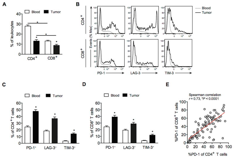
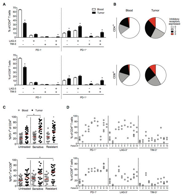
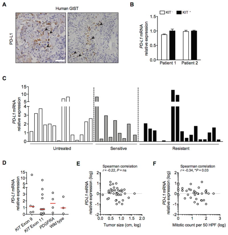
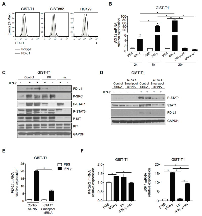
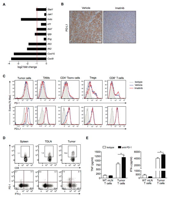
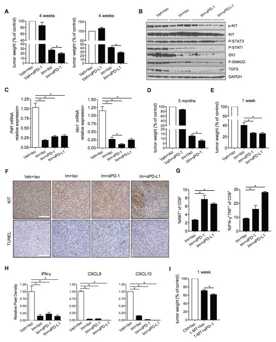
Results: The inhibitory receptors PD-1, lymphocyte activation gene 3, and T-cell immunoglobulin mucin-3 were upregulated on tumor-infiltrating T cells compared with T cells from matched blood. PD-1 expression on T cells was highest in imatinib-treated human GISTs. Meanwhile, intratumoral PD-L1 expression was variable. In human GIST cell lines, treatment with imatinib abrogated the IFNγ-induced upregulation of PD-L1 via STAT1 inhibition. In KitV558Δ/+ mice, imatinib downregulated IFNγ-related genes and reduced PD-L1 expression on tumor cells. PD-1 and PD-L1 blockade in vivo each had no efficacy alone but enhanced the antitumor effects of imatinib by increasing T-cell effector function in the presence of KIT and IDO inhibition.

Conclusions: PD-1/PD-L1 blockade is a promising strategy to improve the effects of targeted therapy in GISTs. Collectively, our results provide the rationale to combine these agents in human GISTs. Clin Cancer Res; 23(2); 454-65. ©2016 AACR.


Conflict of interest statement

The authors have no financial conflict of interest.

Figures

Figure 1
Figure 1. T cells infiltrating human GIST express high levels of inhibitory receptors compared with blood
(A) CD4+ and CD8+ T cells as a percentage of leukocytes (CD45+ cells) in matched blood and tumor specimens (n = 106) from 85 human GIST patients. (B) Histograms of PD-1, LAG-3, and TIM-3 expression on CD4+ and CD8+ T cells in blood (gray), and tumor (black) from a representative sample. PD-1+, LAG-3+, and TIM-3+ cells as percentage of CD4+ (C) and CD8+ T cells (D) from matched peripheral blood and tumor samples. (E) Linear regression analysis between PD-1 expression on CD4+ and CD8+ T cells in 106 human GIST specimens. Data represent mean ± SEM *P < 0.05.
Figure 2
Figure 2. Patterns of PD-1 expression in T cells in human GIST
(A) Expression of individual or the combination of inhibitory receptors (PD-1, LAG-3, TIM-3) on CD4+ and CD8+ T cells in 63 human GIST specimens by Boolean gate analysis. Data represent mean ± SEM *P < 0.05. (B) Total number of inhibitory receptors expressed by CD4+ (top) and CD8+ T cells (bottom) from the blood (left) and tumor (right). (C) Percentage of PD-1+ cells among CD4+ (top) and CD8+ T cells (bottom) in blood and tumor from untreated (n = 36), imatinib-sensitive (n = 38), and imatinib-resistant (n = 32) GIST specimens. Each dot represents a separate specimen. Median values are marked by a horizontal red line. *P < 0.05. (D) Expression of inhibitory receptors on tumor-infiltrating T cells from GIST patients with multiple metastases (n = 10). Each dot represents a separate tumor specimen.
Figure 3
Figure 3. PD-L1 is expressed in a subset of human GISTs
(A) Representative immunohistochemistry with anti-PD-L1 (clone 5H1) shows membranous and cytoplasmic expression in human GIST (indicated by black arrow; scale bar 50 μm). (B) Freshly isolated KIT- (stroma) and KIT+ (tumor) cells from 2 human GISTs were analyzed for PD-L1 mRNA by real-time PCR. Bars represent mean ± SEM. (C) RNA was isolated from fresh frozen untreated (n = 14), imatinib-sensitive (n = 9), and imatinib-resistant (n = 18) human GISTs and analyzed for PD-L1 mRNA using real-time PCR. Bars represent means. (D) PD-L1 mRNA relative expression in 25 GIST samples with confirmed mutation. Median values are marked by a horizontal red line. (E) Scatter plots to analyze the correlation of PD-L1 mRNA with tumor size and (F) mitotic count. Values are log-transformed.
Figure 4
Figure 4. Imatinib abrogates IFN-γ–induced upregulation of PD-L1 on human GIST cell lines
(A) PD-L1 expression in human GIST cell lines as determined by flow cytometry. Gray histograms represent isotype controls. (B) GIST-T1 cells were treated with either PBS, IFN-γ (100 ng/ml), IFN-γ plus the JAK inhibitor tetracyclic pyridine 6 (P6; 1 μM), or IFN-γ plus imatinib (Im; 100 nM). PD-L1 mRNA relative expression was determined at the indicated time points by real-time PCR. (C) Western blot of GIST-T1 treated as described above for 20 h. (D) GIST-T1 cells were transfected with non-target control siRNA or ON-TARGET plus SMARTpool siRNA (STAT1 siRNA) and then treated with PBS or IFN-γ (100 ng/ml) for 6 h. Western blot analysis demonstrating efficiency of STAT1 knockdown and PD-L1 expression. (E) PD-L1 expression in GIST-T1 cells was measured by PCR. (F) GIST-T1 cells were analyzed for IFNGR1 and IRF1 by real-time PCR after 6 h of the indicated treatment. Data are normalized to control. Data represent mean ± SEM *P < 0.05.
Figure 5
Figure 5. Imatinib modulates IFN-γ–related genes and PD-L1 expression in GIST
(A) Bar graph showing mRNA levels of IFN-γ–related genes determined by microarray analysis of tumors from KitV558Δ /+ mice treated with imatinib for 1 week. Data are shown as log2 fold change compared to vehicle-treated KitV558Δ /+ mice (3 per group). Bars represent mean. (B) Representative immunohistochemistry with anti-PD-L1 of tumors from KitV558Δ /+ mice treated with vehicle or imatinib for 4 weeks (scale bar, 100 μm). (C) Histograms of PD-1 and PD-L1 expression on tumor cells (CD45-Kit+), tumor-associated macrophages (TAMs, CD45+F4/80+CD11b+) and T cell subsets (CD4+ Tconv cells: CD45+CD3+CD4+Foxp3-, Tregs: CD45+CD3+CD4+CD25+Foxp3+, and CD8+ T cells: CD45+CD3+CD8+) in GIST-bearing KitV558Δ /+ mice treated with vehicle or imatinib for 1 week (3–5 per group). Representative plots are depicted with isotype control (gray), vehicle (blue) and imatinib (red). (D) Expression of IFN-γ in stimulated CD8+ T cells from spleens, tumor-draining lymph nodes (TdLNs), and tumors from untreated KitV558Δ /+ mice. CD8+ T cells from the indicated tissues were isolated, stimulated in vitro with PMA and ionomycin, then analyzed by flow cytometry. (E) Effect of PD-1 blockade on cytokine production in T cells. T cells were harvested from the mesenteric lymph node (mLN) from B6 mice (WT) or the tumor from untreated KitV558Δ /+ mice and cultured in vitro in the presence of anti-PD-1 (10 μg/ml) or isotype control. After 48 h, culture supernatant was collected and cytokines were measured by cytometric bead array. Data represent mean ± SEM *P < 0.05.
Figure 6
Figure 6. PD-1/PD-L1 blockade enhances the antitumor effects of imatinib in murine GIST
(A) Tumor weights from KitV558Δ /+ mice (3–5 per group) treated with imatinib (Im) or vehicle (Veh) for 4 weeks. Anti-PD-1 or anti-PD-L1 antibodies or their isotypes (Iso) were given on days 0, 4, 8, and 12. Mice were sacrificed at day 28. (B) Western blot of tumor samples from KitV558Δ /+ mice treated as in A. (C) Bulk tumors were analyzed for expression of Pdl1 and Ido1 by real-time PCR. Data are normalized to control. Data represent mean of at least 3 samples ± SEM *P < 0.05. (D) Tumor weights after 3 months of treatment (3–5 mice per group). Antibodies or isotypes were given as in A. Mice were sacrificed at day 90. (E) Tumor weights from KitV558Δ /+ mice (3–5 per group) treated with imatinib for 1 week. Anti-PD-1 or anti-PD-L1 antibodies were given on days 0, 2, 4, and 6. Mice were sacrificed at day 7. (F) Tumor KIT and TUNEL staining after 1 week of treatment. Scale bars, 50μm. (G) Percentages of intratumoral CD8+ T cells that were Ki67+ or IFN-γ/TNF–producing intratumoral CD8+ T cells after in vitro stimulation with PMA and ionomycin. Data represent means of 3–4 samples ± SEM *P < 0.05. (H) Cytokine array densitometry of tumors from KitV558Δ /+ mice after 1 week of treatment. (I) Tumor weights from KitV558Δ /+ mice treated with 1-MT for 1 week (3–5 per group). Anti-PD-1 antibody was given as in E. Mice were sacrificed at day 7. All tumor weights are shown normalized to vehicle control.

Similar articles

Cited by

References

    1. Hirota S, Isozaki K, Moriyama Y, Hashimoto K, Nishida T, Ishiguro S, et al. Gain-of-function mutations of c-kit in human gastrointestinal stromal tumors. Science. 1998;279:577–80. - PubMed
    1. Heinrich MC, Corless CL, Duensing A, McGreevey L, Chen CJ, Joseph N, et al. PDGFRA activating mutations in gastrointestinal stromal tumors. Science. 2003;299:708–10. - PubMed
    1. Joensuu H, DeMatteo RP. The management of gastrointestinal stromal tumors: a model for targeted and multidisciplinary therapy of malignancy. Annu Rev Med. 2012;63:247–58. - PMC - PubMed
    1. Blanke CD, Rankin C, Demetri GD, Ryan CW, von Mehren M, Benjamin RS, et al. Phase III randomized, intergroup trial assessing imatinib mesylate at two dose levels in patients with unresectable or metastatic gastrointestinal stromal tumors expressing the kit receptor tyrosine kinase: S0033. J Clin Oncol. 2008;26:626–32. - PubMed
    1. Verweij J, Casali PG, Zalcberg J, LeCesne A, Reichardt P, Blay JY, et al. Progression-free survival in gastrointestinal stromal tumours with high-dose imatinib: randomised trial. Lancet. 2004;364:1127–34. - PubMed

MeSH terms