. 2016 Sep 23;291(39):20539-50.
doi: 10.1074/jbc.M116.745265. Epub 2016 Jul 28.

Molecular Basis of Chemokine CXCL5-Glycosaminoglycan Interactions

Affiliations

Molecular Basis of Chemokine CXCL5-Glycosaminoglycan Interactions

Krishna Mohan Sepuru et al. J Biol Chem. .

Abstract

Chemokines, a large family of highly versatile small soluble proteins, play crucial roles in defining innate and adaptive immune responses by regulating the trafficking of leukocytes, and also play a key role in various aspects of human physiology. Chemokines share the characteristic feature of reversibly existing as monomers and dimers, and their functional response is intimately coupled to interaction with glycosaminoglycans (GAGs). Currently, nothing is known regarding the structural basis or molecular mechanisms underlying CXCL5-GAG interactions. To address this missing knowledge, we characterized the interaction of a panel of heparin oligosaccharides to CXCL5 using solution NMR, isothermal titration calorimetry, and molecular dynamics simulations. NMR studies indicated that the dimer is the high-affinity GAG binding ligand and that lysine residues from the N-loop, 40s turn, β3 strand, and C-terminal helix mediate binding. Isothermal titration calorimetry indicated a stoichiometry of two oligosaccharides per CXCL5 dimer. NMR-based structural models reveal that these residues form a contiguous surface within a monomer and, interestingly, that the GAG-binding domain overlaps with the receptor-binding domain, indicating that a GAG-bound chemokine cannot activate the receptor. Molecular dynamics simulations indicate that the roles of the individual lysines are not equivalent and that helical lysines play a more prominent role in determining binding geometry and affinity. Further, binding interactions and GAG geometry in CXCL5 are novel and distinctly different compared with the related chemokines CXCL1 and CXCL8. We conclude that a finely tuned balance between the GAG-bound dimer and free soluble monomer regulates CXCL5-mediated receptor signaling and function.

Keywords: NMR; glycobiology; heparin; isothermal titration calorimetry (ITC); molecular dynamics.


Figures

FIGURE 1.
FIGURE 1.
A and B, ribbon representation of the CXCL5 dimer (A) and single monomeric unit (B) of the dimer structures (PDB code 2MGS). The individual monomers in the dimer are shown in dark and light blue for clarity. C-helix, N-loop and 40s-turn residues are highlighted in the monomer structure. C, sequences of ELR chemokines. GAG binding residues in CXCL5 and the corresponding conserved basic residues in related chemokines are highlighted in red. The ELR motif is highlighted in green.
FIGURE 2.
FIGURE 2.
The dimer is the high-affinity GAG ligand. A section of the 1H,15N HSQC spectra showing the overlay of CXCL5 in the free (black) and GAG-bound (red) forms. Dimer (D) and monomer (M) peaks are indicated. The monomer peaks disappear on dp14 binding, indicating that the dimer is the high-affinity GAG ligand. The spectra were collected using an 8 μm CXCL5 sample in 50 mm sodium phosphate (pH 7.5) at 40 °C.
FIGURE 3.
FIGURE 3.
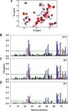
Binding of CXCL5 to heparin oligosaccharides. A, sections of the 1H,15N HSQC spectra showing the overlay of CXCL5 in the free (black) and dp14-bound (red) forms. Arrows indicate the direction of the peak movement. B–D, histograms of chemical shift changes in the CXCL5 dimer on binding heparin dp14 (B), dp8 (C), and dp4 (D). The basic residues Lys and His are shown in blue, and buried/acidic residues are shown in red. The horizontal lines at 0.19 ppm for dp14/dp8 and 0.10 ppm for dp4 represent the cutoff for a residue to be considered perturbed. The spectra were collected using a 100 μm CXCL5 sample in 50 mm sodium phosphate (pH 6.0) at 25 °C.
FIGURE 4.
FIGURE 4.
The molecular surface of the CXCL5 dimer affected on GAG binding. A and B, residues implicated in GAG binding from NMR CSP experiments. C, the location of these residues in one monomer of the dimer structure. Residues that are perturbed on GAG binding are painted on one monomer in dark blue and on the other monomer in red.
FIGURE 5.
FIGURE 5.
Backbone dynamics of the CXCL5-heparin complex. Comparison of 15N,1H NOE values for CXCL5 in the free (black) and dp14-bound (red) forms. The data show that the N-loop and C-helical residues have higher NOE values compared with the free form. The spectra were collected using a 150 μm CXCL5 sample in 50 mm sodium phosphate (pH 6.0) at 25 °C.
FIGURE 6.
FIGURE 6.
Isothermal titration calorimetry. A–C, isotherms corresponding to the binding of dp14 (A), dp8 (B), and dp4 (C) to the CXCL5 dimer. The raw data of the titrations are shown in the top panels, and the bottom panels show the integrated data obtained after subtracting the heat of dilution. The titrations were performed in 50 mm sodium phosphate (pH 6.0) at 25 °C.
FIGURE 7.
FIGURE 7.
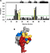
Binding of CXCL5 to the CXCR2 N-terminal domain. A, histogram of chemical shift changes in the CXCL5 dimer on binding the CXCR2 N-terminal domain peptide. The horizontal line at 0.15 ppm represents the cutoff for a residue to be considered perturbed. Residues that are involved in both GAG and receptor binding are highlighted in yellow. The spectra were collected using a 70 μm CXCL5 sample in 50 mm sodium phosphate (pH 5.7) at 25 °C. B, surface representation showing GAG binding (blue), receptor binding (red), and shared binding (yellow) regions. For receptor interactions, residues that showed significant CSP and are solvent-exposed and N-terminal Glu10, Leu11, and Arg12 (of the ELR motif) are considered involved in binding.
FIGURE 8.
FIGURE 8.
CXCL5-GAG binding models. Shown are structural depictions of model I (A–C) and model II (D–F). A and D, the CXCL5 dimer is shown in ribbon representation and GAG as sticks. B and E, GAG-binding residues are shown in light and dark blue and labeled in both monomers of the dimer. C and F, GAG-binding residues are labeled on the electrostatic CXCL5 surface. G, GAG can bind both monomers of the dimer without steric clashes. The CXCL5 dimer is shown in ribbon representation and GAG as sticks. GAG binding residues are highlighted in blue.
FIGURE 9.
FIGURE 9.
Binding properties and energetics from MD simulations. A, snapshots from two time points of CXCL5-GAG simulations showing switching hydrogen-bond partners between Lys25 side chain NH3+ and GAG chain sulfate moieties, illustrating the dynamic interplay. B, occupancy of intermolecular H-bonds for GAG-binding residues. C, water-mediated H-bonding interactions of GAG-binding residues. D, single-residue energy decomposition values for CXCL5-heparin interactions.
FIGURE 10.
FIGURE 10.
Models of CXCL5-heparan sulfate interactions. A, proposed clamp model showing that CXCL5 is sandwiched between NS domains of a single heparan sulfate chain. B, model showing CXCL1 dimer binding to heparan sulfate.

References

    1. Bonecchi R., Galliera E., Borroni E. M., Corsi M. M., Locati M., and Mantovani A. (2009) Chemokines and chemokine receptors: an overview. Front. Biosci. 14, 540–551 - PubMed
    1. Griffith J. W., Sokol C. L., and Luster A. D. (2014) Chemokines and chemokine receptors: positioning cells for host defense and immunity. Annu. Rev. Immunol. 32, 659–702 - PubMed
    1. Fernandez E. J., and Lolis E. (2002) Structure, function, and inhibition of chemokines. Annu. Rev. Pharmacol. Toxicol. 42, 469–499 - PubMed
    1. Zlotnik A., and Yoshie O. (2000) Chemokines: a new classification system and their role in immunity. Immunity 12, 121–127 - PubMed
    1. Stillie R., Farooq S. M., Gordon J. R., and Stadnyk A. W. (2009) The functional significance behind expressing two IL-8 receptor types on PMN. J. Leukocyte Biol. 86, 529–543 - PubMed

MeSH terms