Granular Formation during Apoptosis in Blastocystis sp. Exposed to Metronidazole (MTZ)
- PMID: 27471855
- PMCID: PMC4966910
- DOI: 10.1371/journal.pone.0155390
Granular Formation during Apoptosis in Blastocystis sp. Exposed to Metronidazole (MTZ)
Abstract
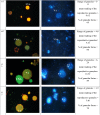
The role and function of the granular life cycle stage in Blastocystis sp, remains uncertain despite suggestions being made that the granules are metabolic, reproductive and lipid in nature. This present study aims to understand granular formation by triggering apoptosis in Blastocystis sp. by treating them with metronidazole (MTZ). Blastocystis sp.cultures of 4 sub-types namely 1, 2, 3 and 5 when treated with 0.01 and 0.0001 mg/ml of metronidazole (MTZ) respectively showed many of the parasites to be both viable and apoptotic (VA). Treated subtype 3 isolates exhibited the highest number of granular forms i.e. 88% (p<0.001) (0.0001 mg/ml) and 69% (p<0.01) (0.01 mg/ml) respectively at the 72 h in in vitro culture compared to other subtypes. These VA forms showed distinct granules using acridine orange (AO) and 4',6-diamino-2-phenylindole (DAPI) staining with a mean per cell ranging from 5 in ST 5 to as high as 16 in ST 3. These forms showed intact mitochondria in both viable apoptotic (VA) and viable non-apoptotic (VNA) cells with a pattern of accumulation of lipid droplets corresponding to viable cells. Granular VA forms looked ultra-structurally different with prominent presence of mitochondria-like organelle (MLO) and a changed mitochondrial trans-membrane potential with thicker membrane and a highly convoluted inner membrane than the less dense non-viable apoptotic (NVA) cells. This suggests that granular formation during apoptosis is a self-regulatory mechanism to produce higher number of viable cells in response to treatment. This study directs the need to search novel chemotherapeutic approaches by incorporating these findings when developing drugs against the emerging Blastocystis sp. infections.
Conflict of interest statement
Figures


Similar articles
-
Comparison of apoptotic responses in Blastocystis sp. upon treatment with Tongkat Ali and Metronidazole.Sci Rep. 2021 Apr 9;11(1):7833. doi: 10.1038/s41598-021-81418-x. Sci Rep. 2021. PMID: 33837230 Free PMC article.
-
Apoptosis in Blastocystis spp. is related to subtype.Trans R Soc Trop Med Hyg. 2012 Dec;106(12):725-30. doi: 10.1016/j.trstmh.2012.08.005. Epub 2012 Nov 8. Trans R Soc Trop Med Hyg. 2012. PMID: 23141370
-
Increase number of mitochondrion-like organelle in symptomatic Blastocystis subtype 3 due to metronidazole treatment.Parasitol Res. 2016 Jan;115(1):391-6. doi: 10.1007/s00436-015-4760-0. Epub 2015 Oct 20. Parasitol Res. 2016. PMID: 26481491
-
Blastocystis sp., Parasite Associated with Gastrointestinal Disorders: An Overview of its Pathogenesis, Immune Modulation and Therapeutic Strategies.Curr Pharm Des. 2018;24(27):3172-3175. doi: 10.2174/1381612824666180807101536. Curr Pharm Des. 2018. PMID: 30084327 Review.
-
Medicinal Plants as Natural Anti-Parasitic Agents Against Blastocystis Species.Recent Adv Antiinfect Drug Discov. 2023;18(1):2-15. doi: 10.2174/2772434418666221124123445. Recent Adv Antiinfect Drug Discov. 2023. PMID: 36424779 Review.
Cited by
-
Comparison of apoptotic responses in Blastocystis sp. upon treatment with Tongkat Ali and Metronidazole.Sci Rep. 2021 Apr 9;11(1):7833. doi: 10.1038/s41598-021-81418-x. Sci Rep. 2021. PMID: 33837230 Free PMC article.
-
In vitro susceptibility of human Blastocystis subtypes to simeprevir.Saudi J Biol Sci. 2021 Apr;28(4):2491-2501. doi: 10.1016/j.sjbs.2021.01.050. Epub 2021 Feb 2. Saudi J Biol Sci. 2021. PMID: 33935570 Free PMC article.
-
Higher amoebic and metronidazole resistant forms of Blastocystis sp. seen in schizophrenic patients.Parasit Vectors. 2022 Sep 5;15(1):313. doi: 10.1186/s13071-022-05418-0. Parasit Vectors. 2022. PMID: 36064639 Free PMC article.
-
Distinct Phenotypic Variation of Blastocystis sp. ST3 from Urban and Orang Asli Population-An Influential Consideration during Sample Collection in Surveys.Biology (Basel). 2022 Aug 12;11(8):1211. doi: 10.3390/biology11081211. Biology (Basel). 2022. PMID: 36009838 Free PMC article.
-
Production of bone mineral material and BMP-2 in osteoblasts cultured on double acid-etched titanium.Med Oral Patol Oral Cir Bucal. 2017 Sep 1;22(5):e651-e659. doi: 10.4317/medoral.22071. Med Oral Patol Oral Cir Bucal. 2017. PMID: 28809380 Free PMC article.
References
-
- Suresh K, Chong S, Howe J, Ho L, Ng G, Yap E, et al. (1995) Tubulovesicular elements in Blastocystis hominis from the caecum of experimentally-infected rats. International journal for parasitology 25: 123–126. - PubMed
-
- Vdovenko AA (2000) Blastocystis hominis: origin and significance of vacuolar and granular forms. Parasitology research 86: 8–10. - PubMed
-
- Ghaffar A (2010) Parasitology chapter one intestinal and luminal protozoa.
MeSH terms
Substances
LinkOut - more resources
Full Text Sources
Other Literature Sources

