. 2016 Jul 27;21(8):975.
doi: 10.3390/molecules21080975.

Corynoline Isolated from Corydalis bungeana Turcz. Exhibits Anti-Inflammatory Effects via Modulation of Nfr2 and MAPKs

Affiliations

Corynoline Isolated from Corydalis bungeana Turcz. Exhibits Anti-Inflammatory Effects via Modulation of Nfr2 and MAPKs

Chunjuan Yang et al. Molecules. .

Abstract

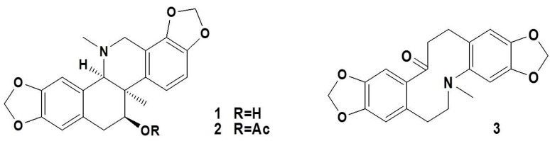
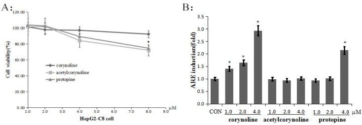
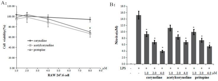
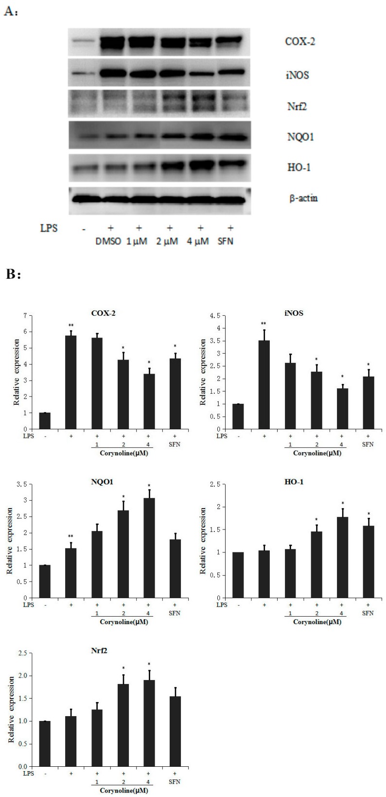
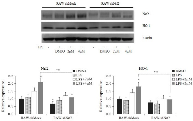
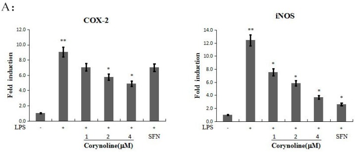
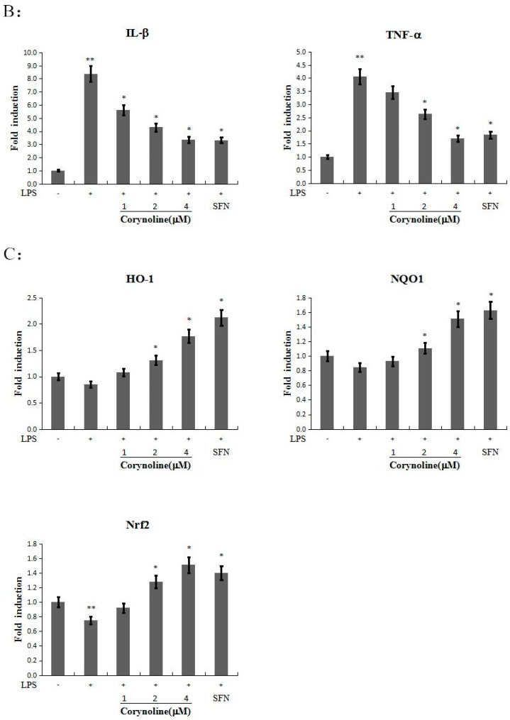
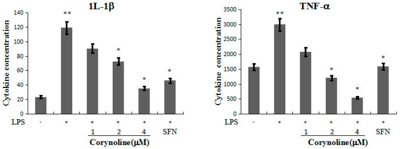
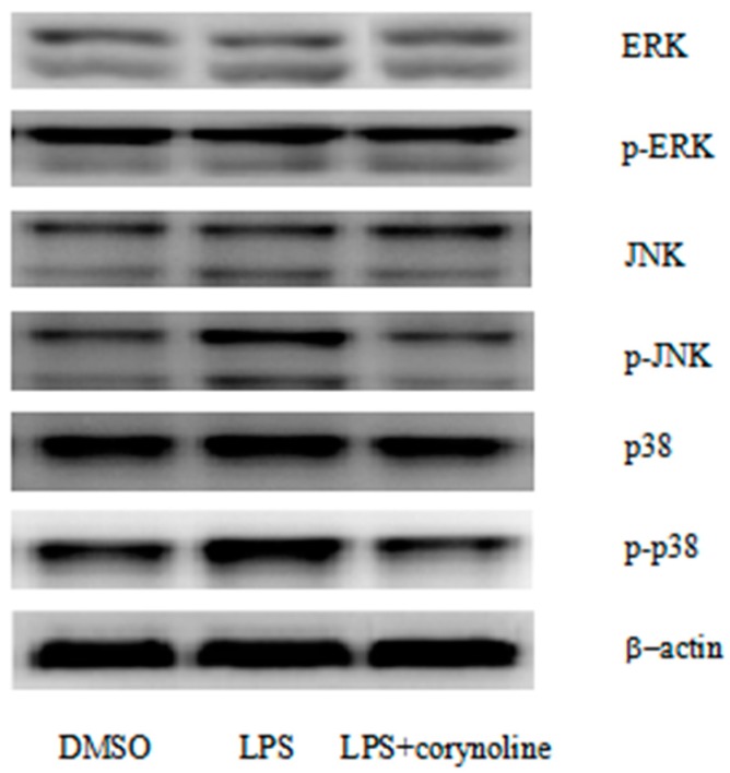
Corydalis bungeana Turcz. is an anti-inflammatory medicinal herb used widely in traditional Chinese medicine for upper respiratory tract infections. It is demonstrated that corynoline is its active anti-inflammatory component. The nuclear factor-erythroid-2-related factor 2 (Nrf2)/antioxidant response element (ARE) pathway and the mitogen-activated protein kinase (MAPK) pathway play important roles in the regulation of inflammation. In this study, we investigated the potential anti-inflammatory mechanism of corynoline through modulation of Nfr2 and MAPKs. Lipopolysaccharide (LPS)-activated RAW264.7 cells were used to explore modulatory role of NO production and the activation of signaling proteins and transcription factors using nitrite assay, Western bloting and qPCR. Treatment with corynoline reduced production of nitric oxide (NO) and the protein and mRNA levels of inducible nitric oxide (iNOS) and cyclooxygenase-2 (COX-2) Treatment also significantly increased the expression of Nrf2, quinone oxidoreductase 1 (NQO1) and hemeoxygenase-1 (HO-1) at the mRNA and protein levels, which demonstrated that corynoline may protect cells from inflammation through the Nrf2/ARE pathway In addition, corynoline suppressed the expression of inflammatory cytokines, such as tumor necrosis factor-α (TNF-α) and interleukin-1β (IL-1β), at the mRNA and protein levels. Furthermore, molecular data revealed that corynoline inhibited lipopolysaccharide-stimulated phosphorylation of c-jun NH2-terminal kinase (JNK) and p38. Taken together, these results suggest that corynoline reduces the levels of pro-inflammatory mediators, such as iNOS, COX-2, TNF-α and IL-1β, by suppressing extracellular signal-regulated kinase 1/2 (ERK) and p38 phosphorylation in RAW264.7 cells, which is regulated by the Nrf2/ARE pathway. These findings reveal part of the molecular basis for the anti-inflammatory properties of corynoline.

Keywords: MAPKs; Nrf2; anti-inflammation; corynoline.


Conflict of interest statement

The authors do not have conflict of interest to declare.

Figures

Figure 1
Figure 1
The structures of corynoline (1), acetylcorynoline (2), and protopine (3).
Figure 2
Figure 2
Corynoline, acetylcorynoline and protopine inhibit cell growth and induce ARE activity in HepG2-C8-ARE luciferase cells. (A) Inhibition of cell growth by corynoline, acetylcorynoline and protopine. For these experiments, HepG2-C8-ARE luciferase cells were seeded onto a 96-well plate and incubated with different concentrations of corynoline, acetylcorynoline or protopine or with DMSO as the vehicle control, for 24 h. MTS reagent was added to each well, and the absorbance of the formazan product was read at 490 nm; (B) Induction of ARE activity in HepG2-C8-ARE luciferase cells. The human hepatoma HepG2-C8-ARE luciferase cells were seeded onto a 96-well plate and treated with different concentrations (1, 2 or 4 μM) of corynoline, acetylcorynoline or protopine for 24 h. The results are expressed as the mean ± SE; * p < 0.05 compared with the vehicle group.
Figure 3
Figure 3
Corynoline, acetylcorynoline and protopine inhibit cell growth and LPS-induced NO production in Raw264.7 cells. (A) Inhibition of cell growth by corynoline, acetylcorynoline and protopine. For these experiments, RAW264.7 cells were seeded onto a 96-well plate and were incubated with different concentrations of corynoline, acetylcorynoline or protopine or with DMSO as the vehicle control, for 24 h. MTS reagent was added to each well, and the absorbance of the formazan product was read at 490 nm; (B) Inhibition of LPS-induced NO production by corynoline, acetylcorynoline and protopine. RAW264.7 cells were seeded onto a 96-well plate and treated with different concentrations of corynoline, acetylcorynoline or protopine with LPS (150 ng/mL) for 24 h. The isolated supernatant fractions were mixed with an equal volume of Griess reagent and incubated at room temperature for 10 min. Nitrite production was measured by reading the absorbance at 540 nm. Each point represents the mean ± SE; * p < 0.05 compared with the LPS-treated group.
Figure 4
Figure 4
Effect of corynoline on the expression of the iNOS, COX-2, HO-1, NQO1 and Nrf2 proteins in LPS-induced RAW264.7 cells. (A) RAW264.7 cells were treated with LPS (150 ng/mL) alone or in combination with corynoline or SFN (each at 1.0 μM). The cells were harvested, and the total protein was extracted at 24 h; the protein levels were measured by Western blotting; (B) Corynoline suppressed the LPS-induced expression of iNOS and COX-2 and increased the LPS-induced expression of HO-1, NQO1 and Nrf2. The bands were densitometrically analyzed using ImageJ software (National Institutes of Health, New York, NY, USA, version 1.50a, http://rsbweb.nih.gov/ij). The relative protein expression levels were calculated and compared with those of the control, which were set to 100%. ** p < 0.05 compared with the vehicle control. * p < 0.05 compared with the LPS-treated group.
Figure 5
Figure 5
Effect of corynoline on the protein expression of Nrf2 and HO-1 in RAW-shMock and RAW-shNrf2 cells. Cells were incubated with various concentrations of corynoline (2 and 4 μM) for 5 days. The protein levels were measured by protein lysate preparation and western blotting, as described in the Materials and Methods section. The relative expression levels were quantified based on the signal intensity of the corresponding bands from three independent experiments and were normalized using β-actin. The graphical data are presented as the mean SD from three independent experiments. * represent p < 0.05, respectively, which indicated significant differences in the target proteins compared with their levels in RAW-shMock cells without corynoline treatment. ** also represent p < 0.05, which indicated statistical significance between RAW-shMock and RAW-shNrf2 cells.
Figure 6
Figure 6
Effect of corynoline on LPS-induced mRNA expression for iNOS, COX-2, IL-1β, TNF-α, HO-1, Nrf2 and NQO1. RAW264.7 cells were treated with LPS (150 ng/mL) either alone or were treated with LPS and corynoline or LPS and SFN (each at 1.0 μM). The cells were harvested for total RNA extraction at 24 h, and the mRNA levels for iNOS, COX-2, IL-1β, TNF-α, HO-1, Nrf-2 and NQO1 were measured using quantitative RT-PCR. Each point represents the mean ± SE; ** p < 0.05 as compared to the vehicle control. * p < 0.05 as compared to the LPS-treated group. (A) Corynoline down-regulates the expression of iNOS, COX-2 at mRNA levels; (B) Corynoline down-regulates the expression of 1L-1β,TNF-α at mRNA levels; (C) Corynoline up-regulates the expression of HO-1,Nrf-2 and NQO1 in LPS-induced RAW264.7 cells at mRNA levels.
Figure 6
Figure 6
Effect of corynoline on LPS-induced mRNA expression for iNOS, COX-2, IL-1β, TNF-α, HO-1, Nrf2 and NQO1. RAW264.7 cells were treated with LPS (150 ng/mL) either alone or were treated with LPS and corynoline or LPS and SFN (each at 1.0 μM). The cells were harvested for total RNA extraction at 24 h, and the mRNA levels for iNOS, COX-2, IL-1β, TNF-α, HO-1, Nrf-2 and NQO1 were measured using quantitative RT-PCR. Each point represents the mean ± SE; ** p < 0.05 as compared to the vehicle control. * p < 0.05 as compared to the LPS-treated group. (A) Corynoline down-regulates the expression of iNOS, COX-2 at mRNA levels; (B) Corynoline down-regulates the expression of 1L-1β,TNF-α at mRNA levels; (C) Corynoline up-regulates the expression of HO-1,Nrf-2 and NQO1 in LPS-induced RAW264.7 cells at mRNA levels.
Figure 7
Figure 7
Inhibitory activity of corynoline on LPS–induced protein expression for 1L-1β, TNF-α. RAW264.7 cells were treated with LPS (150 ng/mL) either alone or were treated with LPS and corynoline or LPS and SFN (each at 1.0 μM). The cytokine concentrations were normalized to the protein concentrations. IL-1β and TNF-α levels were measured using an ELISA kit, and the total protein concentrations were measured using a BCA (bicinchoninic acid) protein assay. Each point represents the mean ± SE; ** p < 0.05 as compared to the vehicle control. * p < 0.05 as compared to the LPS-treated group.
Figure 8
Figure 8
Evaluation of the different cellular signaling pathways affected by corynoline in LPS-induced RAW264.7 cells. Corynoline effectively blocked the activation of p38 MAPK and JNK signaling. RAW264.7 cells were treated with LPS (150 ng/mL) alone or in combination with corynoline (1.0 μM). The cells were harvested, and the proteins were measured after 24 h.

References

    1. Philip M., Rowley D.A., Schreiber H. Inflammation as a tumor promoter in cancer induction. Semin. Cancer Biol. 2004;14:433–439. doi: 10.1016/j.semcancer.2004.06.006. - DOI - PubMed
    1. Aggarwal B.B., Shishodia S., Sandur S.K., Pandey M.K., Sethi G. Inflammation and cancer: How hot is the link? Biochem. Pharmacol. 2006;72:1605–1621. doi: 10.1016/j.bcp.2006.06.029. - DOI - PubMed
    1. Mantovani A. Cancer: Inflammation by remote control. Nature. 2005;435:752–753. doi: 10.1038/435752a. - DOI - PubMed
    1. Coussens L.M., Werb Z. Inflammation and cancer. Nature. 2002;420:860–867. doi: 10.1038/nature01322. - DOI - PMC - PubMed
    1. Balkwill F.R., Mantovani A. Cancer-related inflammation: Common themes and therapeutic opportunities. Semin. Cancer Biol. 2012;22:33–40. doi: 10.1016/j.semcancer.2011.12.005. - DOI - PubMed

MeSH terms