. 2016 Jul 29;11(7):e0159829.
doi: 10.1371/journal.pone.0159829. eCollection 2016.

Novel Method for Differentiating Histological Types of Gastric Adenocarcinoma by Using Confocal Raman Microspectroscopy

Affiliations

Novel Method for Differentiating Histological Types of Gastric Adenocarcinoma by Using Confocal Raman Microspectroscopy

Chih-Wei Hsu et al. PLoS One. .

Abstract

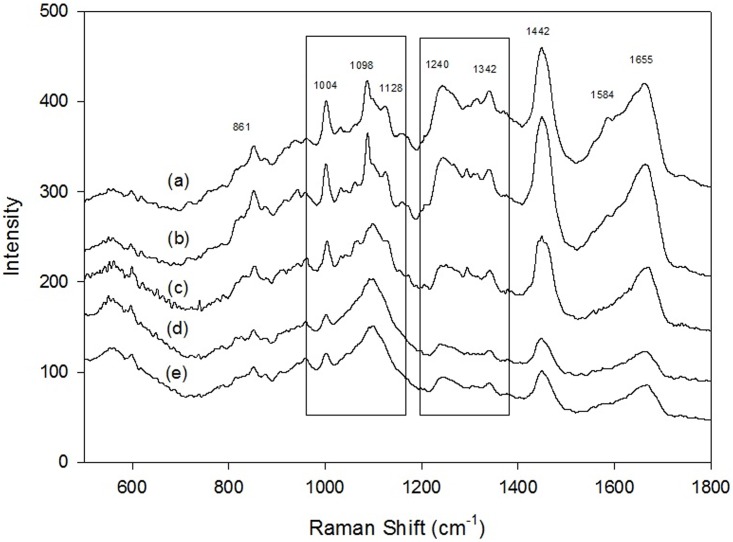
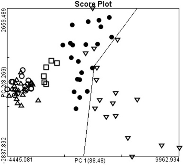
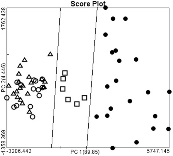
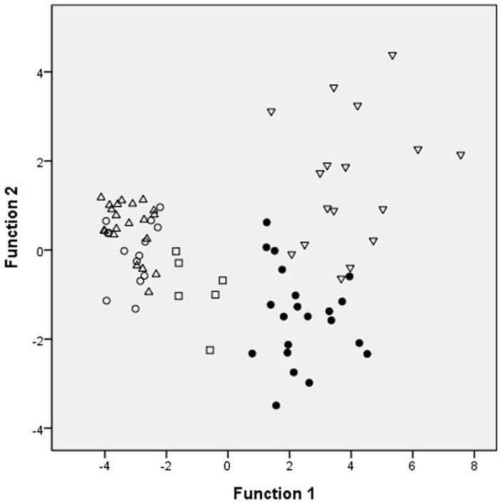
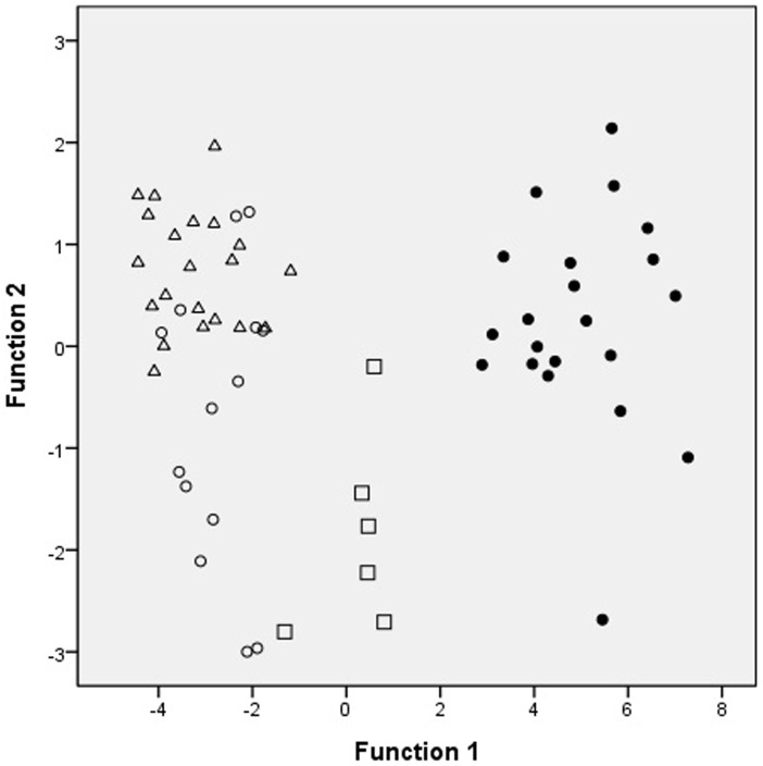
Gastric adenocarcinoma, a single heterogeneous disease with multiple epidemiological and histopathological characteristics, accounts for approximately 10% of cancers worldwide. It is categorized into four histological types: papillary adenocarcinoma (PAC), tubular adenocarcinoma (TAC), mucinous adenocarcinoma (MAC), and signet ring cell adenocarcinoma (SRC). Effective differentiation of the four types of adenocarcinoma will greatly improve the treatment of gastric adenocarcinoma to increase its five-year survival rate. We reported here the differentiation of the four histological types of gastric adenocarcinoma from the molecularly structural viewpoint of confocal Raman microspectroscopy. In total, 79 patients underwent laparoscopic or open radical gastrectomy during 2008-2011: 21 for signet ring cell carcinoma, 21 for tubular adenocarcinoma, 14 for papillary adenocarcinoma, 6 for mucinous carcinoma, and 17 for normal gastric mucosas obtained from patients underwent operation for other benign lesions. Clinical data were retrospectively reviewed from medical charts, and Raman data were processed and analyzed by using principal component analysis (PCA) and linear discriminant analysis (LDA). Two-dimensional plots of PCA and LDA clearly demonstrated that the four histological types of gastric adenocarcinoma could be differentiated, and confocal Raman microspectroscopy provides potentially a rapid and effective method for differentiating SRC and MAC from TAC or PAC.


Conflict of interest statement

Competing Interests: The authors have declared that no competing interests exist.

Figures

Fig 1
Fig 1. Raman spectra, in the range of 550–1800 cm-1, of gastric adenocarcinomas.
(a) normal gastric mucosa; (b) signet ring cell adenocarcinoma; (c) mucinous adenocarcinoma; (d) tubular adenocarcinoma; (e) papillary adenocarcinoma.
Fig 2
Fig 2. The two-dimensional plots of gastric adenocarcinomas and normal gastric mucosa.
The first two calculated principal components (PC1 and PC2), which contained most of the information, were plotted against each other for visualization. The PC1 accounting for the largest Raman spectra variance was 88.48%, and PC2 was 8.26%. The two-dimensional plots showed the separation of gastric adenocarcinoma from normal gastric mucosa (▽: normal gastric mucosa; ●: signet ring cell adenocarcinoma; □: mucinous adenocarcinoma; Δ: tubular tubular adenocarcinoma; ○: papillary adenocarcinoma).
Fig 3
Fig 3. The two-dimensional plots of gastric adenocarcinomas.
The first two calculated principal components (PC1 and PC2), which contained most of the information, were plotted against each other for visualization. The PC1 accounting for the largest Raman spectra variance was 89.85%, and PC2 was 4.45%. The two-dimensional plots showed the separation of signet ring cell adenocarcinoma, mucinous adenocarcinoma, from and tubular carcinoma and papillary adenocarcinoma (●: signet ring cell adenocarcinoma; □: mucinous adenocarcinoma; Δ: tubular adenocarcinoma; ○: papillary adenocarcinoma).
Fig 4
Fig 4. The two-dimensional plots of gastric adenocarcinomas and normal gastric mucosa.
Scatter plots of the first and second important discriminant function plotted against one another. The two-dimensional plots showed the separation of gastric adenocarcinoma from normal gastric mucosa (▽: normal gastric mucosa; ●: signet ring cell adenocarcinoma; □: mucinous adenocarcinoma; Δ: tubular tubular adenocarcinoma; ○: papillary adenocarcinoma).
Fig 5
Fig 5. The two-dimensional plots of gastric adenocarcinomas.
Scatter plots of the first and second important discriminant function plotted against one another. The two-dimensional plots showed the separation of signet ring cell adenocarcinoma, mucinous adenocarcinoma, from and tubular carcinoma and papillary adenocarcinoma (●: signet ring cell adenocarcinoma; □: mucinous adenocarcinoma; Δ: tubular adenocarcinoma; ○: papillary adenocarcinoma).

References

    1. Ferlay J, Bray F, Pisani P, Parkin M. Cancer incidence, Mortality, and Prevalence Worldwide Globocan 2002. Lyon: IARC, 2004.
    1. Shah MA, Khanin R, Tang L, Janjigian YY, Klimstra DS, Gerdes H, et al. Molecular classification of gastric cancer: a new paradigm. Clin Cancer Res. 2011;17:2693–2701. 10.1158/1078-0432.CCR-10-2203 - DOI - PMC - PubMed
    1. Watanabe H, Jass JR, Sobin LH:Histological typing of esophageal and gastric tumors: WHO international histological classification of tumors. 2nd edition Berlin: Springer; 1990.
    1. Lee HH, Song KY, Park CH, Jeon HM. Undifferentiated-type gastric adenocarcinoma: prognostic impact of three histological types. World J Surg Oncol. 2012; 10:254 10.1186/1477-7819-10-254 - DOI - PMC - PubMed
    1. Zheng HC, Zheng YS, Xia P, Xu XY, Xing YN, Takahashi H, et al. The pathobiological behaviors and prognosis associated with Japanese gastric adenocarcinomas of pure WHO histological subtypes. Histol Histopathol. 2010;25:445–452. - PubMed

MeSH terms

LinkOut - more resources