. 2016 Jul;95(30):e4348.
doi: 10.1097/MD.0000000000004348.

Epithelioid hemangioendothelioma in the thorax: Clinicopathologic, CT, PET, and prognostic features

Affiliations

Epithelioid hemangioendothelioma in the thorax: Clinicopathologic, CT, PET, and prognostic features

Jung Han Woo et al. Medicine (Baltimore). 2016 Jul.

Abstract

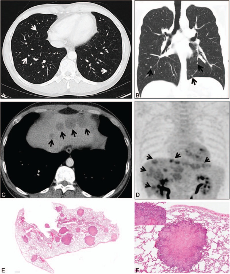
Little is known about prognostic factors in epithelioid hemangioendothelioma (EHE). We aimed to identify prognostic factors among various clinicopathologic and imaging features of thoracic EHEs.Forty-two patients (male:female = 20:22; median age, 49 years) of EHEs with (n = 19) and without (n = 23) thoracic involvement were included. We reviewed electronic medical records for clinical information and computed tomography (CT) features for thoracic involvement. Differences in demographics and survival outcomes of patients with and without thoracic involvement were assessed. We also estimated overall survival.The most common pattern of thoracic involvement was multiple pulmonary nodules (n = 10), followed by parenchymal tumor with pleural invasion (n = 4), reticulonodular opacities (n = 3), and diffuse pleural thickening (n = 2). No significant difference in survival was found between the thoracic EHE group and nonthoracic EHE group (P = 0.68). Among 4 different thoracic involvement types, the lung multinodular pattern tended to demonstrate longer median survival (8.5 months) than other patterns, whereas the shortest median survival (1 month) was observed for the nodule/mass with pleural involvement pattern (P = 0.038).CT manifestations of thoracic EHEs are classified into 4 patterns, of which lung multinodular pattern is associated with longer survival. Survival is not different between patients with and without thoracic involvement.


Conflict of interest statement

The authors have no conflicts of interest to disclose.

Figures

Figure 1
Figure 1
Epithelioid hemangioendothelioma of multiple nodular pattern in a 33-y-old man. (A and B) Lung window and coronal reformatted (2.0-mm section thickness) image shows multiple small well-defined nodules (arrows) in both lungs. (C) Mediastinal window image of enhanced CT scan (5.0-mm section thickness) demonstrates multiple variable-sized liver tumors (arrows) at the dome portion. (D) PET/CT image depicts high FDG uptake within multiple variable-sized liver tumors (arrows). Note scanty FDG uptake in bilateral lung nodules. (E) Low-magnification photomicrograph obtained from a different patient (22-y-old man) reveals multiple well-defined nodules. (F) Another low-magnification photomicrograph reveals nodule showing myxoid or hyalinized central core and cellular periphery. (Inset: High-magnification photomicrograph demonstrating tumor cells containing prominent cytoplasmic vacuoles and extending to alveolar spaces.) CT = computed tomography, FDG = fluorodeoxyglucose, PET = positron emission tomography.
Figure 2
Figure 2
Epithelioid hemangioendothelioma of multifocal areas of reticulonodular pattern in a 63-y-old man. (A) Lung window image of CT scan (2.5-mm section thickness) obtained at the level of aortic arches shows multiple poorly defined nodular lesions (arrows) together with irregular thickening of bronchovascular bundles in both lungs. (B) PET/CT image depicts somewhat higher FDG uptake within bilateral lung nodules (arrows) and liver tumors (arrowheads) than mediastinal blood pool and liver uptake, respectively. CT = computed tomography, FDG = fluorodeoxyglucose, PET = positron emission tomography.
Figure 3
Figure 3
Epithelioid hemangioendothelioma of diffuse pleural pattern in an 18-y-old man. (A) Lung window image of enhanced CT scan (5.0-mm section thickness) obtained at level of left inferior pulmonary vein shows mixed pleural tumor and effusion (arrows) in right pleural space. Also, note marked volume decrease in right hemithorax together with thickened ribs and narrowed intercostal spaces, suggesting the indolent nature of the disease process. (B) PET/CT image obtained at the level of right inferior pulmonary vein demonstrates increased FDG uptake (arrows) in pleural lesions. CT = computed tomography, FDG = fluorodeoxyglucose, PET = positron emission tomography.
Figure 4
Figure 4
Epithelioid hemangioendothelioma of parenchymal tumor(s) with pleural extension pattern in a 53-y-old woman. (A) Mediastinal window images of enhanced CT scan demonstrate subpleural lung tumor (arrow) and tumor pleural seeding (arrowheads). (B and C) PET/CT images obtained at the levels of right inferior pulmonary vein (B) and liver dome (C) depict increased FDG uptake in lung (arrows in B) and pleural (arrows in C) lesions. (D) Low-magnification photomicrograph of parietal (diaphragmatic) pleural biopsy specimen depicts thickened pleura containing areas of tumor cells (arrows). (E) High-magnification photomicrograph reveals epithelioid tumor cells containing prominent cytoplasmic vacuoles or intracytoplasmic lumina (arrows). CT = computed tomography, FDG = fluorodeoxyglucose, PET = positron emission tomography.
Figure 5
Figure 5
Kaplan–Meier survival curve for comparison of survival between patients with different types of pulmonary parenchymal involvement pattern. Although the multinodular pattern tended to show better survival than other patterns, according to Bonferroni correction the P value of 0.038 is considered insignificant for survival difference (<0.013 [0.05/4] required for significance).

References

    1. Dail D, Liebow A. Intravascular bronchioloalveolar tumor. Am J Pathol 1975; 78:A6–A7.
    1. Weiss SW, Enzinger F. Epithelioid hemangioendothelioma a vascular tumor often mistaken for a carcinoma. Cancer 1982; 50:970–981. - PubMed
    1. Corrin B, Manners B, Millard M, et al. Histogenesis of the so-called “intravascular bronchioloalveolar tumour”. J Pathol 1979; 128:163–167. - PubMed
    1. Weldon-Linne C, Victor T, Christ M, et al. Angiogenic nature of the “intravascular bronchioloalveolar tumor” of the lung: an electron microscopic study. Arch Pathol Lab Med 1981; 105:174–179. - PubMed
    1. Van Kasteren M, Van der Wurff A, Palmen F, et al. Epithelioid haemangioendothelioma of the lung: clinical and pathological pitfalls. Eur Respir J 1995; 8:1616–1619. - PubMed

MeSH terms