. 2016 Jul 30;15(1):393.
doi: 10.1186/s12936-016-1447-7.

Integrin αDβ2 (CD11d/CD18) mediates experimental malaria-associated acute respiratory distress syndrome (MA-ARDS)

Affiliations

Integrin αDβ2 (CD11d/CD18) mediates experimental malaria-associated acute respiratory distress syndrome (MA-ARDS)

Isaclaudia G de Azevedo-Quintanilha et al. Malar J. .

Abstract

Background: Malaria-associated acute respiratory distress syndrome (MA-ARDS) is a potentially lethal complication of clinical malaria. Acute lung injury in MA-ARDS shares features with ARDS triggered by other causes, including alveolar inflammation and increased alveolar-capillary permeability, leading to leak of protein-rich pulmonary oedema fluid. Mechanisms and physiologic alterations in MA-ARDS can be examined in murine models of this syndrome. Integrin αDβ2 is a member of the leukocyte, or β2 (CD18), sub-family of integrins, and emerging observations indicate that it has important activities in leukocyte adhesion, accumulation and signalling. The goal was to perform analysis of the lungs of mice wild type C57Bl/6 (a D (+/+) ) and Knockout C57Bl/6 (a D (-/-) ) with malaria-associated acute lung injury to better determine the relevancy of the murine models and investigate the mechanism of disease.

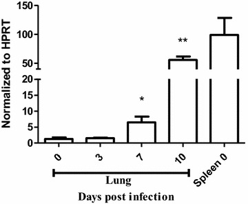
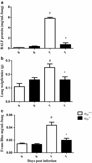
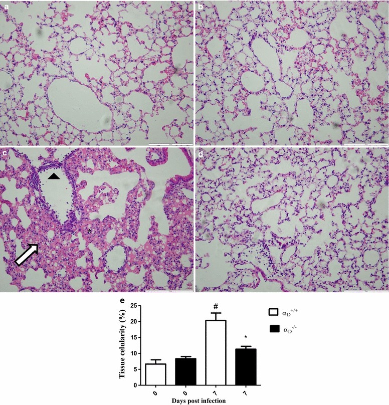
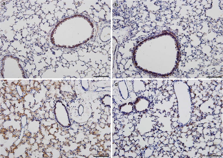
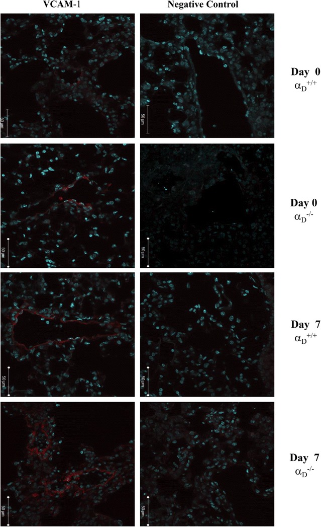
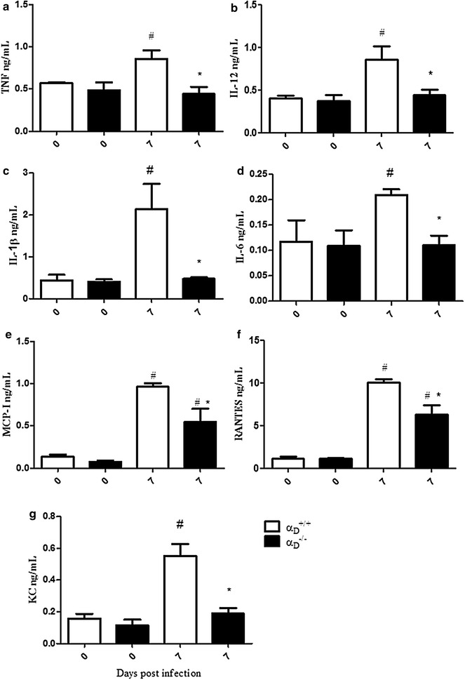
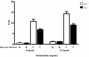
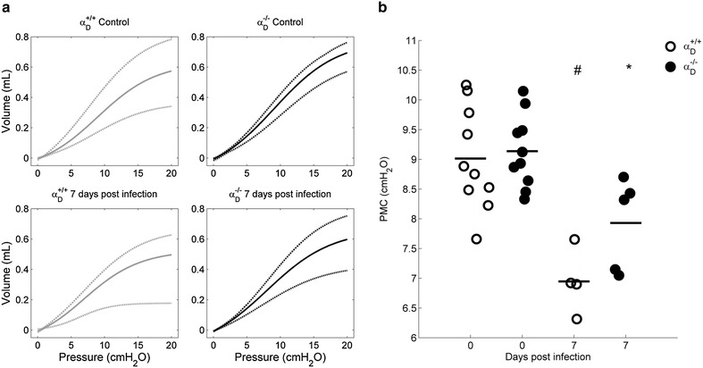
Methods: C57BL/6 wild type (a D (+/+) ) and deficient for CD11d sub-unit (a D (-/-) ) mice were monitored after infection with 10(5) Plasmodium berghei ANKA. CD11d subunit expression RNA was measured by real-time polymerase chain reaction, vascular barrier integrity by Evans blue dye (EBD) exclusion and cytokines by ELISA. Protein and leukocytes were measured in bronchoalveolar lavage fluid (BALF) samples. Tissue cellularity was measured by the point-counting technique, F4/80 and VCAM-1 expression by immunohistochemistry. Respiratory function was analysed by non-invasive BUXCO and mechanical ventilation.

Results: Alveolar inflammation, vascular and interstitial accumulation of monocytes and macrophages, and disrupted alveolar-capillary barrier function with exudation of protein-rich pulmonary oedema fluid were present in P. berghei-infected wild type mice and were improved in αDβ2-deficient animals. Key pro-inflammatory cytokines were also decreased in lung tissue from α D (-/-) mice, providing a mechanistic explanation for reduced alveolar-capillary inflammation and leak.

Conclusions: The results indicate that αDβ2 is an important inflammatory effector molecule in P. berghei-induced MA-ARDS, and that leukocyte integrins regulate critical inflammatory and pathophysiologic events in this model of complicated malaria. Genetic deletion of integrin subunit αD in mice, leading to deficiency of integrin αDβ2, alters lung inflammation and acute lung injury in a mouse model of MA-ARDS caused by P. berghei.

Keywords: Acute lung injury; Acute respiratory distress syndrome; Inflammation; Integrin αDβ2; Malaria.


Figures

Fig. 1
Fig. 1
αD mRNA expression is increased in the lungs of mice infected with Plasmodium berghei ANKA. Infected wild type were sacrificed 7 or 10 days later. Lungs were removed, processed and reverse transcribed. Transcripts for α D were quantified by real-time quantitative PCR and normalized to the levels of hypoxanthine guanine phosphoribosyl transferase (HRPT). Expression of α D in spleen was used as a positive control. Each bar represents the mean ± SEM of determinations in tissue from 5 animals. *P ≤ 0.04, **P ≤ 0.001 by student’s t test
Fig. 2
Fig. 2
Alveolar-capillary barrier disruption and increased permeability pulmonary are ameliorated in αDβ2-deficient mice infected with Plasmodium berghei ANKA. Infected wild type (αD+/+) and αDβ2-deficient (αD−/−) were study 7 days after infection. Bronchoalveolar lavage fluid (BALF) was performed. a Total protein concentration was measured in BALF samples using a BCA protein determination assay. b A second group of animals was sacrificed without BALF and the lungs were removed and weighed immediately after sacrifice. c In a third group of αD+/+ and αD−/− mice, Evans Blue Dye (2 %, 0.2 mL) was injected intravenously. The animals were sacrificed 1 h later, and Evans Blue Dye concentration in the lung tissue. Each bar in panels ac indicates the mean ± SEM. of determinations from 5 animals.#P ≤ 0.05 compared to the respective control group;  P ≤ 0.05 compared to αD+/+ infected mice
Fig. 3
Fig. 3
Vascular and interstitial inflammation are key components of MA-ARDS in Plasmodium berghei ANKA-infected mice. After staining with haematoxylin-eosin, the sections were examined by optical microscopy (×200 magnification). The scale bars indicate 50 µm. a Lung tissue from an uninfected αD+/+ mouse. b Lung section from an uninfected αD−/− animal. c Lung tissue from an infected wild type αD+/+ animal. The arrowhead identifies adherent leukocytes in a pulmonary vessel, indicating vascular inflammation. Diffuse interstitial infiltrates (arrow) and focal haemorrhages (asterisks) were also seen. d Lung tissue section from an infected αD−/− mouse. The features illustrated in ad are representative of those seen in lung tissue from 3 individual mice in each condition. e Lung cellularity was determined by the point counting technique across 20 random, non-coincident microscopic fields at magnification of ×1000, as described in “Methods” section. Each bar represents the mean of determinations in tissue from 3 animals ± SEM.#P ≤ 0.05 compared to respective control
Fig. 4
Fig. 4
F4/80-positive leukocytes accumulate in the alveoli in Plasmodium berghei ANKA-induced experimental MA-ARDS and are reduced in αDβ2-deficient mice. Lung tissue from αD+/+ and αD−/− mice infected with P. berghei was harvested at 7 days after infection. Lung slices were incubated with anti-F4/80 as the primary antibody and HRP-conjugated secondary antibody. The sections were examined and photographed using an Olympus BX41 microscope at ×200 magnification (scale bars 200 mm). a Sections from a control αD+/+ mouse. b Section from a control αD−/− mouse. c Lung tissue from an infected αD+/+ wild type animal. d Section from an infected αD−/− mouse. The staining patterns shown in ad are representative of those in lung tissue from 3 individual mice for each condition
Fig. 5
Fig. 5
VCAM-1 expression is increased in the lungs of wild type and αDβ2-deficient mice after infection. Wild type and αDβ2-deficient mice were infected with PbA and lungs were harvested at sacrifice at 7 days of infection. Lungs from infected wild type animals at day 0 were also obtained and handled in the same fashion. Staining for VCAM-1 was accomplished with a specific fluorescently labelled rat anti-mouse antibody (red staining). A rat anti-mouse IgG2b was used as an irrelevant control antibody. Nuclei were stained with DAPI. The sections were examined and photographed by immunoflourescent microscopy at ×20 magnification (50 μm). The patterns shown in the panels are representative of those seen in samples from 3 individual animals for each condition
Fig. 6
Fig. 6
Inflammatory cytokines are decreased in the lungs of αDβ2-deficient mice in Plasmodium berghei ANKA-induced MA-ARDS. Wild type and αDβ2-deficient mice were infected with PbA and lungs were harvested at 7 days after infection. Cytokine and chemokine levels were measured in the homogenates by ELISA. a TNF. b IL-12. c IL-1b. d IL-6. e MCP-I. f RANTES. g KC. Each bar indicates the mean ± SEM. of determinations in lung samples from 5 individual animals.#P ≤ 0.05 compared to the respective control group; *P ≤ 0.05 compared to infected wild type (αD+/+) mice
Fig. 7
Fig. 7
Plasmodium berghei ANKA infection induces increased airway reactivity that is ameliorated in αDβ2-deficient mice. Infected αDβ2-deficient and wild type mice were studied 7 days after infection. Uninfected αD−/− and αD+/+ animals were also studied as controls. Airway hyperreactivity was evaluated by challenge of the animals with aerosolized phosphate buffered saline (PBS) followed by methacholine (25 mg/mL in PBS) and is expressed as average enhanced pause (Penh). Each bar represents determinations in 10 animals (mean ± SEM).#P ≤ 0.05 compared to the respective control group; *P ≤ 0.05 compared to infected αD+/+ mice
Fig. 8
Fig. 8
Pressure/volume curves are sigmoidal and PMC is decreased in Plasmodium berghei ANKA-infected mice. Wild type and αD−/−animals from infected and control groups were mechanically ventilated and three PV curves were obtained (RR = 6 breaths/min, PEEP = 0 cm H2O, I:E ratio = 4:1, VT = 25–30 mL/kg). The PV curves where peak airway pressure remains stable and near to 20 cm H2O were fitted according to Eq. 1. The curve model gives the pressure value at which the respiratory system has maximum compliance. a Upper (dashdot), mean (solid) and lower (dashed) fitted curves of each group. b The PMC are lower, for infected wild type (P < 0.002) and αD−/− (P < 0.008) mice compared with respective uninfected controls

References

    1. WHO . World malaria report 2015. Geneva: World Health Organization; 2015. pp. 8–21.
    1. Stevenson MM, Riley EM. Innate immunity to malaria. Nat Rev Immunol. 2004;4:169–180. doi: 10.1038/nri1311. - DOI - PubMed
    1. White NJ, Pukrittayakamee S, Hien TT, Faiz MA, Mokuolu OA, Dondorp AM. Malaria. Lancet. 2014;383:723–735. doi: 10.1016/S0140-6736(13)60024-0. - DOI - PubMed
    1. Taylor BM, Kolbasa KP, Chin JE, Richards IM, Fleming WE, Griffin RL, et al. Roles of adhesion molecules ICAM-1 and α4 integrin in antigen-induced changes in microvascular permeability associated with lung inflammation in sensitized brown Norway rats. Am J Respir Cell Mol Biol. 1997;17:757–766. doi: 10.1165/ajrcmb.17.6.2697. - DOI - PubMed
    1. Schofield L, Grau GE. Immunological processes in malaria pathogenesis. Nat Rev Immunol. 2005;5:722–735. doi: 10.1038/nri1686. - DOI - PubMed

MeSH terms