. 2017 Jan;101(1):5-14.
doi: 10.1189/jlb.1HI1115-511R. Epub 2016 Jul 29.

Antigen-presenting cell-derived IL-6 restricts the expression of GATA3 and IL-4 by follicular helper T cells

Affiliations

Antigen-presenting cell-derived IL-6 restricts the expression of GATA3 and IL-4 by follicular helper T cells

Mélanie Hercor et al. J Leukoc Biol. 2017 Jan.

Abstract

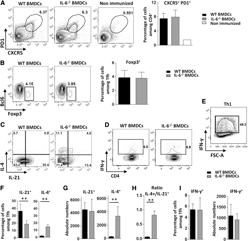
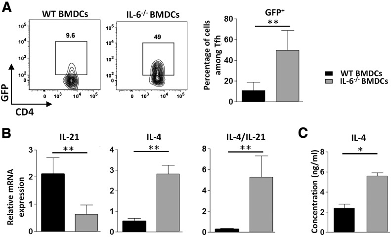
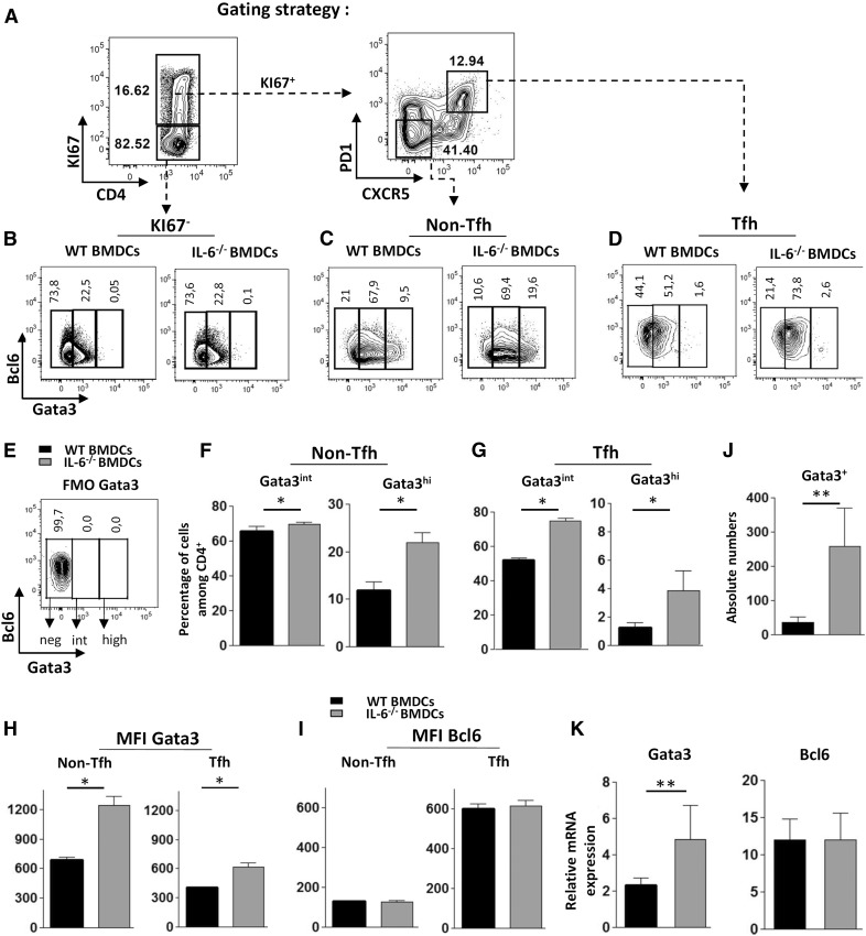
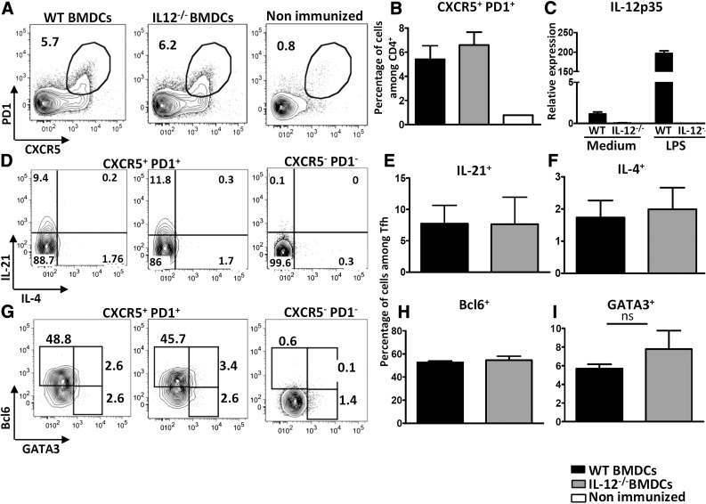
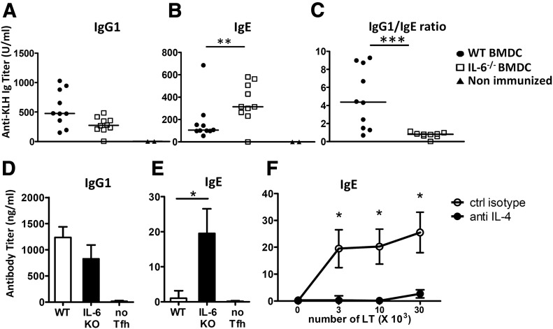
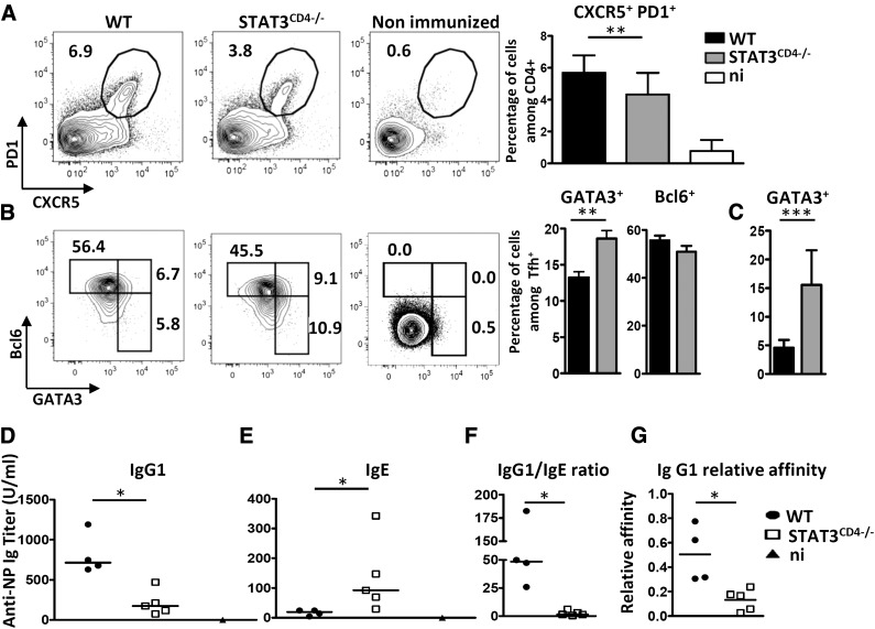
Follicular helper T cells (Tfh) support high-affinity Ab production by germinal center B cells through both membrane interactions and secretion of IL-4 and -21, two major cytokines implicated in B-cell survival and Ab class switch. Tfh-2 cells recently emerged in humans as a strong IL-4 producer Tfh cell subset implicated in both autoimmune and allergic diseases. Although the molecular mechanisms governing Tfh cell differentiation from naive T cells have been widely described, much less is known about the regulation of cytokine secretion by mouse Tfh-2 cells. The purpose of our study was to evaluate the role of dendritic cell-derived IL-6 in fine-tuning cytokine secretion by Tfh cells. Our results demonstrate that priming of Th cells by IL-6-deficient antigen-presenting dendritic cells preferentially leads to accumulation of a subset of Tfh cells characterized by high expression of GATA3 and IL-4, associated with reduced production of IL-21. STAT3-deficient Tfh cells also overexpress GATA3, suggesting that early IL-6/STAT3 signaling during Tfh cell development inhibits the expression of a set of genes associated with the Th2 differentiation program. Overall, our data indicate that IL-6/STAT3 signaling restrains the expression of Th2-like genes in Tfh cells, thus contributing to the control of IgE secretion in vivo.

Keywords: STAT3; Tfh-2; humoral response.


Figures

Figure 1.
Figure 1.. Immunization with IL-6-deficient BMDCs alters cytokine secretion by Tfh cells.
WT mice were immunized by footpad injection of 5 × 10⁵ LPS-stimulated, KLH-pulsed BMDCs derived from WT or IL-6-KO mice. Draining LN cells were recovered on d 7 and (A, B) analyzed for Tfh cell marker expression or (C–I) stimulated with ionomycin and PMA in the presence of monensin to evaluate cytokine production by ICS. Contour plots and histograms in (A) illustrate CXCR5 and PD1 staining profiles of CD4+ live cells within an FSC-A/FSC-W gate, enabling exclusion of doublets and triplets of cells; contour plots in (B–D) represent BCL6 and Foxp3 or IL-4, IFN-γ, and IL-21 staining profiles of Tfh cells (CXCR5+ PD1+) gated as indicated in (A); a positive control for IFN-γ staining (in vitro derived Th1 cells) is also included in (E); (F, G, I) Percentage and absolute number of IL-21-, IL-4- or IFN-γ-secreting Tfh cells; (H) ratio of total number of IL-4+ to IL-21+ Tfh cells. Results represent means ± sd of 5–6 individual mice and are representative of at least 10 (A) or 5 (C–I) independent experiments or are pooled from 4 independent experiments (B, n = 15). Differences between groups were analyzed with the Mann–Whitney U test for 2-tailed data. **P < 0.01. See also Supplemental Fig. S1 for gating strategy and staining controls.
Figure 2.
Figure 2.. IL-6-secreted by BMDCs damps IL-4 mRNA expression and IL-4 secretion by Tfh cells.
(A) The 4-get mice were immunized as in Fig. 1. Contour plots and histograms represent the percentage of GFP+ cells among the gated Tfh cells; (B, C) Tfh cells from C57BL6 mice immunized with WT or IL-6−/− BMDCs were separated by FACS and tested for (B) IL-4 and -21 mRNA expression ex vivo by RT-qPCR (expression was normalized to RPL32) or (C) restimulated with plastic-coated anti-CD3 mAbs for 24 h to allow detection of IL-4 secretion by ELISA. Results are representative of 2 independent experiments and are the means ± sd of 5–6 individual mice (A, B) or means ± SD of 4 individual culture wells. Differences between groups were analyzed with the Mann–Whitney U test for 2-tailed data. *P < 0.05; **P < 0.01.
Figure 3.
Figure 3.. IL-6-deficient BMDCs promote expression of GATA3 in Tfh cells.
WT mice were immunized with KLH-pulsed WT or IL-6−/− BMDCs, as in Fig. 1. Seven days later, GATA3 and BCL6 expression was measured by ICS. (A) Contour plots illustrate the gating strategy to identify CD4+Ki67 and CD4+Ki67+ within viable singlet cells (left) and Tfh and non-Tfh cells among the CD4+Ki67+ gated cells (right); (B–G) contour plots and histograms represent the percentage of GATA3int and GATA3high cells in Ki67, Ki67+ non-Tfh and Ki67+ Tfh+ gated cells; (E) FMO control for GATA3 staining; (H, I) histograms of GATA3 or BCL6 expression intensity (expressed as mean fluorescence intensity) among Tfh and non-Tfh cell subsets; (J) absolute number of GATA3+ (intermediate + high expression) Tfh cells in both experimental groups; and (K) mRNA expression levels of GATA3 and BCL6 (relative to RPL32 mRNA) in Tfh cells sorted as in Fig. 2. Results are representative of 3 (A–J) or 2 (K) independent experiments and are means ± sd of 5–6 individual mice. Differences between groups were analyzed with the Mann–Whitney U test for 2-tailed data. *P < 0.05; **P < 0.01.
Figure 4.
Figure 4.. Immunization with IL-12-deficient BMDCs does not alter Tfh cell differentiation.
WT mice were immunized with KLH-pulsed WT or IL-12p35−/− BMDCs. Draining lymph node cells were recovered on d 7 (A, B, G–I) and analyzed for Tfh cell marker and transcription factor expression or (D–F) stimulated with ionomycin and PMA in the presence of monensin to evaluate IL-4 and -21 production by ICS. Contour plots and histograms illustrate CXCR5 and PD1 staining profiles of CD4+ live singlet cells (A); BCL6, GATA3, IL-4, and IL-21 among Tfh gate (D–I); and (C) mRNA expression of IL-12p35 following a 3 h stimulation of BMDCs with 1 µg/ml LPS [expression was normalized to RPL32 and the results are presented as fold induction compared to WT nonstimulated (NS) BMDC sample set at 1]. Results are means ± sd of 5 individual mice and are representative of at least 3 independent experiments.
Figure 5.
Figure 5.. IL-6-deficient BMDCs promote IgE secretion.
WT mice were immunized with KLH-pulsed WT or IL-6−/−BMDCs, as in Fig. 1. (A–C) Sera were tested on d 14 for KLH-specific IgG1 (A) and IgE (B) contents; (C) relative levels of IgG1 to IgE NP-specific antibodies for each mouse. Results are pooled from 2 independent experiments (n = 10). (D–F) Tfh cells (gate CD4+CXCR5+PD1+) were separated by FACS and cultured for 7 d with purified syngeneic B cells purified from KLH/Alum immunized mice (5 × 10⁴ cells/well) and 10 μg/ml KLH. Histograms in (D, E) represent IgG1 and IgE secretion by B cells cultured alone or with 3 × 10⁴ Tfh cells purified from WT or IL-6−/− BMDC inoculated mice; (F) IgE secretion by B cells cultured with KLH and Tfh cells from IL-6−/− BMDC mice in the presence or absence or anti-IL-4 mAbs. Results are expressed as means ± sd of 4 individual culture wells and are representative of 2 independent experiments. Differences between groups were analyzed with the Mann–Whitney U test for 2-tailed data. *P < 0.05; **P < 0.01; ***P < 0.001.
Figure 6.
Figure 6.. Immunization of STAT3CD4−/− mice results in higher expression of GATA3 in Tfh cells and altered IgG1 and IgE isotype secretion.
STAT3fl/fl and STAT3fl/fl CD4-CRE mice were immunized with NP-KLH in Alum. Draining lymph nodes were analyzed on d 7 for Tfh cells (CXCR5+PD1+) among viable CD4+ cells (A) and GATA3/BCL6 expression among Tfh cells (B). (C) Percentage of GATA3+ cells among Tfh cells in STAT3fl/fl and STAT3fl/fl CD4-CRE mice immunized with KLH-pulsed WT-BMDCs as in Fig. 3. (D–G) Individual sera from mice in (A) were tested on d 14 for NP-specific IgG1 (D) and IgE (E) contents; (F) relative levels of IgG1 to IgE NP-specific antibodies for each mouse; (G) sera from (D) were tested for NP affinity. Relative affinities are expressed as the ratio of 50% binding on NP18- and NP2-BSA. Results in (A, B) are pooled from 2 to 5 independent experiments (n = 23, A; n = 11, B) or are representative of 3 independent experiments (C–G). n.i., nonimmunized. Differences between groups were analyzed with the Mann–Whitney U test for 2-tailed data. *P < 0.05; **P < 0.01; ***P < 0.001.

Comment in

Similar articles

Cited by

References

    1. Crotty S. (2011) Follicular helper CD4 T cells (TFH). Annu. Rev. Immunol. 29, 621–663. - PubMed
    1. Johnston R. J., Poholek A. C., DiToro D., Yusuf I., Eto D., Barnett B., Dent A. L., Craft J., Crotty S. (2009) Bcl6 and Blimp-1 are reciprocal and antagonistic regulators of T follicular helper cell differentiation. Science 325, 1006–1010. - PMC - PubMed
    1. Nurieva R. I., Chung Y., Martinez G. J., Yang X. O., Tanaka S., Matskevitch T. D., Wang Y. H., Dong C. (2009) Bcl6 mediates the development of T follicular helper cells. Science 325, 1001–1005. - PMC - PubMed
    1. Choi Y. S., Kageyama R., Eto D., Escobar T. C., Johnston R. J., Monticelli L., Lao C., Crotty S. (2011) ICOS receptor instructs T follicular helper cell versus effector cell differentiation via induction of the transcriptional repressor Bcl6. Immunity 34, 932–946. - PMC - PubMed
    1. Baumjohann D., Okada T., Ansel K. M. (2011) Cutting edge: distinct waves of BCL6 expression during T follicular helper cell development. J. Immunol. 187, 2089–2092. - PubMed

Publication types

MeSH terms