. 2017 Feb;66(2):483-493.
doi: 10.2337/db16-0051. Epub 2016 Jul 29.

CD40 in Retinal Müller Cells Induces P2X7-Dependent Cytokine Expression in Macrophages/Microglia in Diabetic Mice and Development of Early Experimental Diabetic Retinopathy

Affiliations

CD40 in Retinal Müller Cells Induces P2X7-Dependent Cytokine Expression in Macrophages/Microglia in Diabetic Mice and Development of Early Experimental Diabetic Retinopathy

Jose-Andres C Portillo et al. Diabetes. 2017 Feb.

Abstract

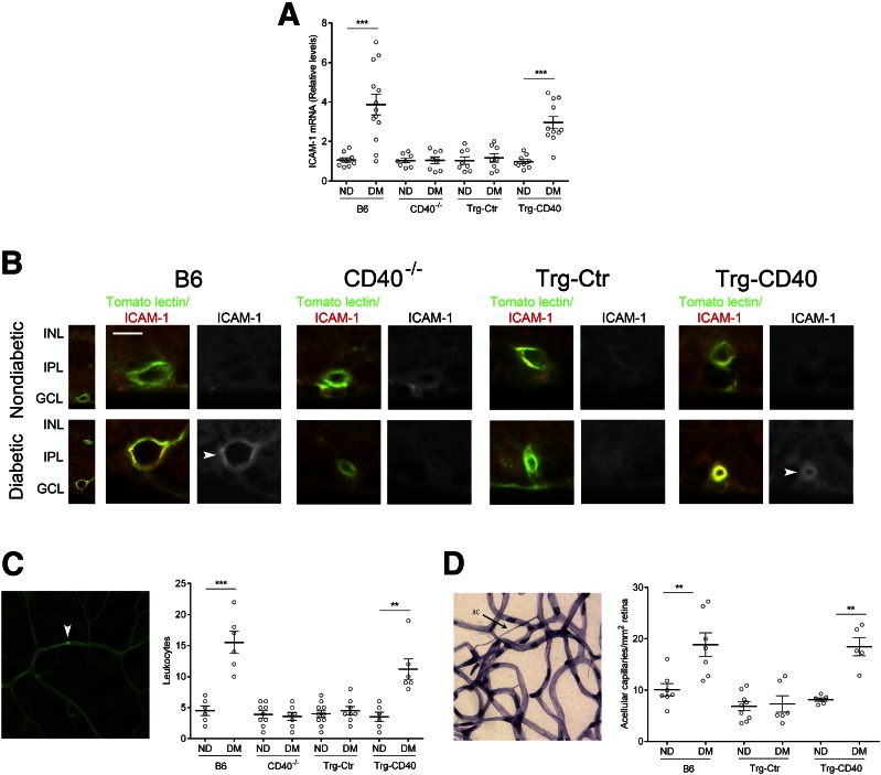
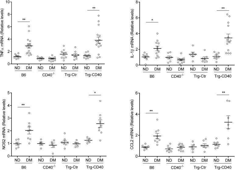
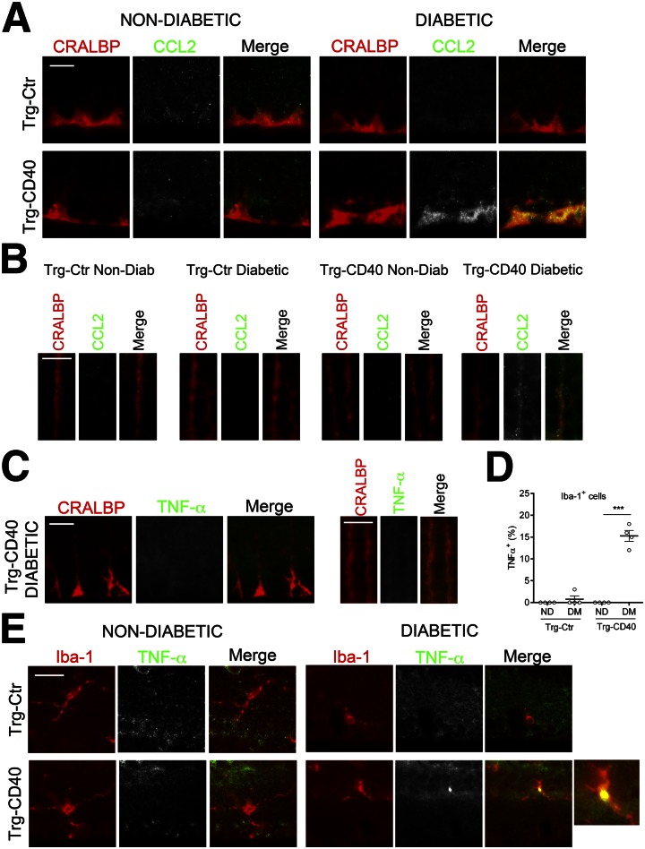
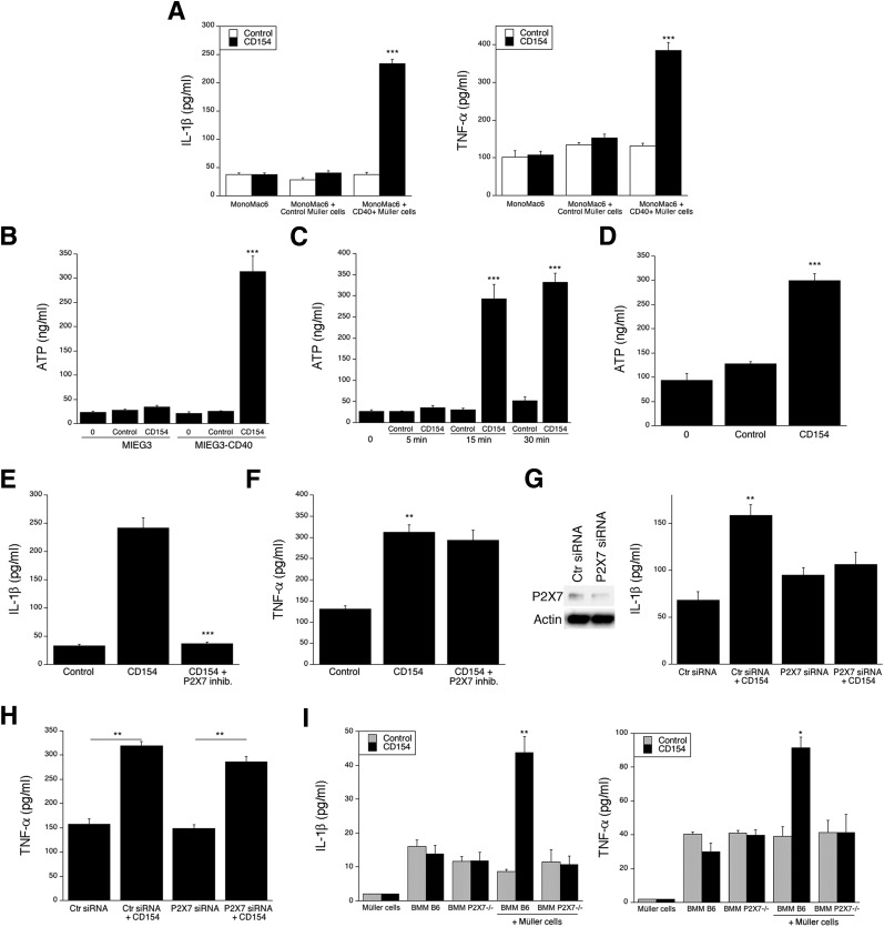
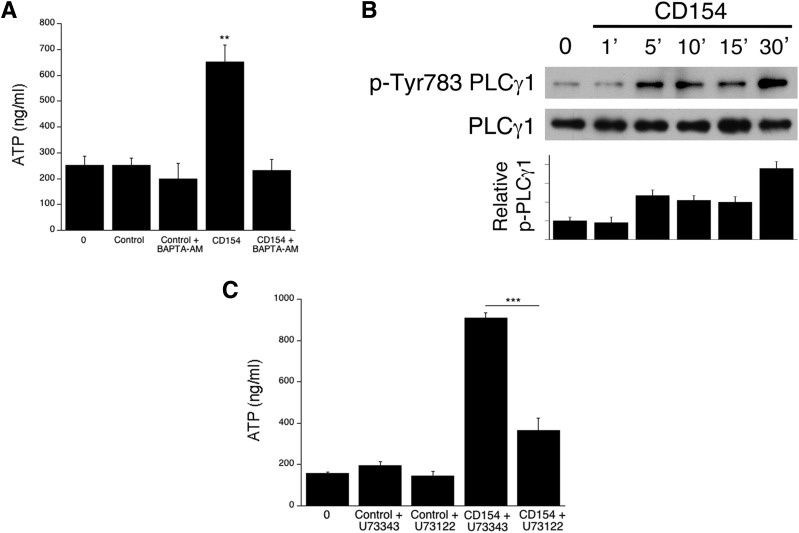
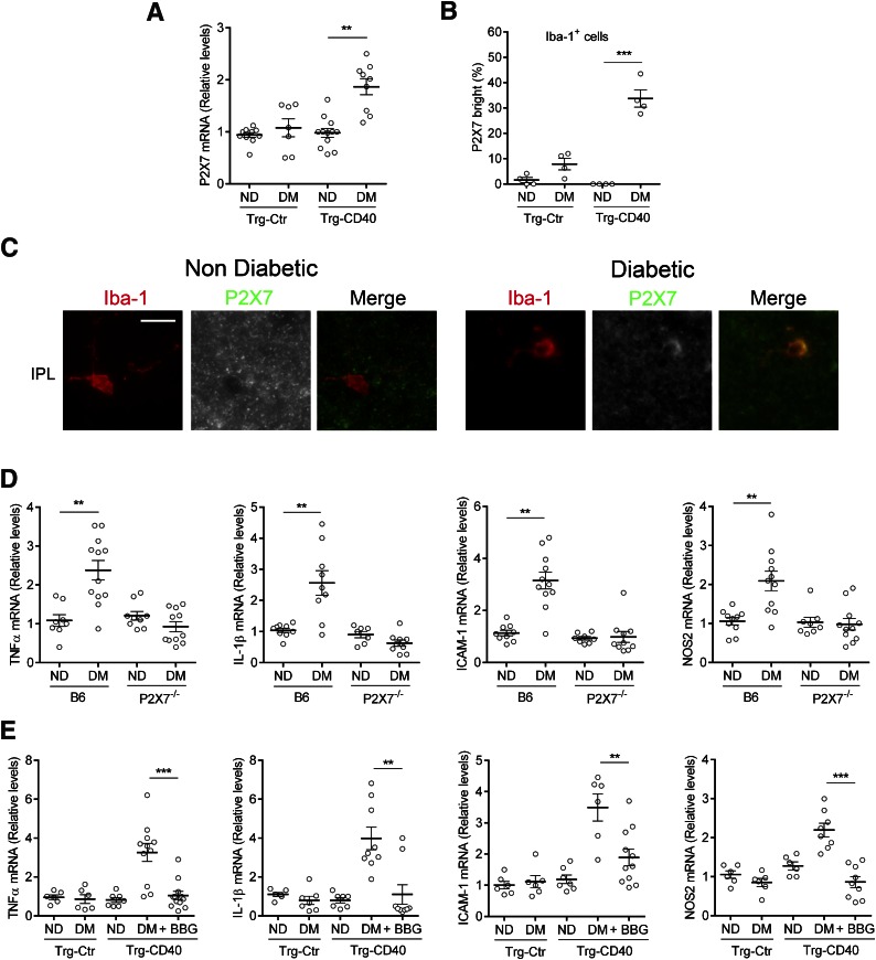
Müller cells and macrophages/microglia are likely important for the development of diabetic retinopathy; however, the interplay between these cells in this disease is not well understood. An inflammatory process is linked to the onset of experimental diabetic retinopathy. CD40 deficiency impairs this process and prevents diabetic retinopathy. Using mice with CD40 expression restricted to Müller cells, we identified a mechanism by which Müller cells trigger proinflammatory cytokine expression in myeloid cells. During diabetes, mice with CD40 expressed in Müller cells upregulated retinal tumor necrosis factor-α (TNF-α), interleukin 1β (IL-1β), intracellular adhesion molecule 1 (ICAM-1), and nitric oxide synthase (NOS2), developed leukostasis and capillary degeneration. However, CD40 did not cause TNF-α or IL-1β secretion in Müller cells. TNF-α was not detected in Müller cells from diabetic mice with CD40+ Müller cells. Rather, TNF-α was upregulated in macrophages/microglia. CD40 ligation in Müller cells triggered phospholipase C-dependent ATP release that caused P2X7-dependent production of TNF-α and IL-1β by macrophages. P2X7-/- mice and mice treated with a P2X7 inhibitor were protected from diabetes-induced TNF-α, IL-1β, ICAM-1, and NOS2 upregulation. Our studies indicate that CD40 in Müller cells is sufficient to upregulate retinal inflammatory markers and appears to promote experimental diabetic retinopathy and that Müller cells orchestrate inflammatory responses in myeloid cells through a CD40-ATP-P2X7 pathway.


Figures

Figure 1
Figure 1
CD40 expression in Müller cells from diabetic mice promotes upregulation of ICAM-1 in the retina and vascular changes of early diabetic retinopathy. A: At 2 months of diabetes, retinas from diabetic B6, CD40−/−, Trg-Ctr, and Trg-CD40 mice (DM) as well as from nondiabetic (ND) control animals were collected and used for mRNA extraction. mRNA levels of ICAM-1 were assessed by real-time quantitative PCR using 18S rRNA as the internal control. One nondiabetic B6 mouse was given an arbitrary value of 1, and data are expressed as the fold-increase compared with this animal. The horizontal bars represent mean ± SEM (n = 8–13 animals per group). B: At 2 months of diabetes, retinal sections were incubated with anti–ICAM-1 monoclonal antibody and tomato lectin (n = 6 mice/group). GCL, ganglion cell layer; INL, inner nuclear layer; IPL, inner plexiform layer. Arrowheads show blood vessels with increased ICAM-1 expression. Scale bar, 10 μm. C: At 2 months of diabetes, adherent leukocytes in the retinal vasculature of diabetic and nondiabetic control mice were quantified by labeling with concanavalin A. Representative image shows an adherent leukocyte (arrowhead) within the vasculature of a diabetic B6 mouse. D: At 8 months of diabetes, retinal digests were examined for the presence of degenerate capillaries. The horizontal bars represent the mean ± SEM (n = 6–10 animals per group). Representative image shows an acellular capillary (arrow) in the retinal digest of a diabetic B6 mouse. **P < 0.01, ***P < 0.001 by ANOVA.
Figure 2
Figure 2
CD40 expression in Müller cells from diabetic mice (DM) restores upregulation of TNF-α, IL-1β, NOS2, and CCL2 mRNA in the retina. At 2 months of diabetes, retinas from diabetic B6, CD40−/−, Trg-Ctr, and Trg-CD40 mice and from nondiabetic (ND) control animals were collected and used for mRNA extraction. mRNA levels were assessed by real-time quantitative PCR using 18S rRNA as the internal control. One nondiabetic B6 mouse was given an arbitrary value of 1, and data are expressed as the fold-increase compared with this animal. The horizontal bars represent the mean ± SEM (n = 7–15 animals per group). *P < 0.05, **P < 0.01 by ANOVA.
Figure 3
Figure 3
Diabetic transgenic mice that express CD40 in retinal Müller cells upregulate CCL2 in Müller cells and TNF-α in microglia/macrophages. A and B: Retinal sections from diabetic Trg-Ctr and Trg-CD40 mice at 2 months of diabetes and from nondiabetic controls were incubated with anti-CCL2 plus anti-CRALBP antibody. CCL2 expression at the level of Müller cell end feet (ganglion cell layer) (A) and at the level of Müller cells stalks (outer plexiform layer) (B). C: Sections were incubated with anti–TNF-α plus anti-CRALBP antibody. Müller cell end feet and stalk areas are shown. D and E: Retinal sections were incubated with anti–TNF-α monoclonal antibody plus anti–Iba-1 antibody. D: Sections were analyzed to determine the percentages of Iba-1+ cells in diabetic mice (DM) and nondiabetic (ND) mice that were stained with anti–TNF-α antibody. ***P < 0.001. E: Images represent microglia/macrophages present in the inner plexiform layer (n = 4 mice/group). Scale bars, 10 μm.
Figure 4
Figure 4
CD40-activated Müller cells secrete ATP and induce P2X7 receptor–dependent production of cytokines by monocytic cells and macrophages. A: Control human Müller cell (transduced with empty retroviral vector MIEG3) and CD40+ human Müller cells (transduced with CD40-encoding retroviral vector MIEG3-CD40) were incubated with CD40 human monocytic cell lines (MonoMac6) with or without CD154. Cytokines were measured in supernatants by ELISA at predetermined optimal time points (24 h for IL-1β and 4 h for TNF-α). B: Human Müller cell line was incubated with or without CD154. Concentrations of extracellular ATP at time 0 and at 15 min of incubation are shown. C: CD40+ Müller cells were incubated with CD154 and concentrations of extracellular ATP were measured at different times. D: Mouse Müller cells transduced with the hmCD40-encoding retroviral vector were incubated with or without human CD154. CD40+ human Müller cells were incubated with MonoMac6 cells with or without CD154 in the presence or absence of the P2X7 receptor inhibitor A-438079, and IL-1β (E) and TNF-α (F) were measured in supernatants by ELISA. CD40+ human Müller cells were incubated with MonoMac6 cells transfected with control or P2X7 receptor siRNA, and IL-1β (G) and TNF-α (H) were measured in supernatants by ELISA. I: Mouse Müller cells that express hmCD40 were incubated with BMM from B6 or P2X7−/− mice, and IL-1β (I) and TNF-α (J) were measured by ELISA. Results are presented as mean ± SEM (n = 3). *P < 0.05, **P < 0.01, ***P < 0.001 by Student t test.
Figure 5
Figure 5
CD40 ligation in Müller cells causes Tyr783 phosphorylation (p) of PLCγ1 and PLC-dependent secretion of ATP. A: CD40+ Müller cells were incubated with or without BAPTA-AM, followed by stimulation with CD154 and measurement of extracellular ATP. Concentrations of extracellular ATP at time 0 and at 15 min of incubation are shown. B: CD40+ human Müller cells were incubated with CD154. Expression of p-Tyr783 PLCγ1 and total PLCγ1 were assessed by immunoblot. The bars represent quantification of relative p-Tyr783 PLCγ1 from three different experiments. C: CD40+ Müller cells were incubated with U73122 or U73343, followed by stimulation with CD154 and measurement of extracellular ATP. Concentrations of extracellular ATP at time 0 and at 15 min of incubation are shown. Results are presented as mean ± SEM (n = 3). **P < 0.01, ***P < 0.001 by Student t test.
Figure 6
Figure 6
Diabetic transgenic mice that express CD40 in retinal Müller cells upregulate P2X7 receptor in microglia/macrophages, and administration of the P2X7 receptor inhibitor BBG impairs upregulation of retinal mRNA levels of TNF-α and IL-1β. A: At 2 months of diabetes, retinas from diabetic (DM) and nondiabetic (ND) Trg-Ctr and Trg-CD40 mice were collected and used for mRNA extraction. mRNA levels of P2X7 receptor were assessed by real-time quantitative PCR using 18S rRNA as the internal control. One nondiabetic Trg-Ctr mouse was given an arbitrary value of 1, and data are expressed as the fold-increase compared with this animal. The horizontal bars represent the mean ± SEM (n = 7–11 animals per group). B and C: Sections from diabetic and nondiabetic Trg-CD40 mice were incubated with anti-P2X7 receptor plus anti–Iba-1 antibodies. B: Sections were analyzed to determine the percentages of Iba-1+ cells that stained brightly with anti-P2X7 receptor anbitody. C: Images represent microglia/macrophages present in the inner plexiform layer (IPL) (n = 4 mice/group). Scale bar, 10 μm. D: At 2 months of diabetes, retinas from diabetic and nondiabetic B6 and P2X7−/− mice were collected and used to measure TNF-α, IL-1β, ICAM-1, and NOS2 mRNA levels. One nondiabetic B6 mouse was given an arbitrary value of 1, and data are expressed as the fold-increase compared with this animal (n = 7–12 animals per group). E: Diabetic Trg-CD40 mice were treated with BBG or vehicle daily for 28 days beginning at 1 month of diabetes. Retinas from these animals and from diabetic and nondiabetic Trg-Ctr were collected and used to measure TNF-α, IL-1β, ICAM-1, and NOS2 mRNA levels (n = 6–11 mice per group.) **P < 0.01, ***P < 0.001 by ANOVA.

Comment in

References

    1. Tang J, Kern TS. Inflammation in diabetic retinopathy. Prog Retin Eye Res 2011;30:343–358 - PMC - PubMed
    1. Antonetti DA, Klein R, Gardner TW. Diabetic retinopathy. N Engl J Med 2012;366:1227–1239 - PubMed
    1. Krady JK, Basu A, Allen CM, et al. . Minocycline reduces proinflammatory cytokine expression, microglial activation, and caspase-3 activation in a rodent model of diabetic retinopathy. Diabetes 2005;54:1559–1565 - PubMed
    1. Yang LP, Sun HL, Wu LM, et al. . Baicalein reduces inflammatory process in a rodent model of diabetic retinopathy. Invest Ophthalmol Vis Sci 2009;50:2319–2327 - PubMed
    1. Joussen AM, Doehmen S, Le ML, et al. . TNF-α mediated apoptosis plays an important role in the development of early diabetic retinopathy and long-term histopathological alterations. Mol Vis 2009;15:1418–1428 - PMC - PubMed

Publication types

MeSH terms