. 2016 Sep 16;291(38):19873-87.
doi: 10.1074/jbc.M116.739557. Epub 2016 Jul 29.

Mechanistic and Evolutionary Insights from the Reciprocal Promiscuity of Two Pyridoxal Phosphate-dependent Enzymes

Affiliations

Mechanistic and Evolutionary Insights from the Reciprocal Promiscuity of Two Pyridoxal Phosphate-dependent Enzymes

Valerie W C Soo et al. J Biol Chem. .

Abstract

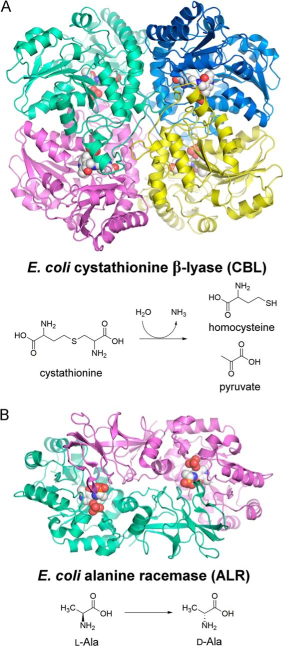
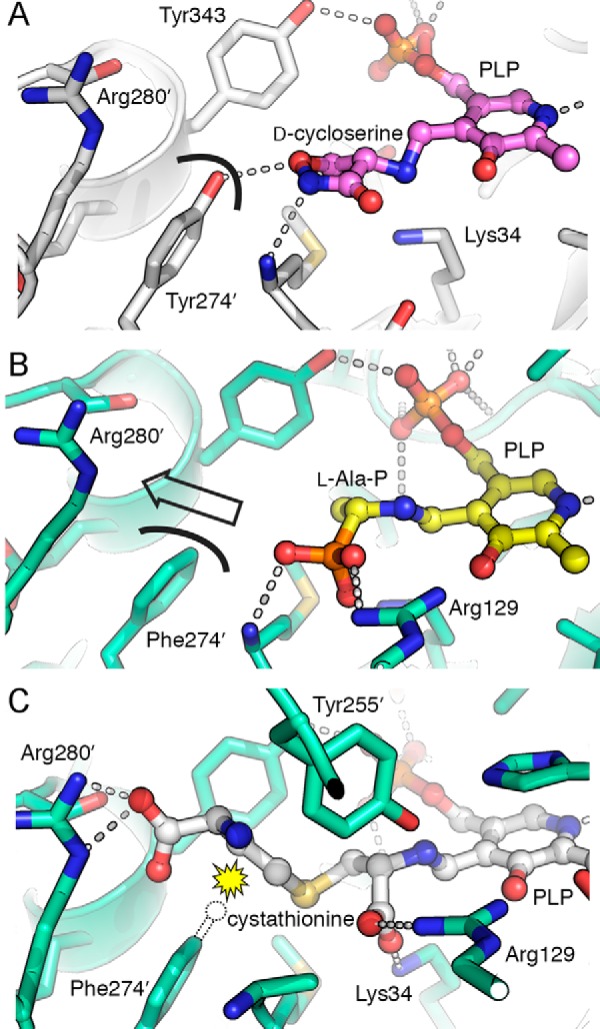
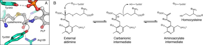
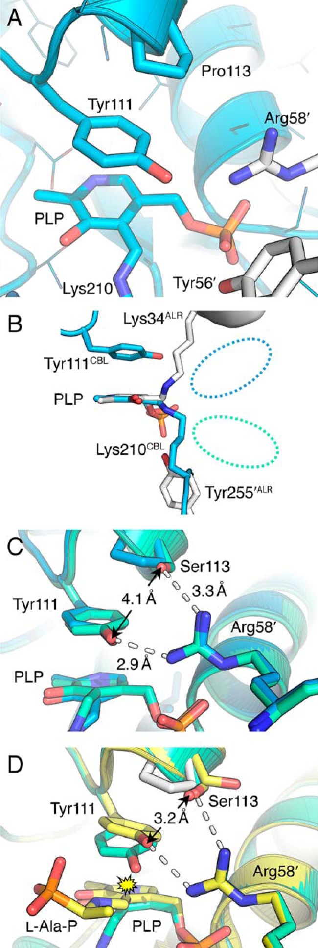
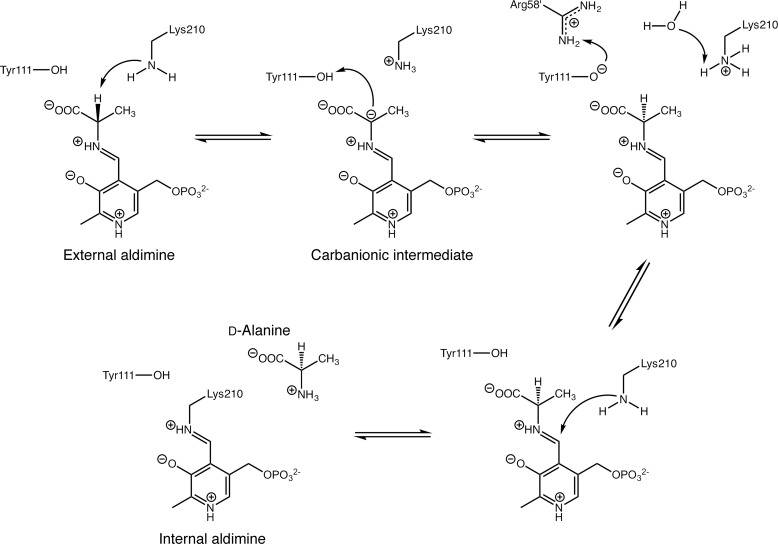
Enzymes that utilize the cofactor pyridoxal 5'-phosphate play essential roles in amino acid metabolism in all organisms. The cofactor is used by proteins that adopt at least five different folds, which raises questions about the evolutionary processes that might explain the observed distribution of functions among folds. In this study, we show that a representative of fold type III, the Escherichia coli alanine racemase (ALR), is a promiscuous cystathionine β-lyase (CBL). Furthermore, E. coli CBL (fold type I) is a promiscuous alanine racemase. A single round of error-prone PCR and selection yielded variant ALR(Y274F), which catalyzes cystathionine β-elimination with a near-native Michaelis constant (Km = 3.3 mm) but a poor turnover number (kcat ≈10 h(-1)). In contrast, directed evolution also yielded CBL(P113S), which catalyzes l-alanine racemization with a poor Km (58 mm) but a high kcat (22 s(-1)). The structures of both variants were solved in the presence and absence of the l-alanine analogue, (R)-1-aminoethylphosphonic acid. As expected, the ALR active site was enlarged by the Y274F substitution, allowing better access for cystathionine. More surprisingly, the favorable kinetic parameters of CBL(P113S) appear to result from optimizing the pKa of Tyr-111, which acts as the catalytic acid during l-alanine racemization. Our data emphasize the short mutational routes between the functions of pyridoxal 5'-phosphate-dependent enzymes, regardless of whether or not they share the same fold. Thus, they confound the prevailing model of enzyme evolution, which predicts that overlapping patterns of promiscuity result from sharing a common multifunctional ancestor.

Keywords: directed evolution; enzyme mechanism; protein evolution; protein structure; pyridoxal phosphate.


Figures

FIGURE 1.
FIGURE 1.
A, structure of the CBL tetramer (PDB entry 1CL1) (11) and the reaction it catalyzes in methionine biosynthesis. B, structure of the ALR dimer (PDB entry 2RJG) (15) and the reaction it catalyzes to provide d-alanine for peptidoglycan biosynthesis. In each structure, the locations of the active sites are highlighted by the PLP cofactor, which is shown in space-filling format.
FIGURE 2.
FIGURE 2.
Structural analysis of the Y274F mutation in ALR. A, in the structure of wild type ALR inhibited by d-cycloserine (PDB entry 2RJH) (15), Tyr-274′ is positioned to form a hydrogen bond with the inhibitor. The curved black line shows the surface at the tyrosine hydroxyl group. B, active site of ALR(Y274F) with the l-Ala-P-PLP external aldimine, shown in the same orientation as A. The curved black line represents the surface of Phe-274′. The lack of a hydroxyl group opens a pathway from the substrate toward Arg-280′ (indicated with an arrow). C, modeled position of l-cystathionine in the ALR(Y274F) active site, docked using GOLD. The distal carboxylate of the substrate is perfectly positioned to interact with Arg-280′. A dotted outline indicates the position that the Tyr-274′ hydroxyl group occupies in the wild type ALR structure. The yellow starburst indicates the steric clash that would occur between this hydroxyl group and cystathionine, disfavoring substrate binding. The Y274F mutation removes this steric clash.
FIGURE 3.
FIGURE 3.
Tyr-255′ is positioned to play a catalytic role in cystathionine β-elimination by ALR(Y274F). A, when l-cystathionine-PLP is docked into the active site using GOLD, the side chain oxygen atom of Tyr-255′ is ∼2.6 Å from the proton that it is poised to abstract from the substrate (black arrow). B, tentative reaction scheme for ALR(Y274F)-catalyzed cystathionine β-elimination. Tyr-255′ has an unusually low pKa (20), so it is assumed to be in the phenolate form for proton abstraction (22). Unlike in CBL, the pyridine nitrogen is not protonated, because of the proximity of Arg-209 in the ALR active site. This results in a resonance-stabilized carbanionic intermediate of unusually high energy (45). By analogy with the CBL mechanism (35), we predict that Tyr-255′ reorients to protonate the leaving group (homocysteine). This would yield the PLP derivative of aminoacrylate, which is the substrate for reverse transaldimination (35), to form iminopropionate and to regenerate the Lys-34-PLP Schiff base. The nature of the carbanionic intermediate and inefficient proton transfer from Tyr-255′ to the leaving group are likely to explain the very poor turnover observed for ALR(Y274F).
FIGURE 4.
FIGURE 4.
Structural analysis of the P113S mutation in CBL. A, in wild type CBL (PDB entry 1CL1) Pro-113 is positioned above Tyr-111, which in turn makes a stacking interaction with the pyridine ring of PLP (11). B, overlay of the catalytic residues in CBL (blue) and ALR (white), with the structures oriented so that their PLP cofactors superimpose. The approximate position occupied by incoming substrate is indicated with a dashed blue oval for CBL and a dashed green oval for ALR. C, CBL(P113S) (green) superimposed upon CBL (blue). Although there are no significant changes in the positions of active site residues, Arg-58′ is now within hydrogen bonding distance of Ser-113 in the mutant. D, overlay of the unliganded CBL(P113S) structure (green) and CBL(P113S) with an l-Ala-P-PLP external aldimine (yellow). Wild type residue Pro-113 is also shown in white to highlight the effect of the P113S substitution. Two rotamers of Ser-113 were observed in the inhibited structure; both are shown. The two hydrogen bonds involving Arg-58′ are the same length as in C. The yellow starburst highlights the steric clash that would occur if Tyr-111 did not pivot away from PLP in the inhibited structure.
FIGURE 5.
FIGURE 5.
Reaction scheme for racemization of l-alanine, catalyzed by CBL(P113S). Estimates from PROPKA suggest that Tyr-111 is likely to be protonated when the l-Ala-PLP external aldimine is formed. When CBL catalyzes cystathionine β-elimination, the carbanionic intermediate is stabilized as the ketamine quinonoid (36). Here, we show instead the resonance structure of the carbanionic intermediate that is consistent with the ALR mechanism (22). Also, as done previously (22), we have shown the proton exchanges at Tyr-111 (involving Arg-58′) and Lys-210 occurring from the same intermediate, but this is not a necessity. Reverse transaldimination to regenerate the Lys-210-PLP Schiff base is assumed to occur as described for the cystathionine β-elimination reaction.

References

    1. Percudani R., and Peracchi A. (2009) The B6 database: a tool for the description and classification of vitamin B6-dependent enzymatic activities and of the corresponding protein families. BMC Bioinformatics 10, 273. - PMC - PubMed
    1. Raboni S., Spyrakis F., Campanini B., Amadasi A., Bettati S., Peracchi A., Mozzarelli A., and Contestabile R. (2010) in Comprehensive Natural Products II: Chemistry and Biology (Mander L. and Liu H.-W., editors in chief) pp. 273–350, Elsevier Ltd., New York
    1. Percudani R., and Peracchi A. (2003) A genomic overview of pyridoxal-phosphate-dependent enzymes. EMBO Rep. 4, 850–854 - PMC - PubMed
    1. Toney M. D. (2011) Controlling reaction specificity in pyridoxal phosphate enzymes. Biochim. Biophys. Acta 1814, 1407–1418 - PMC - PubMed
    1. Richard J. P., Amyes T. L., Crugeiras J., and Rios A. (2011) The PLP cofactor: lessons from studies on model reactions. Biochim. Biophys. Acta 1814, 1419–1425 - PMC - PubMed

MeSH terms