. 2016 Oct 21;291(43):22686-22702.
doi: 10.1074/jbc.M116.746404. Epub 2016 Jul 29.

Accumulation of Peptidoglycan O-Acetylation Leads to Altered Cell Wall Biochemistry and Negatively Impacts Pathogenesis Factors of Campylobacter jejuni

Affiliations

Accumulation of Peptidoglycan O-Acetylation Leads to Altered Cell Wall Biochemistry and Negatively Impacts Pathogenesis Factors of Campylobacter jejuni

Reuben Ha et al. J Biol Chem. .

Abstract

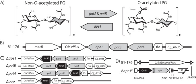
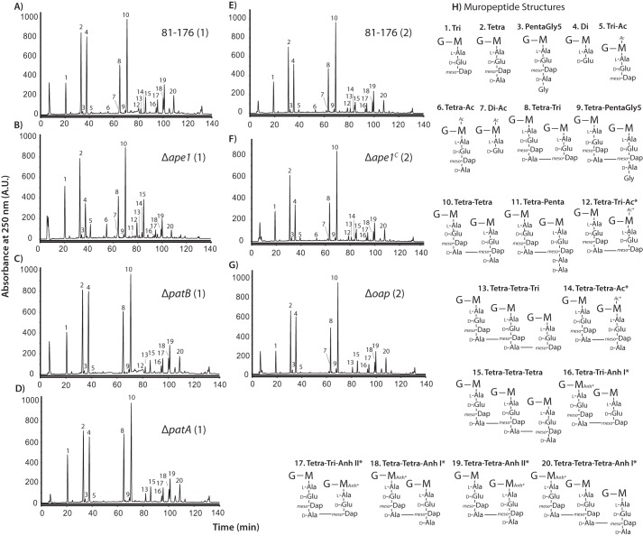
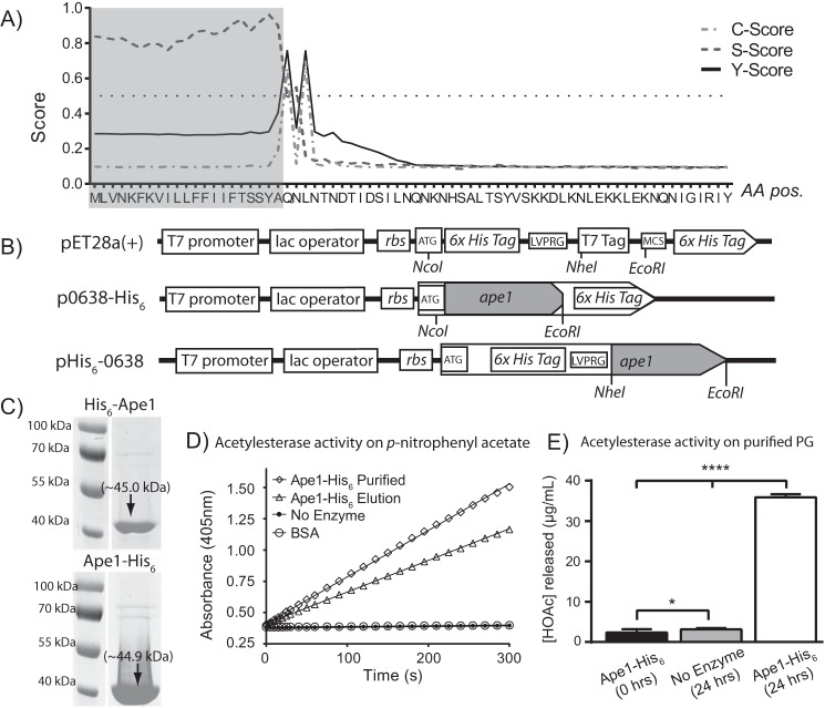
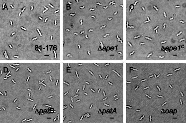
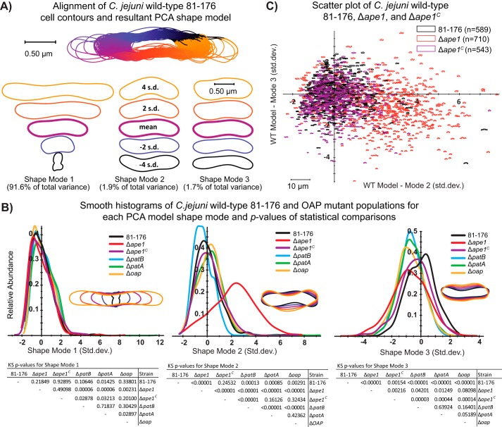
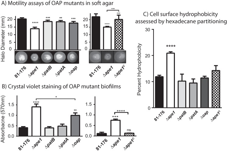
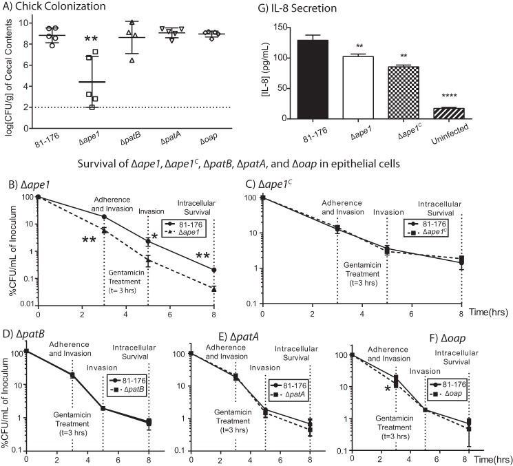
Campylobacter jejuni is a leading cause of bacterial gastroenteritis in the developed world. Despite its prevalence, its mechanisms of pathogenesis are poorly understood. Peptidoglycan (PG) is important for helical shape, colonization, and host-pathogen interactions in C. jejuni Therefore, changes in PG greatly impact the physiology of this organism. O-acetylation of peptidoglycan (OAP) is a bacterial phenomenon proposed to be important for proper cell growth, characterized by acetylation of the C6 hydroxyl group of N-acetylmuramic acid in the PG glycan backbone. The OAP gene cluster consists of a PG O-acetyltransferase A (patA) for translocation of acetate into the periplasm, a PG O-acetyltransferase B (patB) for O-acetylation, and an O-acetylpeptidoglycan esterase (ape1) for de-O-acetylation. In this study, reduced OAP in ΔpatA and ΔpatB had minimal impact on C. jejuni growth and fitness under the conditions tested. However, accumulation of OAP in Δape1 resulted in marked differences in PG biochemistry, including O-acetylation, anhydromuropeptide levels, and changes not expected to result directly from Ape1 activity. This suggests that OAP may be a form of substrate level regulation in PG biosynthesis. Ape1 acetylesterase activity was confirmed in vitro using p-nitrophenyl acetate and O-acetylated PG as substrates. In addition, Δape1 exhibited defects in pathogenesis-associated phenotypes, including cell shape, motility, biofilm formation, cell surface hydrophobicity, and sodium deoxycholate sensitivity. Δape1 was also impaired for chick colonization and adhesion, invasion, intracellular survival, and induction of IL-8 production in INT407 cells in vitro The importance of Ape1 in C. jejuni biology makes it a good candidate as an antimicrobial target.

Keywords: Campylobacter jejuni; O-Acetylation; O-Acetylesterase; acetylation; bacterial pathogenesis; cell wall; gram-negative bacteria; peptidoglycan.


Figures

FIGURE 1.
FIGURE 1.
Location of O-acetyl groups on peptidoglycan subunits, organization of the C. jejuni OAP gene cluster, and description of deletion mutant and complement construction. A, structures of the disaccharide muropeptides showing non-O-acetylated PG and O-acetylated PG, location of O-acetylation (arrow), and the putative involvement of the oap genes. B, genomic organization of the C. jejuni OAP gene cluster in the 81-176 wild-type strain (gray). cjj81176_0638, cjj81176_0639, and cjj81176_0640 are the C. jejuni homologs of Δape1, ΔpatB, and ΔpatA respectively, as identified by BLAST using the N. gonorrhoeae OAP genes sequences. C, OAP mutants were generated by homologous recombination with a mutated copy of the gene (or the entire cluster for Δoap) in which a portion of the gene (or cluster) was deleted and replaced with a non-polar KmR cassette (aphA3) (47). Resistance to Km was used as a selective marker for successful homologous recombination in C. jejuni with the mutated gene. D, complement construction (with Δape1 used as an example, designated Δape1C). Each OAP gene plus upstream sequence containing the ribosomal binding site was cloned into the pRRC vector that contains homologous regions to three ribosomal intergenic regions downstream of the CmR cassette for selection of successful C. jejuni transformants. Complement constructs were transformed into their respective mutant backgrounds. MacB, macrolide-specific efflux pump; OM efflux, outer membrane efflux; ftn, ferritin; 23S, 23S ribosomal RNA (48).
FIGURE 2.
FIGURE 2.
HPLC elution profile of C. jejuni muropeptides and proposed muropeptide structures. Purified PG was digested with cellosyl, and the resulting muropeptides were reduced with sodium borohydride and separated on a Prontosil 120-3-C18 AQ reverse-phase column. HPLC profiles are shown for wild-type strain 81-176 (A and E), Δape1 (B), ΔpatB (C), ΔpatA (D), Δape1C (F), and Δoap (G). Muropeptide profiles were generated in two sets of experiments indicated by (1) for Sample Set 1 and (2) for Sample Set 2. The muropeptide structure represented by each peak was determined previously by mass spectroscopy (18), and proposed muropeptide structures of each peak corresponding to the peak number in the chromatogram are shown in H. The summary of the muropeptide composition is shown in Table 3. G, N-acetylglucosamine; M, reduced N-acetylmuramic acid; l-Ala, l-alanine; d-iGlu, d-isoglutamic acid; meso-Dap, meso-diaminopimelic acid; d-Ala, d-alanine Ac, O-acetyl groups at MurNAc C6 position; Anh, 1,6-anhydro group of MurNAc; *, it is not known on which MurNAc residue the modification occurs.
FIGURE 3.
FIGURE 3.
A, SignalP 4. 1 server (83) output for signal peptide prediction of in-frame translation of Cjj-81-176_0638. C-score (the predicted first amino acid of the mature protein), S-score (the likelihood that a particular amino acid is part of a signal peptide), and Y-score (amino acid with a high C-score exhibiting the greatest change in the S-score) predicted the cleavage site to be between the 21st and 22nd amino acids. B, Cjj81-176_0638 was cloned in-frame without the signal peptide into pET28a(+) protein expression vectors. Top, map of cloning sites in pET28a(+) commercial expression vector. Middle, Ape1-His6 expression construct. NcoI and EcoRI were used to produce a C-terminal His6-tagged Ape1 protein that uses ATG start codon and TGA stop codon encoded in the vector. Bottom, His6-Ape1 expression construct. NheI and EcoRI were used to produce an N-terminal His6-tagged protein that uses AUG start codon encoded by the vector and the original stop codon from Cjj81-176_0638. rbs, ribosome-binding site; LVPRG, thrombin cleavage site; MCS, multiple cloning sites. Gene organization not to scale. C, His6-tagged Ape1 after nickel-nitrilotriacetic acid-agarose purification shows protein of the predicted size (45.0 and 44.9 kDa for His6-Ape1 and Ape1-His6 respectively) in eluted fractions after SDS-PAGE analysis. D, purified Ape1-His6 exhibits acetylesterase/deacetylase activity using pNPAc as a substrate (45). Reactions were monitored over 5 min as a change in the absorbance at 405 nm (formation of p-nitrophenol) after cleavage of the acetyl group. No enzyme control and BSA control are overlapping and show no acetylesterase activity. Results shown are from one protein purification experiment. Results are reproducible for each expression and purification experiment, and activity was routinely assessed before performing enzymatic assays on PG. E, Ape1-His6 has acetylesterase activity using PG muropeptides as a substrate. Determination of acetic acid concentration after treatment of Δape1 PG with Ape1-His6 for 24 h was performed using Megazyme acetic acid assay kit. Treatment and no enzyme control were compared with acetic acid concentration at 0 h of treatment using Student's t test with * and **** indicating p values of <0.05 and <0.0001, respectively. Results are from one representative experiment of two biological replicates performed in triplicate.
FIGURE 4.
FIGURE 4.
C. jejuni Δape1 mutant has a pleomorphic cell shape, and other OAP mutants display unaltered cell morphology. DICM showing the morphology of wild-type strain C. jejuni 81-176 (A), the differentially curved Δape1strain (B), the complemented strain Δape1C with restored morphology (C), ΔpatB (D), ΔpatA (E), and Δoap (F). Cells were harvested from 7 h of growth in MH-TV broth at a mid-exponential phase of growth. Scale is 2 μm (black bar).
FIGURE 5.
FIGURE 5.
CellTool analysis of wild-type strain 81-176, Δape1, Δape1C, ΔpatB, ΔpatA, and Δoap population morphology. Differential interference contrast images were taken of strains grown for 7 h in MH-TV broth at a starting A600 of 0.05 (to mid-exponential phase). Images were converted to binary format (white cells on a black background), and lumps and artifacts were manually removed before processing with CellTool “extract contours function” to generate contours representing each cell (53). A, contour extraction, alignment, and generation of the PCA shape model for C. jejuni wild-type strain 81-176. CellTool “align contours” function was used to align the contours of the wild-type population to one another. B, PCA was performed to generate a wild-type shape model that explains 95% variation in the population in principal components called “shape modes.” Shape modes 1, 2, and 3 represent variation in length, curvature/wavelength, and width, respectively. The extracted contours of the mutant populations were then aligned to the wild-type shape model, and a measurement representing the normalized standard deviation from the wild-type mean in each shape mode was generated and depicted graphically. KS tests were performed for each shape mode between each population and are summarized below the plots. C, measurements of wild type, Δape1, and Δape1C were plotted with shape mode 2 along the x axis and shape mode 3 along the y axis to create a scatterplot showing the variation in the different populations.
FIGURE 6.
FIGURE 6.
Motility in soft agar, biofilm formation, and cell surface hydrophobicity of OAP mutants and wild-type strain 81-176. A, Δape1 exhibits a 30% decrease in halo diameter and abnormal halo formation (rough edges). Motility in soft agar was assessed by measuring the halo diameter after 24 h of strains point-inoculated in 0.4% semi-solid agar. Representative images of halos are shown below each graph. Results shown are representative of one of three independent experiments with 6 replicates. Each strain was compared with wild-type using a paired Student's t test, with **, ***, and **** indicating p < 0.01, p < 0.001, and p < 0.0001. B, Δape1 and Δoap exhibit 5.5- and 2.5-fold enhanced biofilm formation, respectively, at 24 h. Biofilm formation was assessed after 24 h by crystal violet staining of standing cultures in borosilicate tubes and spectrophotometric quantification of dissolved crystal violet at 570 nm. Results shown for the mutants (left) are representative of one of three independent experiments carried out in triplicate. The results for Δape1C (right) are representative of one of two experiments performed in triplicate. ns, not significant. C, Δape1 exhibited a 2.0-fold increase hydrophobicity relative to wild type, as assessed by hexadecane partitioning. Results are representative of one of three independent experiments performed in triplicate. For biofilm and hydrophobicity, strains were compared using an unpaired Student's t test, with *, **, ***, and **** indicating p < 0.05, p < 0.01, p < 0.001, and p < 0.0001. Error bars represent standard deviation.
FIGURE 7.
FIGURE 7.
Effect of OAP levels on C. jejuni host-bacteria interactions. A, Δape1 shows reduced chick colonization compared with wild-type strain 81-176, whereas ΔpatB, ΔpatA, and Δoap mutants display wild-type colonization. Each point represents the recovery of C. jejuni strains in log CFU/g of cecal contents from individual day-old chicks 6 days post-colonization with 1 × 10⁴ CFU/ml of the indicated strain. The geometric mean is denoted by a black bar. Error bars represent 95% confidence intervals. Adherence, invasion, and intracellular survival of C. jejuni in INT407 epithelial cells were assessed by a Gm protection assay and OAP mutant strains. Δape1 (B) shows a reduced ability to adhere to, invade, and survive in INT407 epithelial cells that were restored upon complementation (C). ΔpatB (D), ΔpatA (E), and Δoap (F) exhibit near wild-type adherence, invasion, and intracellular survival properties. INT407 cells were infected with C. jejuni at a multiplicity of infection of ∼80. Adherence and invasion were quantified at 3 h post-infection. At this point, the media in the remaining wells were replaced with MEM containing gentamicin (150 μg/ml) and incubated for 2 h, after which the amount of bacterial cells that had invaded the epithelial cells was measured (5-h invasion time point). The Gm in the remaining wells was washed off, and the cells were incubated with fresh MEM containing 3% FBS and a low dose of Gm (10 μg/ml) for an additional 3 h (8-h intracellular survival time point). CFU/ml was determined for each well by lysing the cells with water and plating the dilutions onto MH-TV plates. Results for B and C are representative of three independent experiments performed in biological triplicate. The data in D, E, and F are representative of two independent experiments performed with three biological replicates. G, INT407 epithelial cells secrete less IL-8 upon infection with Δape1 than wild type. Results are from one representative experiment of three independent experiments performed in triplicate. Error bars represent the standard deviation. *, denotes statistically significant difference using the unpaired Student's t test, with *, **, and **** indicating p < 0.05, p < 0.01, and p < 0.00001 respectively.

Similar articles

Cited by

References

    1. Yuki N., and Hartung H. P. (2012) Guillain-Barre syndrome. N. Engl. J. Med. 366, 2294–2304 - PubMed
    1. Jacobs B. C., Rothbarth P. H., van der Meché F. G., Herbrink P., Schmitz P. I., de Klerk M. A., and van Doorn P. A. (1998) The spectrum of antecedent infections in Guillain-Barre syndrome: a case-control study. Neurology 51, 1110–1115 - PubMed
    1. Man S. M. (2011) The clinical importance of emerging Campylobacter species. Nat. Rev. Gastroenterol. Hepatol. 8, 669–685 - PubMed
    1. Silva J., Leite D., Fernandes M., Mena C., Gibbs P. A., and Teixeira P. (2011) Campylobacter spp. as a foodborne pathogen: a review. Front. Microbiol. 2, 200. - PMC - PubMed
    1. Luangtongkum T., Jeon B., Han J., Plummer P., Logue C. M., and Zhang Q. (2009) Antibiotic resistance in Campylobacter: emergence, transmission and persistence. Future Microbiol. 4, 189–200 - PMC - PubMed

MeSH terms

LinkOut - more resources