. 2017 Feb;65(2):501-514.
doi: 10.1002/hep.28735. Epub 2016 Aug 29.

Extracellular vesicles carry microRNA-195 to intrahepatic cholangiocarcinoma and improve survival in a rat model

Affiliations

Extracellular vesicles carry microRNA-195 to intrahepatic cholangiocarcinoma and improve survival in a rat model

Ling Li et al. Hepatology. 2017 Feb.

Abstract

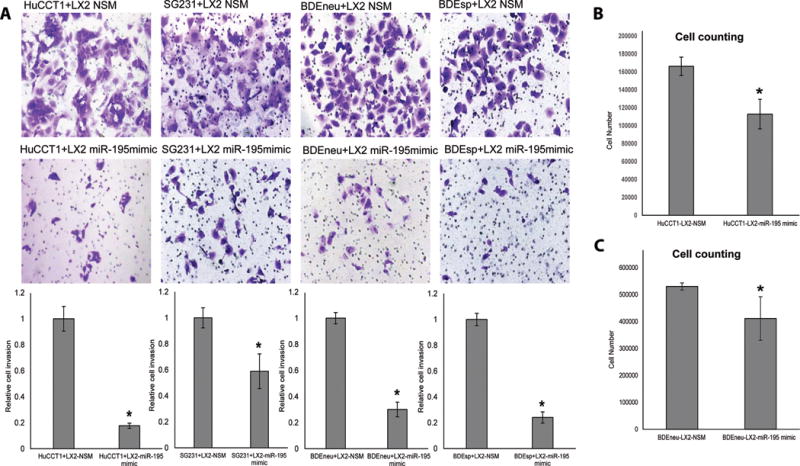
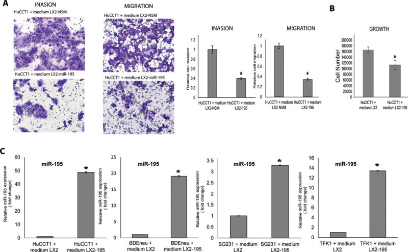
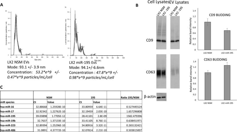
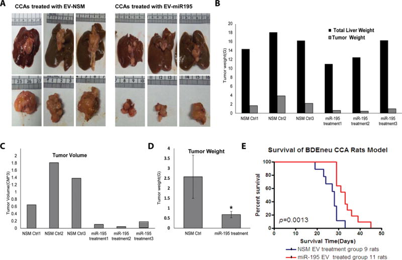
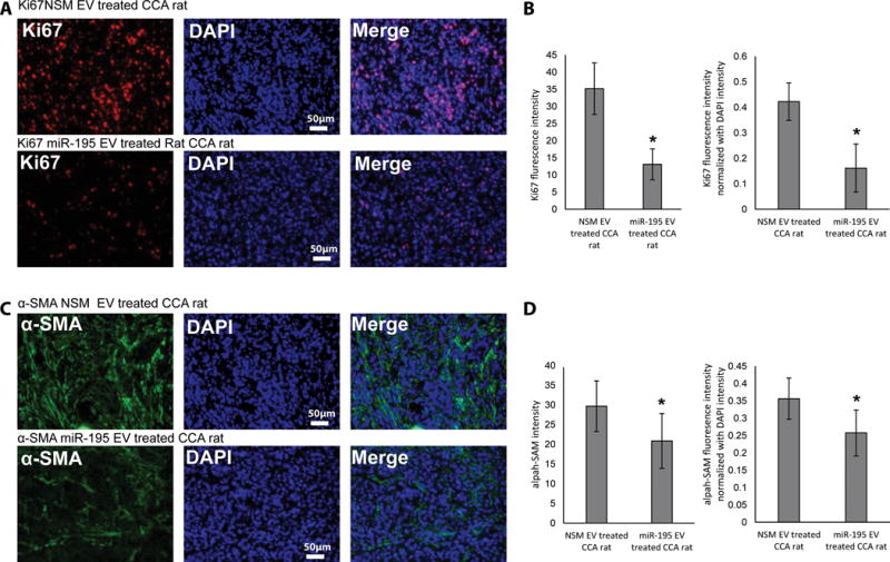
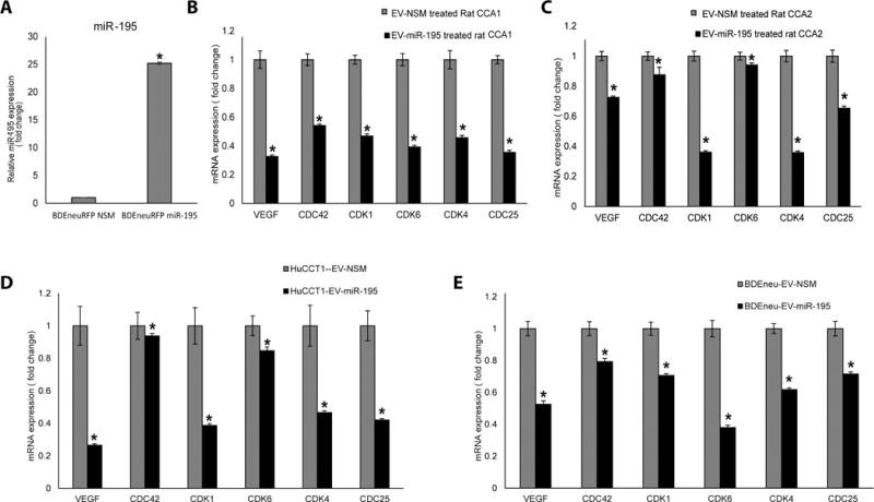
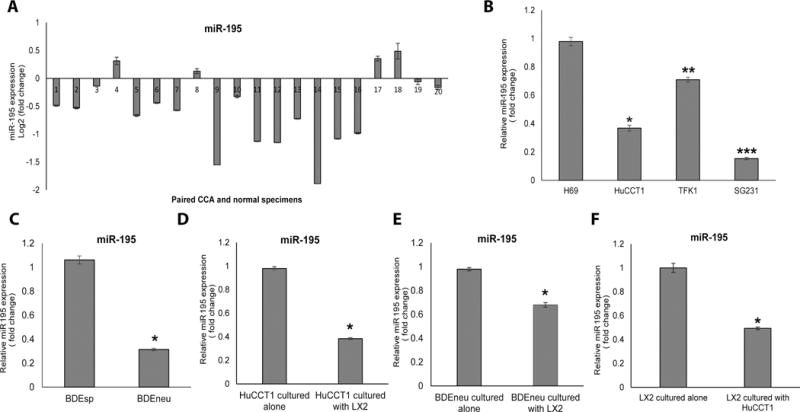
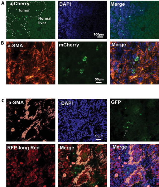
The cancer microenvironment plays a central role in cancer development, growth, and homeostasis. This paradigm suggests that cancer fibroblasts support cancers, probably in response to stimuli received from the cancer cells. We aimed at investigating whether extracellular vesicles (EVs) can shuttle microRNA (miR) species between cancer-associated fibroblasts (CAFs) and cancer cells. To this end, we extracted EVs according to published protocols. EVs were studied for their miR content by quantitative reverse-transcription polymerase chain reaction. EVs were transfected with select miR species and utilized in vitro as well as in vivo in a rat model of cholangiocarcinoma (CCA). We found that miR-195 is down-regulated in CCA cells, as well as in adjoining fibroblasts. Furthermore, we report that EVs shuttle miR-195 from fibroblasts to cancer cells. Last, we show that fibroblast-derived EVs, loaded with miR-195, can be administered in a rat model of CCA, concentrate within the tumor, decrease the size of cancers, and improve survival of treated rats.

Conclusion: EVs play a salient role in trafficking miR species between cancer cells and CAFs in human CCA. Understanding of these mechanisms may allow devising of novel therapeutics. (Hepatology 2017;65:501-514).


Conflict of interest statement

No authors have any potential conflicts in reference to this manuscript.

Figures

Figure 1
Figure 1. Upregulation of miR-195 in co-cultured LX2 fibroblast cells inhibit CCA cell proliferation and invasion
A: Four CCA cell lines were co-cultured with LX2 cells transfected with NSM (upper row) or miR-195 mimic (lower row) and exhibited greatly reduced invasion ability in LX2-miR-195 mimic co-cultured CCA cells. Quantification of invasion is displayed in the lower part of Panel A and is the result of 3 replicates per condition. B–C: Co-culture with LX2-miR 195 mimic reduces CCA cells proliferation (n=3). The statistical significance was analyzed using two-tailed Student’s t-test: *P<0.05
Figure 2
Figure 2. Conditioned medium from LX2-miR-195 upregulates miR-195 within cancer cells and inhibits their growth, invasion and migration
A–C: Conditioned media of LX2-miR-195 inhibit the invasion, migration and growth of CCA cells. D: LX2-miR-195 cells release soluble factors that elevate levels of miR-195 in CCA cancer cells. Levels of miR-195 were measured in 4 different CCA cancer cells after their exposure for 48h to conditioned medium from either LX2 NSM (left bars) or LX2 miR-195 mimic (right bars) cells (n=3). Statistical significance was analyzed using two-tailed Student’s t-test: *P<0.05
Figure 3
Figure 3. Overexpression of miR-195 in LX2 cells does not affect the EV biogenesis or content
A: Same number of cells (2.1*10^6) were plated in triplicate for NSM and miR-195 treatment, respectively. Three days after transfection with NSM and miR-195, respectively, media was collected for EV counting and size measurements. Cells were also counted and the number of EVs was calculated per milliliter of media per cell. We found that LX2 cells treated with miR-195 made 47.8*e^9 +/− 0.98*e^9 particles/mL/cell and LX2 cells treated with NSM made 53.2*e^9 +/− 0.47*e^9 particles/mL/cell. There was no statistical difference between the numbers of EVs produced over 3 days by any of the cell lines. In addition, the mode of EVs was the same in NSM vs. miR-195 treated cells (93.1 +/− 3.9 nm vs. 94.1 +/− 6.6 nm). B: EV budding proteins CD9 and CD63 were analyzed by immunoblotting, performed in 6 replicates per condition. There was no statistically significant difference between budding of EVs from NSM vs. miR-195 treated cells, as measured by CD9 and CD63 blotting. C: EVs collected 3 days after LX2 transfection with NSM or miR-195, respectively, were subjected to RNA extraction followed by qRT-PCR arrays. As shown, a number of 5 miRs were expressed in EVs from NSM-transfected LX2 cells. These miRs were expressed at similar levels in EVs from miR-195 transfected LX2 cells. In contrast, miR-195 was expressed 1,580 fold more in EVs from miR-195 vs. NSM transfected LX2 cells.
Figure 4
Figure 4. miR-195-loaded EVs inhibit CCA growth and increase survival in vivo
A: LX2 EVs were loaded with either (left panels) a non-specific miR mimic NSM (negative control), or (right panels) a miR-195 mimic. Examination of (upper panels) entire liver and (lower panels) excised tumor revealed that tumors were significantly smaller in animals that had been injected with miR-195-loaded EVs. B–D: Tumor weight and volume of cancers from rats treated with miR-195 EVs treated was less vs. EV-NSM treated CCA rats (n=3). Statistical significance was analyzed using two-tailed Student’s t-test: *P<0.05. E: In vivo treatment of CCA with EVs-miR-195 increases the survival by 28.7%. Twenty cancer bearing rats were randomly divided in 2 groups: 11 were treated with EVs-miR-195 and 9 were treated with EVs-NSM (negative control). *P=0.013
Figure 5
Figure 5. In vivo treatment with EVs-miR-195 decreases cancer cell growth as well as tumor fibrous infiltrate
A, B: Ki67 staining was performed on slides cut from cancer blocks (n=3 rats, 4 slides per rat). C, D: a-SMA staining was performed on slides cut from cancer blocks (n=3 rats, 4 slides per rat). Image J was used to quantify fluorescence. Significance was analyzed using two-tailed Student’s t-test: *P<0.05
Figure 6
Figure 6. EVs-miR-195 treatment upregulates miR-195 and downregulates miR-195 targets in cancer cells, in vitro and in vivo
A: miR-195 expression in sorted BDEneu cells is increased 25-fold. B, C: EVs-miR-195 downregulated 6 target genes - VEGF, CDC42, CDK1, CDK6, CDK4, CDC25 – in CCA cells in vivo. Data is shown for 2 pairs of rats. Three tissue samples were obtained from each rat. D, E: EVs-miR-195 downregulated 6 target genes - VEGF, CDC42, CDK1, CDK6, CDK4, CDC25 – in CCA cells in vitro (n=3). Statistical significance was analyzed using two-tailed Student’s t-test: *P<0.05
Figure 7
Figure 7. miR-195 is downregulated in CCA cells
A: qRT-PCR for 20 human CCA tissues and paired normal liver specimens is shown. MiR-195 expression is lower in CCA specimens compared with normal specimens in 16 out of the 20 pairs. B: miR-195 expression in normal biliary epithelial cell line H69 and 3 different human CCA cell lines. C: miR-195 expression in the more aggressive BDEneu cells is downregulated vs. related but less aggressive BDEsp cells. D, E: Co-culture of CCA cells with LX2 cells further downregulates miR-195 within cancer cells. F: Co-culture of CCA cells with LX2 cells reduces miR-195 levels within stellate cells (LX2).
Figure 8
Figure 8. Fibroblast-derived EVs are enriched in CCA tumors in vivo
A, B: Rats with CCA tumors were injected with fibroblast-derived EVs that were labeled with TSG101/mCherry fusion protein. EVs are concentrated within the cancer areas. C: BDEneu cells with a stably integrated loxpP-dsRed-loxp-eGFP construct were injected in the liver of rats in vivo to established tumors. EVs containing Cre-recombinase encoding plasmid were administered via tail vein injections. Staining with anti-EGFP antibodies revealed cancer cells that had switched color as a functional consequence of EVs uptake.

Comment in

References

    1. Mueller MM, Fusenig NE. Friends or foes - bipolar effects of the tumour stroma in cancer. Nat Rev Cancer. 2004;4:839–849. - PubMed
    1. Webber JP, Spary LK, Sanders AJ, Chowdhury R, Jiang WG, Steadman R, Wymant J, et al. Differentiation of tumour-promoting stromal myofibroblasts by cancer exosomes. Oncogene. 2015;34:290–302. - PubMed
    1. Josson S, Gururajan M, Sung SY, Hu P, Shao C, Zhau HE, Liu C, et al. Stromal fibroblast-derived miR-409 promotes epithelial-to-mesenchymal transition and prostate tumorigenesis. Oncogene. 2015;34:2690–2699. - PubMed
    1. Mertens JC, Fingas CD, Christensen JD, Smoot RL, Bronk SF, Werneburg NW, Gustafson MP, et al. Therapeutic effects of deleting cancer-associated fibroblasts in cholangiocarcinoma. Cancer Res. 2013;73:897–907. - PMC - PubMed
    1. Madar S, Goldstein I, Rotter V. ‘Cancer associated fibroblasts’–more than meets the eye. Trends Mol Med. 2013;19:447–453. - PubMed

Publication types

MeSH terms