. 2016 Oct:104:310-22.
doi: 10.1016/j.biomaterials.2016.07.005. Epub 2016 Jul 14.

Dual stimulation of antigen presenting cells using carbon nanotube-based vaccine delivery system for cancer immunotherapy

Affiliations

Dual stimulation of antigen presenting cells using carbon nanotube-based vaccine delivery system for cancer immunotherapy

Hatem A F M Hassan et al. Biomaterials. 2016 Oct.

Abstract

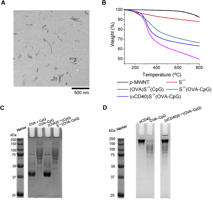
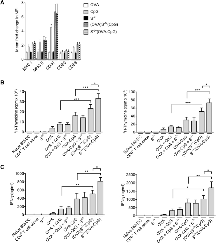
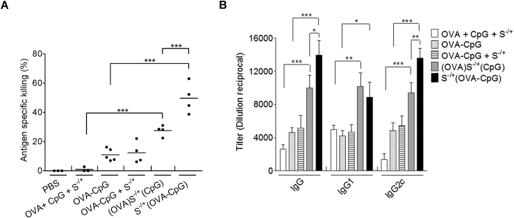
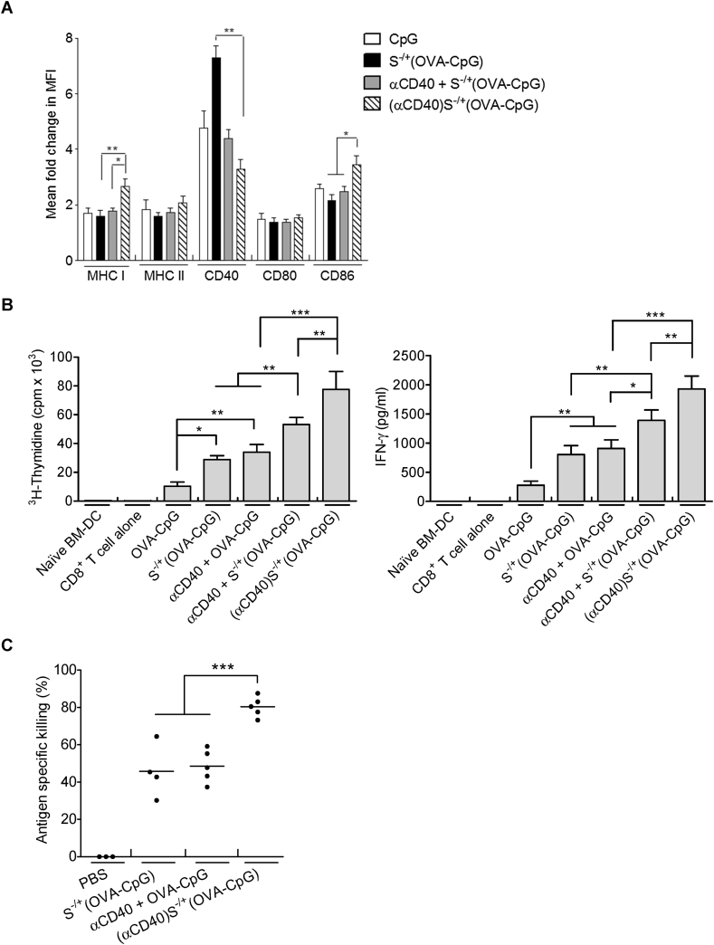
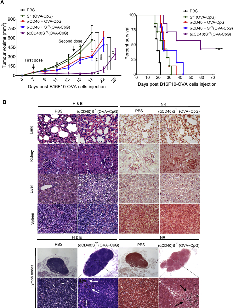
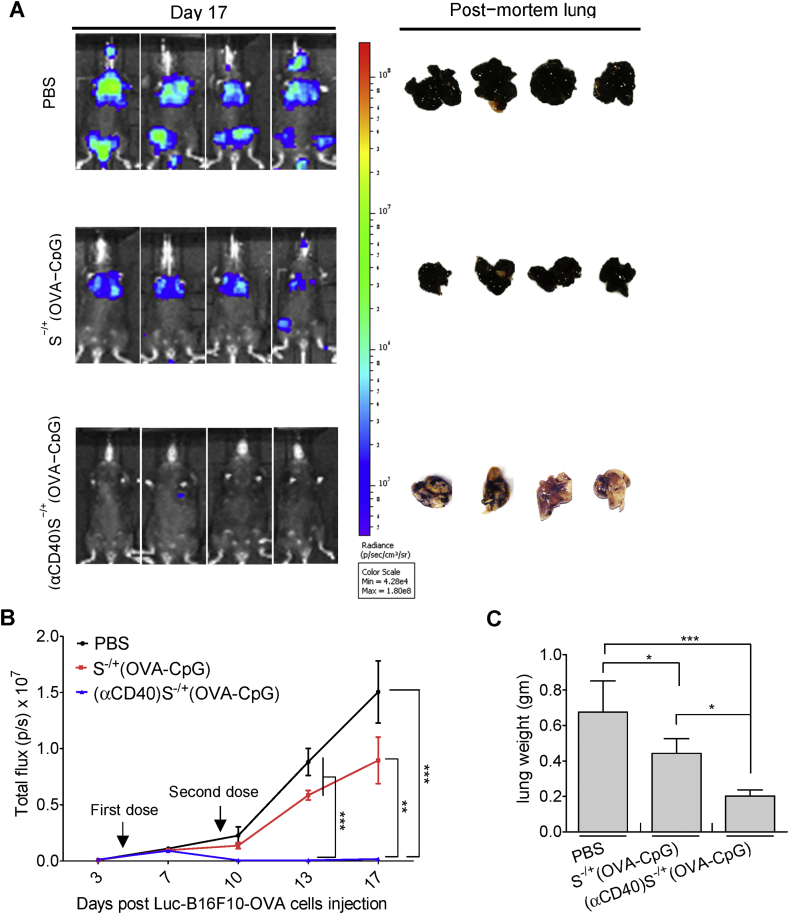
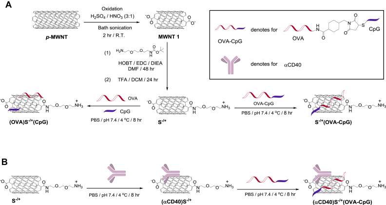
Although anti-cancer immuno-based combinatorial therapeutic approaches have shown promising results, efficient tumour eradication demands further intensification of anti-tumour immune response. With the emerging field of nanovaccinology, multi-walled carbon nanotubes (MWNTs) have manifested prominent potentials as tumour antigen nanocarriers. Nevertheless, the utilization of MWNTs in co-delivering antigen along with different types of immunoadjuvants to antigen presenting cells (APCs) has not been investigated yet. We hypothesized that harnessing MWNT for concurrent delivery of cytosine-phosphate-guanine oligodeoxynucleotide (CpG) and anti-CD40 Ig (αCD40), as immunoadjuvants, along with the model antigen ovalbumin (OVA) could potentiate immune response induced against OVA-expressing tumour cells. We initially investigated the effective method to co-deliver OVA and CpG using MWNT to the APC. Covalent conjugation of OVA and CpG prior to loading onto MWNTs markedly augmented the CpG-mediated adjuvanticity, as demonstrated by the significantly increased OVA-specific T cell responses in vitro and in C57BL/6 mice. αCD40 was then included as a second immunoadjuvant to further intensify the immune response. Immune response elicited in vitro and in vivo by OVA, CpG and αCD40 was significantly potentiated by their co-incorporation onto the MWNTs. Furthermore, MWNT remarkably improved the ability of co-loaded OVA, CpG and αCD40 in inhibiting the growth of OVA-expressing B16F10 melanoma cells in subcutaneous or lung pseudo-metastatic tumour models. Therefore, this study suggests that the utilization of MWNTs for the co-delivery of tumour-derived antigen, CpG and αCD40 could be a competent approach for efficient tumours eradication.

Keywords: Cancer vaccines; Carbon nanotubes; Dendritic cells; Nanomedicine; Vaccine delivery.


Figures

Fig. 1
Fig. 1
Characterization of the conjugates. (A) Representative TEM image of an aqueous dispersion of S−/+. (B) Thermogravimetric profiles. S−/+ or the conjugates, of know weights, were subjected to increasing temperatures and the weight loss was measured at the increased temperature. (C) PAGE of (OVA)S−/+(CpG) or S−/+(OVA−CpG). Free OVA, or OVA contained in the conjugates, each at 10 μg OVA, were loaded in the appropriate lane of 10% native, non−reducing gel. (D) PAGE of (αCD40)S−/+(OVA−CpG). OVA−CpG conjugate containing 3 μg OVA, 10 μg of αCD40 or (αCD40)S−/+(OVA−CpG) containing 3 μg OVA and 10 μg αCD40 were loaded in the appropriate lane of 10% native, non−reducing gel. Bands were detected by gel staining with Coomassie Brilliant blue.
Fig. 2
Fig. 2
Assessment of BM−DC maturation and OVA presentation induced by treatment with (OVA)S−/+(CpG) or S−/+(OVA−CpG) in vitro. (A) Effect of (OVA)S−/+(CpG) or S−/+(OVA−CpG) on BM−DC maturation. BM−DCs were incubated for 24 h with 5 μg/ml CpG, OVA, (OVA)S−/+(CpG) or S−/+(OVA−CpG), each contained 5 μg/ml OVA. BM−DCs were stained with fluorescently labelled specific antibodies against MHC I, MHC II, CD40, CD80 or CD86, and cell analysis was performed using flow cytometry. The mean fluorescence intensity (MFI) of the positive CD11c−expressing BM−DCs was measured to assess the fold change in the expression of each marker with respect to the naïve BM−DCs, results represent the mean ± S.D. (B, C) OVA presentation by (OVA)S−/+(CpG) or S−/+(OVA−CpG) treated BM−DCs. BM−DCs were incubated for 24 h with OVA + CpG, OVA−CpG, (OVA)S−/+(CpG) or S−/+(OVA−CpG), each contained 5 μg/ml OVA. Treated BM−DCs were co−cultured with CD4+ or CD8+ T cells isolated from the spleen of OT−2 or OT−1 C57BL/6 mice, respectively, at 1:4 ratio for 3 days. On the last 18 h of incubation, CD4+ T cells (B, left) or CD8+ T cells (B, right) were pulsed with 1 μCi of 3H−thymidine and the proliferation was measured using 3H−thymidine incorporation assay. The content of IFN−γ in the supernatants of the proliferating CD4+ T cells (C, left) or CD8+ T cells (C, right) was quantified using ELISA. Measurements were performed in triplicates for each condition, results represent the mean ± S.D. *P < 0.05, **P < 0.01, ***P < 0.001.
Fig. 3
Fig. 3
Assessment of immune response induced by (OVA)S−/+(CpG) or S−/+(OVA−CpG) in vivo. (A) Determination of the antigen−specific killing using in vivo CTL assay. C57BL/6 mice (n = 3–5) were immunized with the indicated treatments via footpad injection. Each treatment contained 6 μg of OVA. On day 7 following immunization, a 1:1 splenocytes mixture consisting of target cells pulsed with 200 nM SIINFEKL and labelled with 0.5 μM CFSE and unpulsed control cell labelled with 5 μM CFSE was intravenously administered to the control or immunized mice. Splenocytes were harvested, 18 h later, from the control or immunized mice and analyzed using flow cytometry analysis. Antigen−specific killing induced by each treatment was determined. Each dot represents killing of target cells by each mouse, the mean value for each treatment is shown as a horizontal bar. (B) Quantification of OVA−specific IgG. C57BL/6 mice (n = 3) were immunized with the indicated treatments, via footpad injection, each treatment contained 6 μg of OVA and CpG. On day 21 following injection, control or immunized mice sera were collected. The OVA−specific IgG, IgG1 or IgG2c were determined using ELISA. Data represent the mean value ± S.D. *P < 0.05, **P < 0.01, ***P < 0.001.
Fig. 4
Fig. 4
Assessment of immune response induced by (αCD40)S−/+(OVA−CpG) in vitro and in vivo. (A) Assessment of BM−DC maturation. BM−DCs were incubated for 24 h with S−/+(OVA−CpG), αCD40 + S−/+(OVA−CpG) or (αCD40)S−/+(OVA−CpG)) each contained 0.5 μg/ml OVA, 0.5 μg/ml CpG and/or 1.8 μg/ml αCD40. BM−DCs were stained with fluorescently labelled antibodies and analyzed using flow cytometry. The MFI was measured to assess the fold change in the expression of each marker with respect to naïve BM−DCs. (B) Assessment of OVA presentation. BM−DCs were incubated for 24 h with either 1 μg/ml OVA (contained in OVA−CpG or S−/+(OVA−CpG)) or 0.5 μg/ml OVA (contained in OVA−CpG + αCD40, S−/+(OVA−CpG) + αCD40 or (αCD40)S−/+(OVA−CpG)). S−/+ unconjugated or conjugated αCD40 was used at 1.8 μg/ml. BM−DCs were co−cultured with CD8+ T cells. (Left) CD8+ T cell proliferation. CD8+ T were pulsed with 3H−thymidine and proliferation was measured. (Right) IFN−γ quantification. The content of IFN−γ in the supernatants of the proliferating CD8+ T cell was quantified using ELISA. Measurements were performed in triplicates for each condition, results represent the mean ± S.D. (C) CTL response. C57BL/6 mice (n = 3–5) were immunized, via footpad injection, with either 6 μg OVA (contained in S−/+(OVA−CpG)) or 3 μg OVA (contained in OVA−CpG + αCD40 or (αCD40)S−/+(OVA−CpG)). The S−/+ unconjugated or conjugated αCD40 was 10 μg. Each dot represents killing of target cells by each mouse, the mean value for each treatment is shown as a horizontal bar. *P < 0.05, **P < 0.01, ***P < 0.001.
Fig. 5
Fig. 5
Assessment of anti−tumour response in subcutaneous tumour models. C57BL/6 mice (n = 7) were subcutaneously injected with 2.5 × 105 OVA−B16F10−Luc cells. On the 7th and 14th days post tumour cells injection, tumour−inoculated mice were immunized via footpad injection with the indicated treatments, each contained 6 μg OVA. (A) Tumour growth curve and mice survival. (Left) Tumour growth monitored by calliper measurement. Values are expressed as mean value ± SEM. (Right) Tumour−inoculated mice survival. *P < 0.05, **P < 0.01, ***P < 0.001. (B) Histological analysis. The main organs and lymph nodes excised from scarified subcutaneous tumour inoculated mice stained with haematoxylin and eosin (H & E) (left) or neutral red (NR) (right). Images were captured at ×40 magnification. S−/+ appeared as dark black aggregates (arrows). (For interpretation of the references to colour in this figure legend, the reader is referred to the web version of this article.)
Fig. 6
Fig. 6
Assessment of anti−tumour response in lung pseudo−metastatic tumour models. C57BL/6 mice (n = 6–8) were intravenously injected with 2.5 × 105 OVA−B16F10−Luc cells. On the 4th and 9th days post tumour cells injection, tumour−inoculated mice were immunized via footpad injection with the indicated treatments, each contained 6 μg OVA. (A) Lung pseudo−metastatic tumour model. Tumour growth was monitored by whole body imaging. Representative images for in vivo bioluminescent imaging and the corresponding post−mortem lung photographs are shown. (B) Quantification of photon flux, expressed as number of photons per second (p/s). Values are expressed as mean value ± SEM. (C) The weights of the lung excised from scarified tumour inoculated mice. Values are expressed as mean value ± S.D. *P < 0.05, **P < 0.01, ***P < 0.001.
Scheme 1
Scheme 1
Synthesis of the conjugates. (A) Synthesis of (OVA)S−/+(CpG) or S−/+(OVA−CpG) conjugates. p−MWNT was oxidized using acidic mixture yielding MWNT 1. The carboxylic acid moieties of MWNT 1 were reacted with Boc−protected amine−terminated spacer via amide coupling reaction yielding S−/+. (OVA)S−/+(CpG) was synthesized by the simultaneous addition of OVA and CpG to S−/+, while S−/+(OVA−CpG) was synthesized by reacting the OVA−CpG with S−/+. (B) Synthesis of (αCD40)S−/+(OVA−CpG). αCD40 was first conjugated with S−/+ yielding (αCD40)S−/+ that following conjugation with OVA−CpG yielded (αCD40)S−/+(OVA−CpG).

References

    1. Palucka K., Banchereau J. Cancer immunotherapy via dendritic cells. Nat. Rev. Cancer. 2012;12:265–277. - PMC - PubMed
    1. Quail D.F., Joyce J.A. Microenvironmental regulation of tumor progression and metastasis. Nat. Med. 2013;19:1423–1437. - PMC - PubMed
    1. Smyth M.J., Ngiow S.F., Ribas A., Teng M.W. Combination cancer immunotherapies tailored to the tumour microenvironment. Nat. Rev. Clin. Oncol. 2016;13:143–158. - PubMed
    1. Kam N.W., Liu Z., Dai H. Carbon nanotubes as intracellular transporters for proteins and DNA: an investigation of the uptake mechanism and pathway. Angew. Chem. Int. Ed. Engl. 2006;45:577–581. - PubMed
    1. Bianco A., Kostarelos K., Partidos C.D., Prato M. Biomedical applications of functionalised carbon nanotubes. Chem. Commun. 2005:571–577. - PubMed

Publication types

MeSH terms