

Review of Prostate Anatomy and Embryology and the Etiology of Benign Prostatic Hyperplasia
- PMID: 27476121
- PMCID: PMC4968575
- DOI: 10.1016/j.ucl.2016.04.012
Review of Prostate Anatomy and Embryology and the Etiology of Benign Prostatic Hyperplasia
Abstract
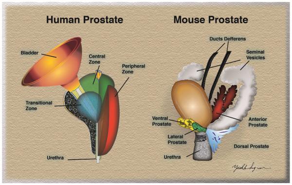
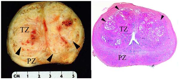
Prostate development follows a common pattern between species and depends on the actions of androgens to induce and support ductal branching morphogenesis of buds emerging from the urogenital sinus. The human prostate has a compact zonal anatomy immediately surrounding the urethra and below the urinary bladder. Rodents have a lobular prostate with lobes radiating away from the urethra. The human prostate is the site of benign hyperplasia, prostate cancer, and prostatitis. The rodent prostate has little naturally occurring disease. Rodents can be used to model aspects of human benign hyperplasia, but care should be taken in data interpretation and extrapolation to the human condition.
Keywords: BPH; LUTS; Prostate anatomy; Prostate embryology.
Copyright © 2016 Elsevier Inc. All rights reserved.
Figures
References
-
- Vesalius A, O'Malley CD, Calcar JSv, Saunders JBdCM. The illustrations from the works of Andreas Vesalius of Brussels : with annotations and translations, a discussion of the plates and their background, authorship and influence, and a biographical sketch of Vesalius. World Publishing Company; 1950.
-
- Geller J. Pathogenesis and medical treatment of benign prostatic hyperplasia. Prostate Suppliment. 1989;2:95–104. - PubMed
-
- Price D, Williams-Ashman H. The accessory reproductive glands of mammals. In: Young W, editor. Sex and internal secretions. 3 Vol. 1. Williams and Wilkins; Baltimore: 1961. pp. 366–448.
-
- Shapiro E, Hartanto V, Lepor H. Quantifying the smooth muscle content of the prostate using double-immunoenzymatic staining and color assisted image analysis. J. Urol. 1992;147:1167–1170. - PubMed
-
- Josso N. Physiology of Sex Differentiation. Pediat. Adolesc. Endocr. 1981;8:1–13.
Publication types
MeSH terms
Grants and funding
LinkOut - more resources
Full Text Sources
Other Literature Sources
Medical
