. 2016 Dec;229(6):871-891.
doi: 10.1111/joa.12526. Epub 2016 Aug 1.

Dynamic expression of the mouse orthologue of the human amyotropic lateral sclerosis associated gene C9orf72 during central nervous system development and neuronal differentiation

Affiliations

Dynamic expression of the mouse orthologue of the human amyotropic lateral sclerosis associated gene C9orf72 during central nervous system development and neuronal differentiation

Ross Ferguson et al. J Anat. 2016 Dec.

Abstract

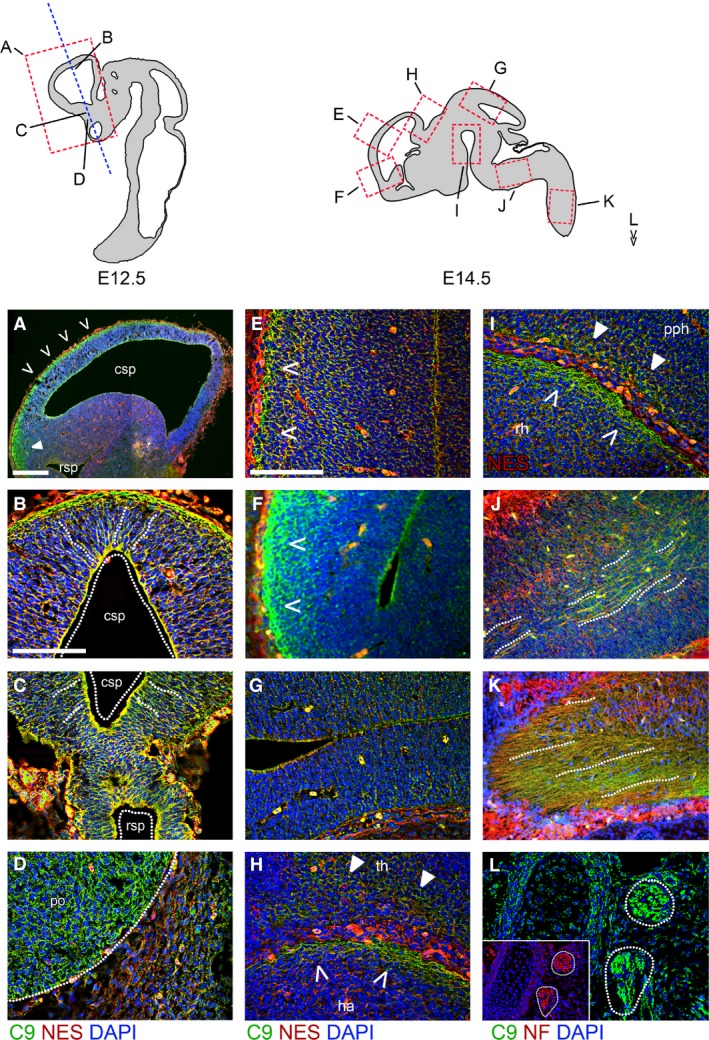
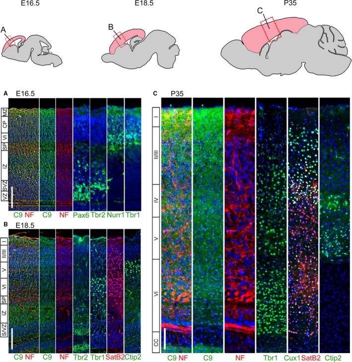
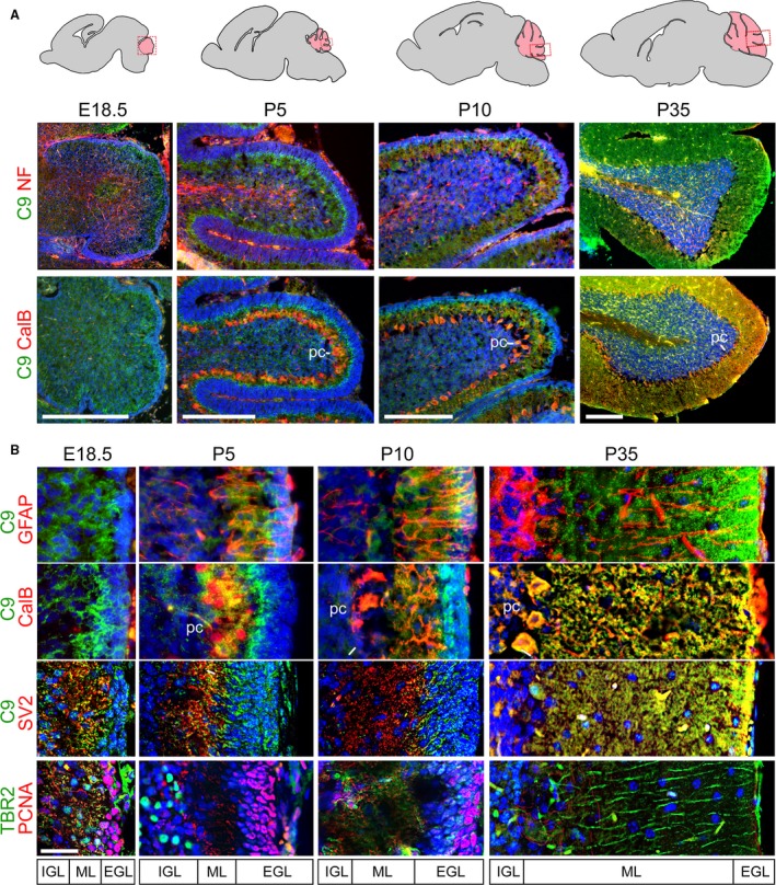
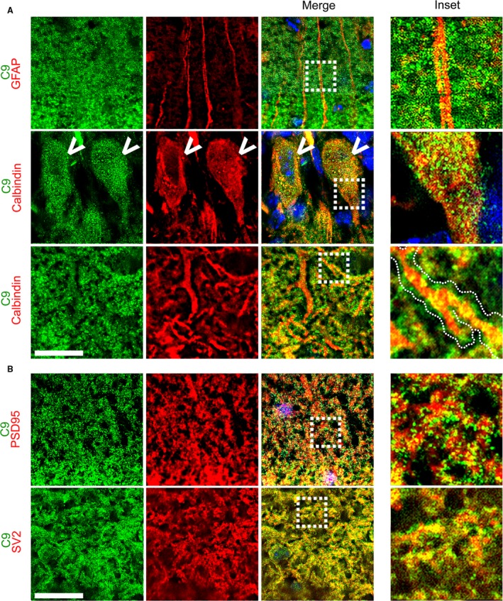
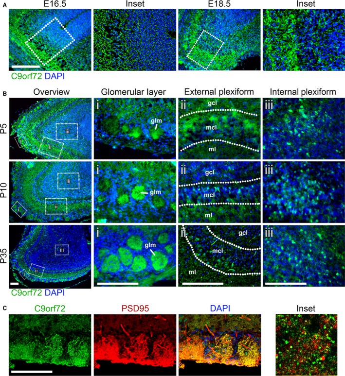
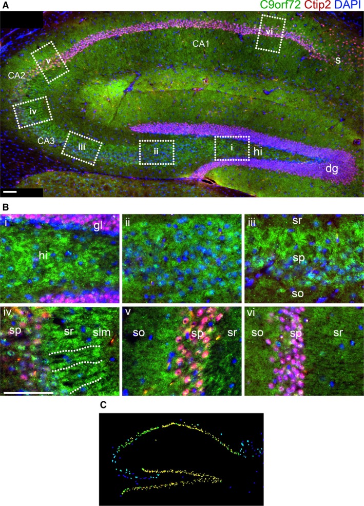
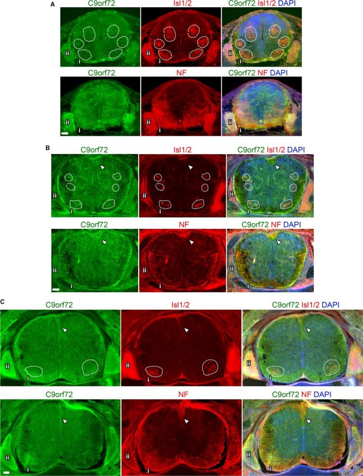
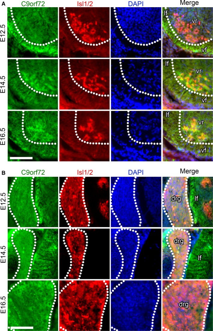
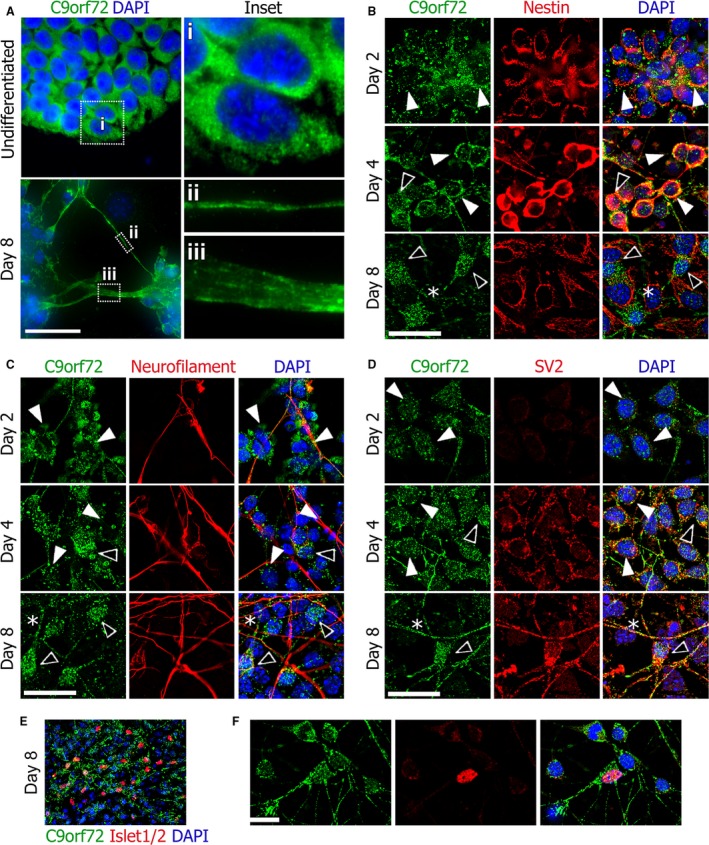
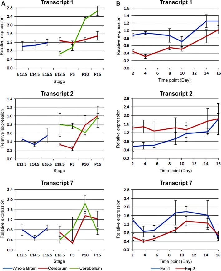
The hexanucleotide repeat in the first intron of the C9orf72 gene is the most significant cause of amyotropic lateral sclerosis as well as some forms of fronto-temporal dementia. The C9orf72 protein has been previously reported to be expressed in post-mortem human brain as well as in late embryonic and some postnatal stages in mice. Herein, we present a detailed study of the distribution of C9orf72 protein in the embryonic, postnatal and adult mouse brain, spinal cord as well as during the differentiation of P19 embryonal carcinoma cells to neurons including motor neurons. We show that the expression levels of the C9orf72 transcripts in the developing and adult mouse brain as well as in differentiating neurons, are dynamic. Besides the strong expression in the cerebellum and motor cortex reported previously, we show for the first time that C9orf72 is expressed strongly in the olfactory bulb and also in the hippocampus. Our immunostaining data also reveal a hitherto unreported switch in the cellular distribution of C9orf72 from a predominantly cytoplasmic to a nucleo-cytoplasmic distribution during corticogenesis. This switch in distribution was also observed during differentiation of the pluripotent embryonal carcinoma P19 cell line to mature neurons. Our findings have implications for interpreting the pathophysiology caused by the repeat expansions in C9orf72 in mouse models.

Keywords: C9orf72; brain development; expression; neuronal differentiation; spinal cord.


Figures

Figure 1
Figure 1
C9orf72 is present in the early embryonic brain and spinal cord. (A–D) Immunostaining for C9orf72 and nestin in E12.5 embryos in sagittal (A) and coronal (B–D) orientations. Strong C9orf72 staining can be seen in the dorsal pallium of the telecephalon (A), in the superficial layer of the rostral pallium (B) as well as in regions adjacent to the caudal secondary proencephalic ventricle (csp) in a radial arrangement spanning outwards (dotted lines). A similar pattern is seen in the caudal subpallium (C) adjacent to the rostral secondary proencephalic ventricle (rsp). C9orf72 is strongest in neuronal tissues, as shown by expression in the pre‐optic area (D, po) adjacent to non‐neuronal tissue. Immunostaining for C9orf72 and nestin (E–I) or neurofilament (J–L) in E14.5 brain. Expression is strongest in the superficial layers of the caudal rostral pallium (E) and pre‐olfactory pallium (F). This pattern of expression is not seen in the caudal midbrain tectum (G). Strong staining again is found in the superficial layer of the hippocampal allocortex (H, ha, open arrows) and spread in deeper layers of the adjacent thalamic tissue (th, closed arrows). Strong staining is also seen in the superficial layer of the rostral hypothalamus (rh) and prepontine hindbrain (pph) (I). C9orf72 can be found aligned with neurofilament‐positive tracts in the medulla (J) and spinal cord (K). C9orf72 and neurofilament are found together in the spinal ganglia (L). Scale bars: 100 μm.
Figure 2
Figure 2
Layer‐specific C9orf72 distribution changes in the developing and adult cortex. Immunostaining for C9orf72, neurofilament and layer specific markers in adjacent sections of the brain. At E16.5 (A) the cortex is divided into the Pax6+ ventricular zone (vz), Tbr2+ subventricular zone (svz), intermediate zone (iz), Nurr1+ subplate (SP), Tbr1+ layer VI/cortical plate (CP) and finally the marginal zone (mz) at the pial surface. C9orf72 expression appears throughout but is strongest in the marginal and intermediate zones. At E18.5 (B), further layers have formed in the cortical plate (layers II/IIVI, marked by SatB2 and Ctip2). Strong expression continues in the marginal zone and in a narrower region of the intermediate zone. By P35 all the layers of the cortex have formed (C) and the intermediate zone is absent. C9orf72 staining remains strongest in layer I at the pial surface and is also seen strongly in upper portions of layer II, throughout layer IV and the middle of layer VI. C9orf72 has also switched from a mainly cytoplasmic distribution at embryonic stages to a more uniform distribution, with staining seen throughout the cytoplasm and nucleus. Scale bars: 100 μm.
Figure 3
Figure 3
C9orf72 is highly expressed in primary cortical neurons in comparison with glia. Strong expression of C9orf72 is seen in the cell body and neurites of neurofilament‐positive cultured primary cortical neurons isolated at P0 (A). C9orf72 expression in GFAP‐positive glial cells is largely undetectable (A, arrows). C9orf72 expression is found consistently in all cortical neuron subtypes identified (B). Scale bars: 50 μm.
Figure 4
Figure 4
C9orf72 is strongly expressed in the molecular layer of the developing and adult cerebellum. Expression of C9orf72 in the developing cerebellum from E18.5 to P35 (A). C9orf72 staining is not seen in the neurofilament‐positive internal structures at any stage, but is found strongly at the inner edge of the developing external granule layer (EGL) at E18.5, which then goes onto to expand as the molecular layer (ML) expands, between the EGL and calbindin (CalB)‐positive Purkinje cell bodies (pc) adjacent to the forming inner granule layer (IGL). Scale bars: 500 μm. Throughout cerebellar development (B) C9orf72 expression in the molecular layer is not associated with Bergmann glia (GFAP) or proliferating granule cells (PCNA) but does associate with Purkinje dendrites (CalB) and synapses (SV2). C9orf72 appear strongest at the most superficial part of the molecular layer. Scale bars: 50 μm.
Figure 5
Figure 5
C9orf72 in the adult cerebellar molecular layer is pre‐synaptic. The molecular layer in the adult cerebellum (p35) shows no association of C9orf72 with Bergman glia fibres (GFAP), but clustered staining can be found directly adjacent to calbindin‐positive Purkinje dendrites (A, inset, dotted outline). C9orf72 expression can also be seen within the Purkinje cell body (arrow). C9orf72 shows no overlap with the post‐synaptic marker PSD95 but co‐localises with the synaptic vesicle marker SV2 (B). Scale bars: 20 μm.
Figure 6
Figure 6
C9orf72 is strongly expressed in the olfactory bulb. Between E16.5 and E18.5, expression is seen in the superficial layer of the olfactory bulb (A). Scale bars: 200 μm. In post‐natal mice from P5 onwards, C9orf72 expression is found in three distinct olfactory layers (B); the glomerular (i), internal (ii) and external plexiform areas (iii). Between P5 and P10, the distribution of C9orf72 in the mitral cell layer of the external plexiform area (mcl) changes from a predominantly cytoplasmic to one with increased nuclear staining. C9orf72 staining in the olfactory glomeruli (glm) becomes more distinct as the size and frequency of glomeruli increases with age. C9orf72 in the glomeruli (C) is strongly co‐incident with PSD95 but without overlap similar to the cerebellum. Scale bars: 250 μm.
Figure 7
Figure 7
C9orf72 in the hippocampus is found in areas associated with the mossy fibre tracts. An intense band of C9orf72 can be seen extending from the hilus (hi) of the dentate gyrus (dg) to the stratum radiatum (sr) of the CA3 field and part of CA2 (A). It does not appear as tracts between the granule layer (gl) and CA3sr (B, i–iii), but can be seen organised radially from the CA3 stratum pyramidale (sp) (B, iv). Further C9orf72 staining can be seen at the efferent stratum oriens (so) adjacent to the CA2 sp (B, v). C9orf72 staining is not as distinct in the CA1 or the subiculum (s) (B, vi). Allen Brain Atlas expression data (C) show strong expression in the dg but not CA3 granule layers. Scale bars: 100 μm.
Figure 8
Figure 8
Overview of C9orf72 expression in the developing spinal cord. Transverse sections of mid‐thoracic spinal cord from the indicated embryonic stages. Comparison of adjacent sections immunostained with either Isl1/2 or neurofilament (NF) and C9orf72, counterstained with DAPI at E12.5 (A), E14.5 (B) and E16.5 (C). Motor pools highlighted by dashed boxes. Ventral spinal cord (i) shown in detail in Fig. 8A and Supporting Information Fig. S4A. Dorsal root ganglion shown in detail in Figures 8B and S4B. Arrows indicate dorsal corticospinal tracts. Scale bars: 100 μm.
Figure 9
Figure 9
C9orf72 expression increases in ventral root motor‐neurons and dorsal root ganglion neurons during development. Transverse sections of mid‐thoracic region of indicated embryonic stages, stained with anti‐C9orf72 and and anti‐Islet1/2 (Isl1/2) antibodies, counterstained with DAPI. C9orf72 expression in the ventral spinal cord is strongest in the transverse tracts of the lateral (lf) and ventral funiculus (vf) at E12.5. Increased expression can be seen in the motor neuron pool in the ventral root (vr) from E14.5 to E16.5 (A). C9orf72 expression is also found in the dorsal root ganglia with expression increasing from E12.5 to E16.5. Scale bars: 100 μm.
Figure 10
Figure 10
Neurons differentiated from pluripotent P19 cell line including motor neurons show a nucleo‐cytoplasmic change in C9orf72 distribution during differentiation. Undifferentiated P19 cells show a predominantly cytoplasmic distribution of C9orf72 but after differentiation to neurons, distribution is more nuclear and C9orf72 can also be seen on the cell membranes in neurites (A). The early cytoplasmic distribution of C9orf72 is found in nestin‐positive neuronal precursors seen on days 2 and 4, whereas nestin‐negative mature neurons display increased nuclear C9orf72 (B). C9orf72 is found weakly within nestin‐positive processes (B, asterisk). Neurofilament expression follows nestin and, as expected, increased nuclear C9orf72 is seen in mature neurofilament‐positive neurons (C). C9orf72 expression also appears to be found at higher levels in neurofilament‐positive neurites (C, asterisk). The presence of synaptic vesicle marker SV2 indicates mature axons (D); the strongest C9orf72 staining is found in SV2‐positive neurites (D, asterisk). The motor neuron (MN) marker Islet1/2 expression can be seen on day 8 (E) and C9orf72 expression in MNs is similar to other neurons present in the culture (F). Scale bars: 25 μm.
Figure 11
Figure 11
C9orf72 expression levels increase throughout development and differentiation in the CNS. Expression levels of mC9ORF79 transcripts relative to Actb determined by qRTPCR. (A) C9orf72 expression in a developmental series of mouse brains; E12.5 head; E14.5 and E16.5 brains; E18.5 fore and mid/hind brain; P5, 10 and 15 cerebrum and cerebellum. RNA from four siblings pooled in duplicate. Levels of all three transcripts increase over development, particularly transcript 1 in the cerebellum. (B) A time course of C9orf72 expression in P19 embryonal carcinoma cells differentiated on matrigel as described in Materials and Methods at the indicated time points. As with transcripts in brain tissue, all three C9orf72 transcripts appear to increase steadily during differentiation. RNA from two independent experiments and all reactions performed in triplicate.

Similar articles

Cited by

References

    1. Arrant AE, Patel AR, Roberson ED (2015) Effects of exercise on progranulin levels and gliosis in progranulin‐insufficient mice. eNeuro 2, pii: 0061‐14,1‐12. - PMC - PubMed
    1. Atkinson RAK, Fernandez‐Martos CM, Atkin JD, et al. (2015) C9ORF72 expression and cellular localization over mouse development. Acta Neuropathol Commun 3, 59. - PMC - PubMed
    1. Bede P, Bokde ALW, Byrne S, et al. (2013) Multiparametric MRI study of ALS stratified for the C9orf72 genotype. Neurology 81, 361–369. - PMC - PubMed
    1. Bede P, Elamin M, Byrne S, et al. (2015) Patterns of cerebral and cerebellar white matter degeneration in ALS. J Neurol Neurosurg Psychiatry 86, 468–470. - PMC - PubMed
    1. van Blitterswijk M, DeJesus‐Hernandez M, Rademakers R (2012) How do C9ORF72 repeat expansions cause amyotrophic lateral sclerosis and frontotemporal dementia: can we learn from other noncoding repeat expansion disorders? Curr Opin Neurol 25, 689–700. - PMC - PubMed

MeSH terms