. 2016 Dec;229(6):778-790.
doi: 10.1111/joa.12527. Epub 2016 Aug 1.

Innervation of taste buds revealed with Brainbow-labeling in mouse

Affiliations

Innervation of taste buds revealed with Brainbow-labeling in mouse

Faisal N Zaidi et al. J Anat. 2016 Dec.

Abstract

Nerve fibers that surround and innervate the taste bud were visualized with inherent fluorescence using Brainbow transgenic mice that were generated by mating the founder line L with nestin-cre mice. Multicolor fluorescence revealed perigemmal fibers as branched within the non-taste epithelium and ending in clusters of multiple rounded swellings surrounding the taste pore. Brainbow-labeling also revealed the morphology and branching pattern of single intragemmal fibers. These taste bud fibers frequently innervated both the peripheral bud, where immature gemmal cells are located, and the central bud, where mature, differentiated cells are located. The fibers typically bore preterminal and terminal swellings, growth cones with filopodia, swellings, and rounded retraction bulbs. These results establish an anatomical substrate for taste nerve fibers to contact and remodel among receptor cells at all stages of their differentiation, an interpretation that was supported by staining with GAP-43, a marker for growing fibers and growth cones.

Keywords: geniculate ganglion; innervation; morphology; mouse; plasticity; taste bud.


Figures

Figure 1
Figure 1
Illustration of the regions within each bud section for which the degree of nerve fiber fluorescence was quantified (see data in Fig. 7). Numbers refer to locations around the bud as if viewing a superimposed clock: 12 o'clock at the taste pore, 3 and 9 o'clock at the bud sides, etc. Letters refer to subregions within the bud: central apical region (a, yellow); central basal region (b, cyan); peripheral regions (p1, red) and (p2, green). See Materials and methods for full description.
Figure 2
Figure 2
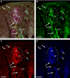
(A) Brainbow‐labeling of geniculate ganglion neurons demonstrating their multicoloration resulting from random recombination; they constitute a subset of all ganglion cells (unlabeled cells are not evident in the images). (B–D) Labeled ganglion cells viewed with each of the three fluorescence settings that are merged in (A): GFP/green (B); RFP/red (C); CFP/blue (D). Note that some cells express mainly green fluorescence (arrowheads), other cells express mainly red (thick arrows), and other cells express mainly blue (thin arrows). Additionally, some cells express significant mixtures of fluorescence: green + red (arrowhead + thick arrow); red + blue (thick arrow + thin arrow); green + blue (arrowhead + thin arrow); green + red + blue (asterisks). Thick labeled fibers in cross‐section, upper right, are axons of the motor facial nerve. Thin multicolored profiles between the labeled somata are fibers of the geniculate neurons. Scale bar: 10 μm.
Figure 3
Figure 3
Brainbow‐labeling of intragemmal and perigemmal innervation of a fungiform taste bud and papilla.demonstrating random multicoloration of the fibers. (A) Merged fluorescence illustrates perigemmal fibers and endings expressing predominantly GFP (green) (arrowheads), perigemmal fibers expressing predominantly CFP (blue) (thin arrows), and perigemmal endings expressing a mixture of green (GFP), blue (CFP), and red (RFP) (asterisk). Two intragemmal fibers expressing predominantly green fluorescence (double arrowheads) enter the base of the bud. One intragemmal fiber ends as a blunt ending in the bud periphery (triple arrowheads) and the other expands with filopodia at its ending in the central bud; the expansion expresses green and blue fluorescence (arrowhead + thin arrow) (compare the coloration of this structure in A, B, C, D). (B–D) Labeling viewed with each of the three fluorescence settings that are merged in (A). Scale bar: 10 μm.
Figure 4
Figure 4
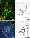
Brainbow‐labeled intragemmal and perigemmal innervation of a fungiform papilla, the taste bud of which also contains Brainbow‐fluorescent cells. (A) A thin intragemmal fiber that expresses green and red fluorescence only (double arrowheads + thick arrow) ascends the papillary core and enters the base of the taste bud. Fluorescent perigemmal fibers include a thick one ascending the papillary core and perigemmal endings, both expressing predominantly green and blue (arrowhead + thin arrow), and perigemmal fibers and endings expressing predominantly green and red (arrowhead + thick arrow). Two adjoined, spindle‐shaped Brainbow‐fluorescent gemmal cells with non‐fluorescent nuclei express green, red and blue in the central bud (asterisks). Immunostaining for KCNQ1 delineates the bud (pink) (dashed lines). (B–D) Labeling viewed with each of the three fluorescence settings that are merged in (A). Scale bar: 10 μm.
Figure 5
Figure 5
Intragemmal and perigemmal fibers labeled with Brainbow fluorescence in merged images and corresponding drawings (fiber drawings were arbitrarily colored digitally for clarity). (A) Thin intragemmal fibers (1,2) ascend to the taste bud and become expanded within the bud (dashed lines). Fiber 1 (see also drawing in B) is unbranched, bears preterminal swellings (arrowheads), and ends bluntly. Fiber 2 (see also drawing in B), by contrast, is irregularly shaped, branches widely in the basal half of the bud and bears thin terminal filaments and swellings (e.g. at arrowheads), one of which appears as a retraction bulb (small double arrows) below the bud. A thick perigemmal fiber (5) (see also drawing in B) ascends the papillary core and crosses over the intragemmal fibers to terminate as lumpy enlargements in the epithelium surrounding the bud. Red fluorescence within the bud appears to indicate faintly labeled gemmal cells. Small yellow fluorescent blobs (see text) are seen in and below the bud (inset: higher magnification). (C) Intragemmal fibers (3,4) ascending to the taste bud (dashed lines) become expanded as elaborately branched endings within the bud (see also drawings in D). Fiber 3 enters the central bud at its base, then branches abruptly, entering the peripheral bud before recurving centrally to end in the apical half of the bud; its changes of direction are marked by swellings (arrowheads). Fiber 4 thickens after entering the peripheral bud before trifurcating into apical and centrally directed branches; the branch point bears a large swelling (arrowhead). The endings of fiber 4 appear, morphologically, as growth cones (small arrows). Fibers 5 and 6 are perigemmal fibers; they bear lumpy endings in the non‐taste epithelium. Scale bars: 10 μm (3 μm for inset).
Figure 6
Figure 6
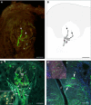
Brainbow‐labeling of intragemmal and perigemmal fibers associated with fungiform and circumvallate taste buds. (A) A single intragemmal fiber, seen in its entirety in the plane of this oblique section, exhibits shape irregularities immediately below and within the base of the bud (the basal lamina is indicated by dashed lines) where it branches to reach a broad region within the central bud. The endings are expanded (small arrows). (B) Drawing of the fiber in A. (C) Perigemmal fibers (arrows) ascend in the epithelium adjacent to the sides of the taste bud (TB), ending as rounded swellings in the non‐taste surface epithelium (asterisks). Intragemmal fibers (arrowheads) enter and terminate, often with swellings (small arrow) or ball‐on‐stalk endings (double small arrows) within the taste bud. Inset: small yellow/white fluorescent blobs (see text) are present in the central bud. (D) Vallate taste buds contain intragemmal fibers that reach all regions of each bud. The fibers are predominantly oriented vertically, toward the surface of the epithelium. They are branched and bear preterminal and terminal swellings. Preterminal gustatory fibers course parallel to the epithelial basal lamina and turn at right angles to enter the buds. Brainbow‐fluorescent gemmal cells (arrows) within vallate buds; the apical process of one terminates at the taste pore (P). Inset: There are few perigemmal fibers (asterisk). Scale bars: (A–C) 10 μm, (inset to A–C) 3 μm, (D) 20 μm, (inset to D) 40 μm.
Figure 7
Figure 7
Degree of fluorescence‐nerve fiber labeling within the subregions of the fungiform taste bud (a = apical region, b = basal region, p1 and p2 = peripheral regions) (see Fig. 1). The central basal region (b) has the highest percentage of total bud fluorescence (36.9 ± 15.4%), and the central apical region (a) has an intermediate percentage (27.6 ± 15.3%). Left and right peripheral regions (p1 and p2) contain the least amount of labeling (21.3 ± 15.0%, 16.1 ± 13.7 SD). Total central (a + b) labeling (64.5 ± 17.3 SD) is ~ twofold higher than total peripheral (p1 + p2; 35.5 ± 17.3% SD) labeling (P < 0.0001).
Figure 8
Figure 8
(A) GAP‐43 immunoreactivity in developing and mature fungiform taste buds. GAP‐43 associated with fibers is present subepithelially, below the taste bud, in the fungiform core, between the base of the bud and the muscle layer, and within the basal taste bud proper (arrow). (B) Intense GAP‐43 immunoreactivity in a developing fungiform taste bud in a 1‐day‐old mouse, associated with growing GG cell fibers, the timing and nature of which are established for the mouse (Ma et al. 2009). Scale bars: 20 μm. (C) Confocal micrograph demonstrating co‐localization of GAP‐43 and P2X2 immunoreactivity. GAP‐43 (green) entirely overlaps P2X2 nerve fiber staining (red) in this merged image (yellow). Immunoreactive double staining is particularly dense at the base of the fungiform papilla (boxed area). Double staining of punctate profiles is also present in the taste bud (TB) proper (arrowheads). Some fibers located subepithelially or ascending to the taste bud are single‐stained for P2X2 (arrows). Scale bars: 20 μm.

References

    1. Allegra Mascaro AL, Sacconi CP, Mandolesi GG, et al. (2013) In vivo single branch axotomy induces GAP‐43‐dependent sprouting and synaptic remodeling in cerebellar cortex. PNAS 110, 10824–10829. - PMC - PubMed
    1. Beidler LM, Smallman RL (1965) Renewal of cells within taste buds. J Cell Biol 27, 263–272. - PMC - PubMed
    1. Benowitz LI, Routtenberg A (1997) GAP‐43: an intrinsic determinant of neuronal development and plasticity. Trends Neurosci 20, 84–91. - PubMed
    1. Bo X, Alavi A, Xiang Z, Oglesby I, Ford A, Burnstock G (1999) Localization of ATP‐gated P2X2 and P2X3 receptor immunoreactive nerves in rat taste buds. Neuroreport 10, 1107–1111. - PubMed
    1. Bovolenta P, Mason C (1987) Growth cone morphology varies with position in the developing mouse visual pathway from retina to first targets. J Neurosci 7, 1447–1460. - PMC - PubMed

Publication types

LinkOut - more resources